Pneumocystis murina

Last updated

Pneumocystis murina
Scientific classification OOjs UI icon edit-ltr.svg
Domain: Eukaryota
Kingdom: Fungi
Division: Ascomycota
Class: Pneumocystidomycetes
Order: Pneumocystidales
Family: Pneumocystidaceae
Genus: Pneumocystis
Species:
P. murina
Binomial name
Pneumocystis murina
Keely, Fischer, Cushion & Stringer (2004)

Pneumocystis murina is a species of fungus, first isolated from laboratory mice, hence its name. [1] . Pneumocystis murina is a species of fungi belonging to the genus Pneumocystis , characterized by its unique cyst-like multinucleate morphology and association with the respiratory tract of mammals. [2] P. murina is particularly notable for its exclusive infection of laboratory mice and its role as an important model for studying human diseases, specifically Pneumocystis jirovecii infections in immunocompromised patients. [3] Pneumocystis murina was first phylogenetically identified and described as a distinct species by Keely, S. P., Fischer, J. M., Cushion, M. T., and Stringer, J. R. in 2012. [4]

Contents

Morphology

Pneumocystis murina is characterized by its cyst-like multinucleate form, which stains strongly with silver and is found alongside a uninucleate pleiomorphic 'trophic' form in the alveolar spaces of infected mammalian lungs. [2] In comparison to other mammalian strains, P. murina exhibits thinner and more abundant filopodia. [5]

Ecology

P. murina is a fungal pathogen that exclusively infects laboratory mice, causing the formation of cysts in the respiratory tract of immunocompromised hosts. [6] It is not found in nature and has not been reported in wild mice populations. [7]

Habitat

Pneumocystis murina inhabits the respiratory tract of laboratory mice, particularly the alveolar spaces within the lungs. [6]

Geographical Distribution

P. murina has no known geographical distribution, as it is exclusively found in laboratory settings. [7]

Unique Aspects of the Fungus

Pneumocystis murina is unique for several reasons. Firstly, it is only found in laboratory mice, not in natural environments. [7] Secondly, it is a pathogen that causes cyst formation in the respiratory tract of immunocompromised lab mice, leading to clinical signs of infection such as dyspnea, weight loss, hunched posture, and scaly skin. [6] P. murina is not edible or cultivatable, and it cannot be cultured in vitro. [8]

The fungus is transmitted between mice via inhalation or through the shedding of P. murina from one host to another. It serves as an important model for studying human diseases, particularly Pneumocystis jirovecii infections, which are a significant cause of morbidity and mortality in immunocompromised individuals, such as those with HIV/AIDS or patients receiving immunosuppressive therapies. [3]

The unique aspects of Pneumocystis murina make it an invaluable model for studying the human pathogen Pneumocystis jirovecii, as both pathogens share similar biological and molecular characteristics. [3] Research on P. murina has contributed to the development of novel treatment and prevention strategies for P. jirovecii infections in immunocompromised patients. [9]

One of the critical challenges in studying P. murina and P. jirovecii is their inability to be cultured in vitro, which has hindered the understanding of their life cycles and the development of targeted therapies. [8] However, recent advances in molecular biology and imaging techniques have allowed researchers to gain more insight into the biology and pathogenesis of these organisms. [4]

P. murina infections in laboratory mice serve as an essential tool for understanding the host-pathogen interaction, immune response, and potential treatments for P. jirovecii pneumonia (PJP) in humans. [10] As the incidence of PJP continues to rise due to the increasing number of immunocompromised individuals, the need for effective treatment and prevention strategies is paramount. [11] Research on P. murina and its unique characteristics holds great promise for addressing this significant public health challenge.

Related Research Articles

<span class="mw-page-title-main">Opportunistic infection</span> Infection caused by pathogens that take advantage of an opportunity not normally available

An opportunistic infection is an infection caused by pathogens that take advantage of an opportunity not normally available. These opportunities can stem from a variety of sources, such as a weakened immune system, an altered microbiome, or breached integumentary barriers. Many of these pathogens do not necessarily cause disease in a healthy host that has a non-compromised immune system, and can, in some cases, act as commensals until the balance of the immune system is disrupted. Opportunistic infections can also be attributed to pathogens which cause mild illness in healthy individuals but lead to more serious illness when given the opportunity to take advantage of an immunocompromised host.

AIDS-defining clinical conditions is the list of diseases published by the Centers for Disease Control and Prevention (CDC) that are associated with AIDS and used worldwide as a guideline for AIDS diagnosis. CDC exclusively uses the term AIDS-defining clinical conditions, but the other terms remain in common use.

<span class="mw-page-title-main">Atovaquone</span> Antimicrobial and antiprotozoan drug

Atovaquone, sold under the brand name Mepron, is an antimicrobial medication for the prevention and treatment of Pneumocystis jirovecii pneumonia (PCP).

<i>Stenotrophomonas maltophilia</i> Species of bacterium

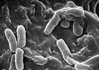
Stenotrophomonas maltophilia is an aerobic, nonfermentative, Gram-negative bacterium. It is an uncommon bacterium and human infection is difficult to treat. Initially classified as Bacterium bookeri, then renamed Pseudomonas maltophilia, S. maltophilia was also grouped in the genus Xanthomonas before eventually becoming the type species of the genus Stenotrophomonas in 1993.

<span class="mw-page-title-main">Sputum culture</span> Medical test to detect & identify bacteria or fungi in lung airways

A sputum culture is a test to detect and identify bacteria or fungi that infect the lungs or breathing passages. Sputum is a thick fluid produced in the lungs and in the adjacent airways. Normally, fresh morning sample is preferred for the bacteriological examination of sputum. A sample of sputum is collected in a sterile, wide-mouthed, dry, leak-proof and break-resistant plastic-container and sent to the laboratory for testing. Sampling may be performed by sputum being expectorated, induced, or taken via an endotracheal tube with a protected specimen brush in an intensive care setting. For selected organisms such as Cytomegalovirus or "Pneumocystis jiroveci" in specific clinical settings a bronchoalveolar lavage might be taken by an experienced pneumologist. If no bacteria or fungi grow, the culture is negative. If organisms that can cause the infection grow, the culture is positive. The type of bacterium or fungus is identified by microscopy, colony morphology and biochemical tests of bacterial growth.

<span class="mw-page-title-main">Pneumocystosis</span> Medical condition

Pneumocystosis is a fungal infection that most often presents as Pneumocystis pneumonia in people with HIV/AIDS or poor immunity. It usually causes cough, difficulty breathing and fever, and can lead to respiratory failure. Involvement outside the lungs is rare but, can occur as a disseminated type affecting lymph nodes, spleen, liver, bone marrow, eyes, kidneys, thyroid, gastrointestinal tract or other organs. If occurring in the skin, it usually presents as nodular growths in the ear canals or underarms.

Pneumococcal pneumonia is a type of bacterial pneumonia that is caused by Streptococcus pneumoniae (pneumococcus). It is the most common bacterial pneumonia found in adults, the most common type of community-acquired pneumonia, and one of the common types of pneumococcal infection. The estimated number of Americans with pneumococcal pneumonia is 900,000 annually, with almost 400,000 cases hospitalized and fatalities accounting for 5-7% of these cases.

Otto Jírovec was a Czechoslovak professor of parasitology and protozoology.

Chlamydia felis is a Gram-negative, obligate intracellular bacterial pathogen that infects cats. It is endemic among domestic cats worldwide, primarily causing inflammation of feline conjunctiva, rhinitis and respiratory problems. C. felis can be recovered from the stomach and reproductive tract. Zoonotic infection of humans with C. felis has been reported. Strains FP Pring and FP Cello have an extrachromosomal plasmid, whereas the FP Baker strain does not. FP Cello produces lethal disease in mice, whereas the FP Baker does not. An attenuated FP Baker strain, and an attenuated 905 strain, are used as live vaccines for cats.

<span class="mw-page-title-main">Hospital-acquired pneumonia</span>

Hospital-acquired pneumonia (HAP) or nosocomial pneumonia refers to any pneumonia contracted by a patient in a hospital at least 48–72 hours after being admitted. It is thus distinguished from community-acquired pneumonia. It is usually caused by a bacterial infection, rather than a virus.

<i>Pneumocystis jirovecii</i> Species of fungus

Pneumocystis jirovecii is a yeast-like fungus of the genus Pneumocystis. The causative organism of Pneumocystis pneumonia, it is an important human pathogen, particularly among immunocompromised hosts. Prior to its discovery as a human-specific pathogen, P. jirovecii was known as P. carinii.

<i>Pneumocystis</i> pneumonia Medical condition

Pneumocystis pneumonia (PCP), also known as Pneumocystis jirovecii pneumonia (PJP), is a form of pneumonia that is caused by the yeast-like fungus Pneumocystis jirovecii.

<i>Enterobacter cloacae</i> Species of bacterium

Enterobacter cloacae is a clinically significant Gram-negative, facultatively-anaerobic, rod-shaped bacterium.

<span class="mw-page-title-main">Pathogenic bacteria</span> Disease-causing bacteria

Pathogenic bacteria are bacteria that can cause disease. This article focuses on the bacteria that are pathogenic to humans. Most species of bacteria are harmless and are often beneficial but others can cause infectious diseases. The number of these pathogenic species in humans is estimated to be fewer than a hundred. By contrast, several thousand species are part of the gut flora present in the digestive tract.

Pathogenic fungi are fungi that cause disease in humans or other organisms. Although fungi are eukaryotic, many pathogenic fungi are microorganisms. Approximately 300 fungi are known to be pathogenic to humans; their study is called "medical mycology". Fungal infections kill more people than either tuberculosis or malaria—about 2 million people per year.

<i>Pseudomonas</i> infection Medical condition

Pseudomonas infection refers to a disease caused by one of the species of the genus Pseudomonas.

Antonio Carini (1872–1950) was an Italian physician, bacteriologist and professor. He worked in the public health services of São Paulo, Brazil for over forty years. Carini showed that rabies of herbivores could be transmitted by bats, and discovered a parasitic fungus, which causes pneumocystosis.

<span class="mw-page-title-main">Classification of pneumonia</span> Medical condition

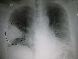
Pneumonia can be classified in several ways, most commonly by where it was acquired, but may also by the area of lung affected or by the causative organism. There is also a combined clinical classification, which combines factors such as age, risk factors for certain microorganisms, the presence of underlying lung disease or systemic disease and whether the person has recently been hospitalized.

<span class="mw-page-title-main">T cell deficiency</span> Medical condition

T cell deficiency is a deficiency of T cells, caused by decreased function of individual T cells, it causes an immunodeficiency of cell-mediated immunity. T cells normal function is to help with the human body's immunity, they are one of the two primary types of lymphocytes(the other being B cells).

Pneumocystis wakefieldiae is a parasitic fungus isolated from rats.

References

  1. Keely, S. P. (2004). "Phylogenetic identification of Pneumocystis murina sp. nov., a new species in laboratory mice". Microbiology. 150 (5): 1153–1165. doi: 10.1099/mic.0.26921-0 . ISSN   1350-0872. PMID   15133075.
  2. 1 2 Keely, S. P., Stringer, J. R., Baughman, R. P., Linke, M. J., Walzer, P. D., & Smulian, A. G. (2005). Genetic variation among Pneumocystis carinii hominis isolates in recurrent pneumocystosis. The Journal of infectious diseases, 192(7), 1261-1269.
  3. 1 2 3 Cushion, M. T., & Stringer, J. R. (2010). Stealth and opportunism: alternative lifestyles of species in the fungal genus Pneumocystis. Annual Review of Microbiology, 64, 431-452.
  4. 1 2 Keely, S. P., Fischer, J. M., Cushion, M. T., & Stringer, J. R. (2012). Phylogenetic identification of Pneumocystis murina sp. nov., a new species in laboratory mice. Microbiology, 158(Pt 10), 2457-2466.
  5. Cushion, M. T., Walzer, P. D., & Collins, M. S. (1990). Unique ultrastructure of Pneumocystis carinii. Journal of Protozoology, 37(1), 25-31.
  6. 1 2 3 Gigliotti, F., & Wright, T. W. (2012). Pneumocystis: where does it live? PLoS Pathogens, 8(11), e1003025.
  7. 1 2 3 Cushion, M. T., & Linke, M. J. (2010). Pneumocystis: unraveling the cloak of obscurity. Trends in Microbiology, 18(6), 243-251.
  8. 1 2 Cushion, M. T. (2010). Are members of the fungal genus Pneumocystis (a) commensals; (b) opportunists; (c) pathogens; or (d) all of the above? PLoS Pathogens, 6(9), e1001009.
  9. McAllister, F., Steele, C., Zheng, M., Young, E., Shellito, J. E., Marrero, L., & Kolls, J. K. (2004). T cytotoxic-1 CD8+ T cells are effector cells against Pneumocystis in mice. The Journal of Immunology, 172(2), 1132-1138.
  10. Roths, J. B., Marshall, J. D., Allen, R. D., Carlson, G. A., & Sidman, C. L. (1991). Spontaneous Pneumocystis carinii pneumonia in immunodeficient mutant scid mice. Natural history and pathobiology. American Journal of Pathology, 138(6), 1379-1387.
  11. Thomas, C. F. Jr., & Limper,A. H. (2004). Pneumocystis pneumonia. New England Journal of Medicine, 350(24), 2487-2498.


Further reading