Related Research Articles

The hair follicle is an organ found in mammalian skin. It resides in the dermal layer of the skin and is made up of 20 different cell types, each with distinct functions. The hair follicle regulates hair growth via a complex interaction between hormones, neuropeptides, and immune cells. This complex interaction induces the hair follicle to produce different types of hair as seen on different parts of the body. For example, terminal hairs grow on the scalp and lanugo hairs are seen covering the bodies of fetuses in the uterus and in some newborn babies. The process of hair growth occurs in distinct sequential stages. The first stage is called anagen and is the active growth phase, telogen is the resting stage, catagen is the regression of the hair follicle phase, exogen is the active shedding of hair phase and lastly kenogen is the phase between the empty hair follicle and the growth of new hair.

Hyperkeratosis is thickening of the stratum corneum, often associated with the presence of an abnormal quantity of keratin, and also usually accompanied by an increase in the granular layer. As the corneum layer normally varies greatly in thickness in different sites, some experience is needed to assess minor degrees of hyperkeratosis.

The arrector pili muscles, also knows as hair erector muscles, are small muscles attached to hair follicles in mammals. Contraction of these muscles causes the hairs to stand on end, known colloquially as goose bumps (piloerection).

Hair transplantation is a surgical technique that removes hair follicles from one part of the body, called the 'donor site', to a bald or balding part of the body known as the 'recipient site'. The technique is primarily used to treat male pattern baldness. In this minimally invasive procedure, grafts containing hair follicles that are genetically resistant to balding are transplanted to the bald scalp. Hair transplantation can also be used to restore eyelashes, eyebrows, beard hair, chest hair, pubic hair and to fill in scars caused by accidents or surgery such as face-lifts and previous hair transplants. Hair transplantation differs from skin grafting in that grafts contain almost all of the epidermis and dermis surrounding the hair follicle, and many tiny grafts are transplanted rather than a single strip of skin.
Follicular dysplasia is a genetic disease of dogs causing alopecia, or hair loss. It is caused by hair follicles that are misfunctioning due to structural abnormality. There are several types, some affecting only certain breeds. Diagnosis is achieved through a biopsy, and treatment is rarely successful. Certain breeds, such as the Mexican Hairless Dog and Chinese Crested Dog, are bred specifically for alopecia.
Lichen spinulosus is a rare skin disorder characterized by follicular keratotic papules that are grouped into large patches. It is a variant of keratosis pilaris named for its resemblance to a patch of lichen.
Central centrifugal cicatricial alopecia (CCCA), is a type of alopecia first noticed in African Americans in the 1950s and reported by LoPresti et al. in 1968 as a result of application of petrolatum followed by a stove-heated iron comb. The original theory was that the hot petrolatum would travel down to the hair root, burn the follicle, and after repetitive injury scarring would result. Later CCCA was realized to affect men and women without a history significant for use of such styling techniques. Consequently, the terms "follicular degeneration syndrome" per Sperling and Sau in 1992 and then CCCA per Olsent et al. in 2003 were evolved. Plausible contributing factors may include other African-American styling techniques such as relaxers, tight braids, heavy extensions, certain oils, gels or pomades.
Pruritic folliculitis of pregnancy is a dermatosis of pregnancy characterized by small follicular pustules scattered widely over the trunk, appearing during the second or third trimester, and resolving by 2 or 3 weeks after delivery.
Perforating folliculitis is a skin condition in humans characterized by discrete follicular keratotic eruptions involving mainly the hairy parts of the extremities.
Acne necrotica presents with a primary lesion that is a pruritic or painful erythematous follicular-based papule that develops central necrosis and crusting and heals with a varioliform scar.
Keratosis pilaris atrophicans faciei begins in infancy as follicular papules with perifollicular erythema. Initially, the lesions are restricted to the lateral eyebrows, but with time spread to involve the cheeks and forehead, and may also be associated with keratosis pilaris on the extremities and buttocks.
Atrophodermia vermiculata presents with erythematous follicular papules on the cheeks in childhood and, with time, the lesions develop into pit-like depressions.
Disseminate and recurrent infundibulofolliculitis, also called disseminate and recurrent infundibular folliculitis or Hitch and Lund disease, is a rare follicular skin condition that presents with irregularly shaped papules pierced by hair, is mildly itchy at times, and is chronic with recurrent exacerbations.
Traumatic anserine folliculosis is a curious gooseflesh-like follicular hyperkeratosis that may result from persistent pressure and lateral friction of one skin surface against another.
Alopecia mucinosa is a skin disorder that generally presents, but not exclusively, as erythematous plaques or flat patches without hair primarily on the scalp, neck and face. This can also be present on the body as a follicular mucinosis and may represent a systemic disease.
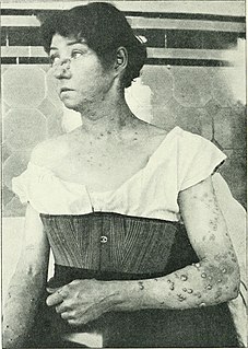
Iododermas are caused by iodides, with the most common sources of exposure being oral and intravenous contrast materials used to treat thyroid disease. The most common type of eruption is an acneiform eruption with numerous acutely inflamed follicular pustules, each surrounded by a ring of hyperemia.

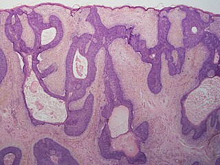
Warty dyskeratoma, also known as an Isolated dyskeratosis follicularis, is a benign epidermal proliferation with distinctive histologic findings that may mimic invasive squamous cell carcinoma and commonly manifests as an umbilicated lesion with a keratotic plug, WD have some histopathologic similarities to viral warts but it's not caused by HPV and the majority of these lesions display overall histopathologic features consistent with a follicular adnexal neoplasm. Usually limited to the head, neck, scalp or face and vulva. Lesions are generally solitary and sporadic and may be associated with a follicular unit. Oral involvement, particularly the hard palate, and genital involvement have been reported. it can also be thought of as one of the manifestations of focal acantholytic dyskeratosis, an epidermal reaction pattern that can be seen in several disorders, including Darier's disease and Grover's disease. But the main Difference between Darier disease and Warty dyskeratoma, is that Darier disease inherited dermatosis consisting of multiple keratotic papules on the face, trunk, and extremities, while WD occurs as an isolated, noninherited, single keratotic nodule mainly confined to the head and neck as mentioned earlier.
Follicular atrophoderma is a skin condition consisting of follicular indentations without hairs, notably occurring on extensor surfaces of the hands, legs and arms.
A basaloid follicular hamartoma is a cutaneous condition characterized as distinctive benign adnexal tumor that has several described variants.

Isthmicoma are a cutaneous condition characterized by flat, keratotic papules of the head and neck, skin lesions that are usually solitary.
References
- ↑ James, William D.; Berger, Timothy G.; et al. (2006). Andrews' Diseases of the Skin: clinical Dermatology. Saunders Elsevier. ISBN 0-7216-2921-0.