Ribeiroia ondatrae

Last updated

Ribeiroia ondatrae
Scientific classification OOjs UI icon edit-ltr.svg
Domain: Eukaryota
Kingdom: Animalia
Phylum: Platyhelminthes
Class: Trematoda
Order: Plagiorchiida
Family: Psilostomatidae
Genus: Ribeiroia
Species:
R. ondatrae
Binomial name
Ribeiroia ondatrae
(Price, 1931)
Synonyms
  • Psilostomum ondatraePrice, 1931
  • Cercaria thomasiMcMullen, 1938
  • Ribeiroia insignisTravassos, 1939
  • Pseudopsilostoma ondatraeYamaguti, 1958
  • Ribeiroia thomasiYamaguti, 1958

Ribeiroia ondatrae, or the frog-mutating flatworm is a parasite in the genus Ribeiroia which is believed to be responsible for many of the recent increases in amphibian limb malformations, particularly missing, malformed, and additional hind legs.

Contents

It was first reported from livers of Ondatra , hence its specific name. [1] In recent studies, it was found that in areas infected with R. ondatrae, the population of amphibian limb malformations was much higher than populations in which this trematode was not present. Each species studied showed varying results. For example, amphibians of species Pseudacris regilla , Rana aurora and Taricha torosa were found to physically display a higher frequency in the number of abnormalities. [2]

The exact mechanism of deformation has not been determined but it has been theorized that deformation results from mechanical disruption of the cells involved in limb bud formation during the amphibian larval stage.

Life cycle

R. ondatrae is a digenetic trematode that occurs in lentic aquatic environments and has a complex life cycle that requires the infection of three different host species. The life cycle commences once an avian or mammalian host, containing the adult worms, releases eggs contained in feces into lakes, farm ponds, or streams. It takes around 2-3 weeks for the fertilized eggs to develop. The newly hatched miracidia will swim rapidly to search for its primary intermediate host (Planorbella trivolvis). [3]

The first intermediate host is the ram's horn snail. The second intermediate hosts are fish and larval amphibians including frogs and salamanders. For second intermediate hosts, the free-swimming cercariae will begin to locate a suitable area for encystment where cercarial bodies will enclose themselves within cyst walls (metacercariae). These cysts are freed when the definitive host consumes the second intermediate host, and they will begin to migrate up the ileum and attach to the mucosa of the definitive host for sexual maturity. [3] Inside amphibians, cercariae are attracted to limb bud regions where the hind limbs form. As a result, large numbers of metacercariae encyst near the base of the hind legs. The definitive hosts are predators such as hawks, herons, ducks, and badgers.

The abundance of the teratogenic trematode Ribeiroia ondatre has been found to increase in eutrophic (nutrient rich) waters.

Enhanced transmission

An important factor to the R. ondatrae infections is the exposure to run off nutrients, i.e. eutrophication. Fertilizers have phosphates in them which is also a predictor of larval trematode abundance in amphibians. The herbicide atrazine has proven to weaken amphibians’ immune systems which causes frogs to become more prone to R. ondatrae infections which in turn causes predators such as birds to attack the multiple or missing limbed frogs. Since herbicides and pesticides affect the prevalence of R. ondatrae in frogs, they tend to increase mortality and pathology due to extra or missing limbs. [4]

Morphology of cercariae (species identification)

The cercariae body (tail tip to oral sucker) of R. ondatrae measures to be 800 µm long and 200 µm wide and is truncated posteriorly and tapered anteriorly. Like Trifolium spp., the defining morphological structure of R. ondatrae is the appearance of two laterally projecting esophageal diverticula, which are located halfway along the length of the esophagus. Aside from these features, the cercariae possess a pair of main excretory collecting tubes that emerge from the excretory bladder medially, running laterally and anteriorly. Also, these tubes are filled with large refractive granules (approximately 150). Another feature is the presence of a rose-colored organ or pink colored tissue that resides between the oral sucker and pharynx of freshly shed living specimens. However, this detail should be used cautiously because not all R. ondatrae cercariae have this feature and the color visibility along with intensity can vary. [3]

Location of infection/possible mechanisms

R. ondatrae’s mechanism of causing malformations is still unknown, but there seems to be evidence in which areas of the body it infects more. Studies show that when frogs or toads are affected with R. ondatrae it seems that the most common spot for any deformity is on the hind limbs. These malformations are characterized by the following: skin webbings, missing digits/limbs, cutaneous fusions and bony bridges. The general thought is that it is believed that the forelimb is not as likely to be malformed as the hind limbs, which develop in unceasing proximity to the environment. The amount of exposure to R. ondatrae cercariae appears to determine where a deformity will occur. For instance, a moderate amount of R. ondatrae can affect the forelimbs of amphibians, but a heavy parasite load does not affect the forelimbs and only causes deformities in hind limbs. [5]

There are 2 proposals for the mechanism of how R. ondatrae metacercariae cause malformations. The first explanation for these limb deformities proposes that abnormal cellular outgrowth is caused by mechanical disruption of the spatial organization of cells that stems from metacercariae that encyst within tissues of the developing limb buds. The second proposal for the mechanism that serves as an alternative to the parasite-induced mechanical perturbation is that the malformations are directly caused by the parasite itself. The parasite's production/release of growth factors act as a source of confusion for the molecular signals directing limb development. [3]

Species infected

Related Research Articles

<span class="mw-page-title-main">Trematoda</span> Class of parasitic flatworms

Trematoda is a class of flatworms known as flukes or trematodes. They are obligate internal parasites with a complex life cycle requiring at least two hosts. The intermediate host, in which asexual reproduction occurs, is usually a snail. The definitive host, where the flukes sexually reproduce, is a vertebrate. Infection by trematodes can cause disease in all five traditional vertebrate classes: mammals, birds, amphibians, reptiles, and fish.

<span class="mw-page-title-main">Decline in amphibian populations</span> Ongoing mass extinction of amphibian species worldwide

Since the 1980s, decreases in amphibian populations, including population decline and localized mass extinctions, have been observed in locations all over the world. These declines are known as one of the most critical threats to global biodiversity.

<span class="mw-page-title-main">Trematode life cycle stages</span>

Trematodes are parasitic flatworms of the class Trematoda, specifically parasitic flukes with two suckers: one ventral and the other oral. Trematodes are covered by a tegument, that protects the organism from the environment by providing secretory and absorptive functions.

Ribeiroia is a genus of trematode parasites that sequentially infect freshwater snails in the family Planorbidae as first intermediate hosts, fish and larval amphibians as second intermediate hosts, and birds and mammals as definitive hosts. In North America, infection by Ribeiroia has been linked to amphibians with limb malformations. The connection between parasitic infection and limb malformations has generated questions about (a) whether parasite-induced malformations in amphibians are increasing, and (b) the consequences of such abnormalities for amphibian population conservation.

<i>Paragonimus westermani</i> Species of fluke

Paragonimus westermani is the most common species of lung fluke that infects humans, causing paragonimiasis. Human infections are most common in eastern Asia and in South America. Paragonimiasis may present as a sub-acute to chronic inflammatory disease of the lung. It was discovered by Coenraad Kerbert (1849–1927) in 1878.

<i>Echinostoma</i> Genus of flukes

Echinostoma is a genus of trematodes (flukes), which can infect both humans and other animals. These intestinal flukes have a three-host life cycle with snails or other aquatic organisms as intermediate hosts, and a variety of animals, including humans, as their definitive hosts.

<span class="mw-page-title-main">Paragonimiasis</span> Medical condition

Paragonimiasis is a food-borne parasitic disease caused by several species of lung flukes belonging to genus Paragonimus. Infection is acquired by eating crustaceans such as crabs and crayfishes which host the infective forms called metacercariae, or by eating raw or undercooked meat of mammals harboring the metacercariae from crustaceans.

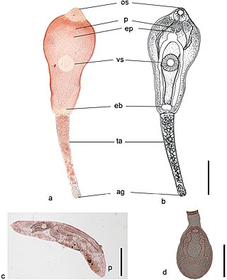
Metagonimoides oregonensis is a trematode, or fluke worm, in the family Heterophyidae. This North American parasite is found primarily in the intestines of raccoons, American minks, frogs in the genus Rana, and freshwater snails in the genus Goniobasis. It was first described in 1931 by E. W. Price. The parasite has a large distribution, from Oregon to North Carolina. Adult flukes vary in host range and morphology dependent on the geographical location. This results in different life cycles, as well as intermediate hosts, across the United States. On the west coast, the intermediate host is freshwater snails (Goniobasis), while on the east coast the intermediate host is salamanders (Desmognathus). The parasites on the west coast are generally much larger than on the east coast. For example, the pharynx as well as the body of the parasite are distinctly larger in Oregon than in North Carolina. The reverse pattern is observed on the east coast for uterine eggs, which are larger on the west coast. In snails, there is also a higher rate of infection in female snails than in males. Research on the life history traits of the parasites have been performed with hamsters and frogs as model species.

<i>Fasciolopsis</i> Genus of flukes

Fasciolopsis is a genus of trematodes. They are also known as giant intestinal flukes.

<i>Nanophyetus</i> Genus of flukes

Nanophyetus salmincola is a food-borne intestinal trematode parasite prevalent on the Pacific Northwest coast. The species may be the most common trematode endemic to the United States.

<i>Bucephalus polymorphus</i> Species of fluke

Bucephalus polymorphus is a type of flatworm. This species is within the Bucephalidae family of Digenea, which in turn is a subclass of Trematodes within the phylum Platyhelminthes. It is characterized by having a mouth near the middle of its body, along with a sac-like gut. The mouth opening is located in the centre of the ventral surface. This is a specific body type of cecaria known as a gastrostome.

Halipegus eccentricus is a monoecious, digenea parasitic trematode commonly found in true frogs in North America. It was first described in 1939.

<i>Echinostoma revolutum</i> Species of fluke

Echinostoma revolutum is a trematode parasites, of which the adults can infect birds and mammals, including humans. In humans, it causes echinostomiasis.

<i>Telogaster</i> Genus of flukes

Telogaster opisthorchis is an endoparasite in the class Trematoda within the phylum Platyhelminthes. This fluke is known for causing tumor like malformations in fishes by attaching onto its spinal region in the metacercariae form. Malformations cause fish to become more susceptible to fish eating predators allowing T. opisthorchis to continue with its lifecycle.

<i>Clinostomum marginatum</i> Species of fluke

Clinostomum marginatum is a species of parasitic fluke. It is commonly called the "yellow grub". It is found in many freshwater fish in North America, and no fish so far is immune to this parasite. It is also found in frogs. Clinostomum marginatum can also be found in the mouth of aquatic birds such as herons and egrets. They are commonly present in the esophagus of fish-eating birds and reptiles. Eggs of these trematodes are shed in the feces of aquatic birds and released into water. Aquatic birds become hosts of this parasite by ingesting infected freshwater fish. The metacercariae are found right beneath the skin or in the muscles of host fish.

Megalodiscus temperatus is a Digenean in the phylum Platyhelminthes. This parasite belongs to the Cladorchiidae family and is a common parasite located in the urinary bladder and rectum of frogs. The primary host is frogs and the intermediate hosts of Megalodiscus temeperatus are freshwater snails in the genus Helisoma.

<i>Philophthalmus gralli</i> Species of fluke

Philophthalmus gralli, commonly known as the Oriental avian eye fluke, parasitises the conjunctival sac of the eyes of many species of birds, including birds of the orders Galliformes and Anseriformes. In Brazil this parasite was reported in native Anseriformes species. It was first discovered by Mathis and Leger in 1910 in domestic chickens from Hanoi, Vietnam. Birds are definitive hosts and freshwater snail species are intermediate hosts. Human cases of philophthalmosis are rare, but have been previously reported in Europe, Asia, and America.

Philophthalmus lacrimosus is a species of trematodes in the family Philophthalmidae.

<i>Metagonimus yokogawai</i> Species of fluke

Metagonimus yokogawai, or the Yokogawa fluke, is a species of a trematode, or fluke worm, in the family Heterophyidae.

<span class="mw-page-title-main">Gastropod-borne parasitic disease</span> Medical condition

Gastropod-borne parasitic diseases (GPDs) are a group of infectious diseases that require a gastropod species to serve as an intermediate host for a parasitic organism that can infect humans upon ingesting the parasite or coming into contact with contaminated water sources. These diseases can cause a range of symptoms, from mild discomfort to severe, life-threatening conditions, with them being prevalent in many parts of the world, particularly in developing regions. Preventive measures such as proper sanitation and hygiene practices, avoiding contact with infected gastropods and cooking or boiling food properly can help to reduce the risk of these diseases.

References

  1. Price, E. W. (1931). "Four new species of trematode worms from the muskrat, Ondatra zibethica, with a key to the trematode parasites of the muskrat". Proceedings of the United States National Museum. 79 (4): 2870. doi:10.5479/si.00963801.79-2870.1.
  2. Johnson, Pieter T. J.; Lunde, Kevin B.; Thurman, E. Michael; Ritchie, Euan G.; Wray, Simon N.; Sutherland, Daniel R.; Kapfer, Joshua M.; Frest, Terrence J.; Bowerman, Jay; Blaustein, Andrew R. (2002). "Parasite (Ribeiroia Ondatrae) Infection Linked to Amphibian Malformations in the Western United States". Ecological Monographs. 72 (2): 151–168. doi:10.1890/0012-9615(2002)072[0151:PROILT]2.0.CO;2.
  3. 1 2 3 4 Richardson, Jean M L; Szuroczki, Dorina (2009). "The role of trematode parasites in larval anuran communities: an aquatic ecologist's guide to the major players". Oecologia. 161 (2): 371–385.
  4. 1 2 Koprivnikar, Janet; Marcogliese, David J.; Rohr, Jason R.; Orlofske, Sarah A.; Raffel, Thomas R.; Johnson, Pieter T. J. (2012). "Macroparasite Infections of Amphibians: What Can They Tell Us?". EcoHealth. 9 (3): 342–360. doi:10.1007/s10393-012-0785-3. PMID   22810498.
  5. Johnson, Pieter T.J.; Lunde, Kevin B.; Haight, Ryan W.; Bowerman, Jay; Blaustein, Andrew R. (2001). "Ribeiroia ondatrae (Trematoda: Digenea) infection induces severe limb malformations in western toads (Bufo boreas)". Canadian Journal of Zoology. 79 (3): 370–379. doi:10.1139/cjz-79-3-370.