
The cell nucleus is a membrane-bound organelle found in eukaryotic cells. Eukaryotic cells usually have a single nucleus, but a few cell types, such as mammalian red blood cells, have no nuclei, and a few others including osteoclasts have many. The main structures making up the nucleus are the nuclear envelope, a double membrane that encloses the entire organelle and isolates its contents from the cellular cytoplasm; and the nuclear matrix, a network within the nucleus that adds mechanical support.

The centromere links a pair of sister chromatids together during cell division. This constricted region of chromosome connects the sister chromatids, creating a short arm (p) and a long arm (q) on the chromatids. During mitosis, spindle fibers attach to the centromere via the kinetochore.
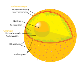
The nucleolus is the largest structure in the nucleus of eukaryotic cells. It is best known as the site of ribosome biogenesis, which is the synthesis of ribosomes. The nucleolus also participates in the formation of signal recognition particles and plays a role in the cell's response to stress. Nucleoli are made of proteins, DNA and RNA, and form around specific chromosomal regions called nucleolar organizing regions. Malfunction of nucleoli can be the cause of several human conditions called "nucleolopathies" and the nucleolus is being investigated as a target for cancer chemotherapy.

A karyotype is the general appearance of the complete set of chromosomes in the cells of a species or in an individual organism, mainly including their sizes, numbers, and shapes. Karyotyping is the process by which a karyotype is discerned by determining the chromosome complement of an individual, including the number of chromosomes and any abnormalities.

Ribosomal DNA (rDNA) is a DNA sequence that codes for ribosomal RNA. These sequences regulate transcription initiation and amplification, and contain both transcribed and non-transcribed spacer segments.

Ribosomal ribonucleic acid (rRNA) is a type of non-coding RNA which is the primary component of ribosomes, essential to all cells. rRNA is a ribozyme which carries out protein synthesis in ribosomes. Ribosomal RNA is transcribed from ribosomal DNA (rDNA) and then bound to ribosomal proteins to form small and large ribosome subunits. rRNA is the physical and mechanical factor of the ribosome that forces transfer RNA (tRNA) and messenger RNA (mRNA) to process and translate the latter into proteins. Ribosomal RNA is the predominant form of RNA found in most cells; it makes up about 80% of cellular RNA despite never being translated into proteins itself. Ribosomes are composed of approximately 60% rRNA and 40% ribosomal proteins, though this ratio differs between prokaryotes and eukaryotes.
RNA polymerase 1 is, in higher eukaryotes, the polymerase that only transcribes ribosomal RNA, a type of RNA that accounts for over 50% of the total RNA synthesized in a cell.

Secondary constrictions are the constricted or the narrow region found at any point of the chromosome other than that of centromere. The difference between the two constrictions can be noticed during anaphase, as chromosomes can only bend at the site of primary constriction. Secondary constrictions are useful in identifying a chromosome from a set. There are either 0, 1, 2, 3, or 4 secondary constriction sites in a cell at anaphase. Some parts of these constrictions indicate sites of nucleolus formation and are called "nucleolar organizing regions" (NORs). The nucleolus in the nucleus remains associated with the NOR of the secondary constriction area. In humans, the number of NORs is equal to the number of nucleoli, which is ten. However, not all secondary constrictions are NORs. The formations of nucleoli takes place around the NOR region. The secondary constriction also contains the genes for rRNA synthesis. Genes for 5S rRNA are present on chromosome 1. Due to secondary constriction, a knob-like structure is formed at the end called a satellite chromosome . DNA in a secondary constriction which forms rRNA is called rDNA..

Treacle protein is a protein that studies suggest is involved in the production of a molecule called ribosomal RNA (rRNA) within cells. Treacle is active in the nucleolus, which is a small region inside the nucleus where rRNA is produced. As a major component of cell structures called ribosomes, rRNA is essential for the assembly of proteins. This protein is active during early embryonic development in structures that become bones and other tissues in the face. Although the precise function of this protein is unknown, researchers believe that it plays a critical role in the development of facial bones and related structures.

Nucleolus organizer regions (NORs) are chromosomal regions crucial for the formation of the nucleolus. In humans, the NORs are located on the short arms of the acrocentric chromosomes 13, 14, 15, 21 and 22, the genes RNR1, RNR2, RNR3, RNR4, and RNR5 respectively. These regions code for 5.8S, 18S, and 28S ribosomal RNA. The NORs are "sandwiched" between the repetitive, heterochromatic DNA sequences of the centromeres and telomeres. The exact sequence of these regions is not included in the human reference genome as of 2016 or the GRCh38.p10 released January 6, 2017. On 28 February 2019, GRCh38.p13 was released, which added the NOR sequences for the short arms of chromosomes 13, 14, 15, 21, and 22. However, it is known that NORs contain tandem copies of ribosomal DNA (rDNA) genes. Some sequences of flanking sequences proximal and distal to NORs have been reported. The NORs of a loris have been reported to be highly variable. There are also DNA sequences related to rDNA that are on other chromosomes and may be involved in nucleoli formation.

In molecular biology, U3 snoRNA is a non-coding RNA found predominantly in the nucleolus. U3 has C/D box motifs that technically make it a member of the box C/D class of snoRNAs; however, unlike other C/D box snoRNAs, it has not been shown to direct 2'-O-methylation of other RNAs. Rather, U3 is thought to guide site-specific cleavage of ribosomal RNA (rRNA) during pre-rRNA processing.
Nucleophosmin (NPM), also known as nucleolar phosphoprotein B23 or numatrin, is a protein that in humans is encoded by the NPM1 gene.

Upstream binding transcription factor (UBTF), or upstream binding factor (UBF), is a protein that in humans is encoded by the UBTF gene.
RNA, ribosomal 4, also known as RNR4, is a human gene.

40S ribosomal protein S8 is a protein that in humans is encoded by the RPS8 gene.
RNA, ribosomal 1, also known as RNR1, is a human gene.
RNA, ribosomal 3, also known as RNR3, is a human gene. It is a minor isoform of large subunit of ribonucleotide-diphosphate reductase; the RNR complex catalyzes the rate-limiting step in dNTP synthesis, regulated by DNA replication and DNA damage checkpoint pathways via localization of small subunits; RNR3 has a paralog, RNR1, that arose from the whole genome duplication.
RNA, ribosomal 5, also known as RNR5, is a human gene. Genes for ribosomal RNA are clustered on the short arms of chromosomes 13, 14, 15, 20, 21. The gene for RNR5 exists in multiple copies on chromosome 22. Each gene cluster contains 30–40 copies and encodes a 45S RNA product that is then cleaved to form 18S, 5.8S and 28S rRNA subunits. In general, genes for RNA remain poorly annotated in most large public databases.
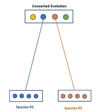
Concerted evolution is the phenomenon where paralogous genes within one species are more closely related to one another than to members of the same gene family in closely related species. In other terms, when specific members of a family are investigated, a greater amount of similarity is found within a species rather than between species. This is suggesting that members within this family do not in fact evolve independently of one another.
RNA, ribosomal 2, also known as RNR2, is a human gene coding for ribosomal RNA. Genes for ribosomal RNA are clustered on the short arms of chromosomes 13 (RNR1), 14 (RNR2), 15 (RNR3), 20 (RNR4), 21 (RNR5). The gene for RNR2 exists in multiple copies on chromosome 14. Each gene cluster contains 30–40 copies and encodes a 45S RNA product that is then processed to form 18S, 5.8S and 28S rRNA.
