
Ophthalmology is a surgical subspecialty within medicine that deals with the diagnosis and treatment of eye disorders. A former term is oculism.
Diabetic retinopathy, is a medical condition in which damage occurs to the retina due to diabetes mellitus. It is a leading cause of blindness in developed countries.

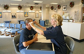
Optometry is a specialized health care profession that involves examining the eyes and related structures for defects or abnormalities. Optometrists are health care professionals who typically provide comprehensive primary eye care.
An optician is an individual who fits eyeglasses or contact lenses by filling a refractive prescription from an optometrist or ophthalmologist. They are able to translate and adapt ophthalmic prescriptions, dispense products, and work with accessories. There are several specialties within the field.

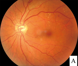
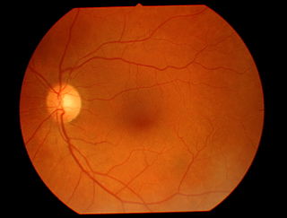
The optic disc or optic nerve head is the point of exit for ganglion cell axons leaving the eye. Because there are no rods or cones overlying the optic disc, it corresponds to a small blind spot in each eye.
In ophthalmology and optometry, a slit lamp is an instrument consisting of a high-intensity light source that can be focused to shine a thin sheet of light into the eye. It is used in conjunction with a biomicroscope. The lamp facilitates an examination of the anterior segment and posterior segment of the human eye, which includes the eyelid, sclera, conjunctiva, iris, natural crystalline lens, and cornea. The binocular slit-lamp examination provides a stereoscopic magnified view of the eye structures in detail, enabling anatomical diagnoses to be made for a variety of eye conditions. A second, hand-held lens is used to examine the retina.
Retinal hemorrhage is a disorder of the eye in which bleeding occurs in the retina, the light sensitive tissue, located on the back wall of the eye. There are photoreceptor cells in the retina called rods and cones, which transduce light energy into nerve signals that can be processed by the brain to form visual images. Retinal hemorrhage is strongly associated with child abuse in infants and young children and often leaves such abused infants permanently blind. In older children and adults, retinal hemorrhage can be caused by several medical conditions such as hypertension, retinal vein occlusion, anemia, leukemia or diabetes.

Sankara Nethralaya is a not-for-profit missionary institution for ophthalmic care headquartered in Chennai, India. In the name "Sankara Nethralaya", "Sankara" is a reference to Lord Shiva and "Nethralaya" means "The Temple of the Eye". Sankara Nethralaya receives patients from India and abroad. Sankara Nethralaya has over 1000 employees and serves around 1500 patients per day, performing over 100 surgeries per day. The annual revenue as per the taxes is close to US$100 million.
An eye care professional is an individual who provides a service related to the eyes or vision. It is any healthcare worker involved in eye care, from one with a small amount of post-secondary training to practitioners with a doctoral level of education.

The Illinois College of Optometry (ICO) is a private optometry college in Chicago, Illinois. Graduating approximately 160 optometrists a year, it is the largest optometry college in the United States and the oldest continually operating educational facility dedicated solely to the teaching of optometrists. The college complex incorporates more than 366,000 square feet (34,000 m2) including an on-site eye care clinic, electronically enhanced lecture center, library, computerized clinical learning equipment, cafeteria, fitness center, and living facilities.

Fundus photography involves photographing the rear of an eye, also known as the fundus. Specialized fundus cameras consisting of an intricate microscope attached to a flash enabled camera are used in fundus photography. The main structures that can be visualized on a fundus photo are the central and peripheral retina, optic disc and macula. Fundus photography can be performed with colored filters, or with specialized dyes including fluorescein and indocyanine green.
Marshall B. Ketchum University is a private university focused on graduate programs in healthcare and located in Fullerton, California. MBKU expanded from the Southern California College of Optometry which was founded in 1904. The university was officially established as a multidisciplinary university with the addition of School of PA Studies in 2011 and College of Pharmacy in 2013. Along with Hope International University, the campus bookends the north and south sides of the Cal State Fullerton campus respectively.

The Herbert Wertheim School of Optometry & Vision Science at the University of California, Berkeley is an optometry school at the University of California, Berkeley. It offers a graduate-level, four-year professional program leading to the Doctor of Optometry degree (OD), and a one-year, ACOE-accredited residency program in clinical optometry specialties. It is also the home department for the multidisciplinary Vision Science Group at UC Berkeley, whose graduate students earn either MS or PhD degrees.

The eye care system in Ghana can be said to be one in its infant or growing stages. Today there are less than 300 eye care professionals taking care of the eye needs of over 23 million Ghanaians.
Bascom Palmer Eye Institute is the University of Miami School of Medicine's ophthalmic care, research, and education center. The institute is based in the Health District of Miami, Florida, and has been ranked consistently as the best eye hospital and vision research center in the nation.

The Australian College of Optometry (ACO) is an Australian non-profit working to improving the eye health and well-being of various Australian communities. Established in 1940, the ACO's goal is to deliver public health optometry, vision research and professional education.
Teleophthalmology is a branch of telemedicine that delivers eye care through digital medical equipment and telecommunications technology. Today, applications of teleophthalmology encompass access to eye specialists for patients in remote areas, ophthalmic disease screening, diagnosis and monitoring; as well as distant learning.
Eye care in the United Kingdom is available through the National Health Service. Eye care in the community is almost entirely provided by optometrists in private practice. Specialist NHS services are provided from a small number of eye hospitals, and their staff often run clinics in general hospitals in their region.
Sickle cell retinopathy can be defined as retinal changes due to blood vessel damage in the eye of a person with a background of sickle cell disease. It can likely progress to loss of vision in late stages due to vitreous hemorrhage or retinal detachment. Sickle cell disease is a structural red blood cell disorder leading to consequences in multiple systems. It is characterized by chronic red blood cell destruction, vascular injury, and tissue ischemia causing damage to the brain, eyes, heart, lungs, kidneys, spleen, and musculoskeletal system.
Polypoidal choroidal vasculopathy (PCV) is an eye disease primarily affecting the choroid. It may cause sudden blurring of vision or a scotoma in the central field of vision. Since Indocyanine green angiography gives better imaging of choroidal structures, it is more preferred in diagnosing PCV. Treatment options of PCV include careful observation, photodynamic therapy, thermal laser, intravitreal injection of anti-VEGF therapy, or combination therapy.
