
The endoplasmic reticulum (ER) is, in essence, the transportation system of the eukaryotic cell, and has many other important functions such as protein folding. It is a type of organelle made up of two subunits – rough endoplasmic reticulum (RER), and smooth endoplasmic reticulum (SER). The endoplasmic reticulum is found in most eukaryotic cells and forms an interconnected network of flattened, membrane-enclosed sacs known as cisternae, and tubular structures in the SER. The membranes of the ER are continuous with the outer nuclear membrane. The endoplasmic reticulum is not found in red blood cells, or spermatozoa.

The endomembrane system is composed of the different membranes that are suspended in the cytoplasm within a eukaryotic cell. These membranes divide the cell into functional and structural compartments, or organelles. In eukaryotes the organelles of the endomembrane system include: the nuclear membrane, the endoplasmic reticulum, the Golgi apparatus, lysosomes, vesicles, endosomes, and plasma (cell) membrane among others. The system is defined more accurately as the set of membranes that form a single functional and developmental unit, either being connected directly, or exchanging material through vesicle transport. Importantly, the endomembrane system does not include the membranes of chloroplasts or mitochondria, but might have evolved from the latter.
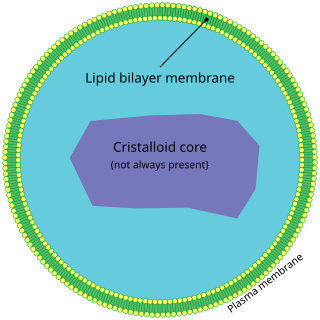
A peroxisome (IPA: [pɛɜˈɹɒksɪˌsoʊm]) is a membrane-bound organelle (formerly known as a microbody), found in the cytoplasm of virtually all eukaryotic cells. Peroxisomes are oxidative organelles. Frequently, molecular oxygen serves as a co-substrate, from which hydrogen peroxide (H2O2) is then formed. Peroxisomes owe their name to hydrogen peroxide generating and scavenging activities. They perform key roles in lipid metabolism and the conversion of reactive oxygen species. Peroxisomes are involved in the catabolism of very long chain fatty acids, branched chain fatty acids, bile acid intermediates (in the liver), D-amino acids, and polyamines, the reduction of reactive oxygen species – specifically hydrogen peroxide – and the biosynthesis of plasmalogens, i.e., ether phospholipids critical for the normal function of mammalian brains and lungs. They also contain approximately 10% of the total activity of two enzymes (Glucose-6-phosphate dehydrogenase and 6-Phosphogluconate dehydrogenase) in the pentose phosphate pathway, which is important for energy metabolism. It is vigorously debated whether peroxisomes are involved in isoprenoid and cholesterol synthesis in animals. Other known peroxisomal functions include the glyoxylate cycle in germinating seeds ("glyoxysomes"), photorespiration in leaves, glycolysis in trypanosomes ("glycosomes"), and methanol and/or amine oxidation and assimilation in some yeasts.
Ribosomes are macromolecular machines, found within all living cells, that perform biological protein synthesis. Ribosomes link amino acids together in the order specified by the codons of messenger RNA (mRNA) molecules to form polypeptide chains. Ribosomes consist of two major components: the small and large ribosomal subunits. Each subunit consists of one or more ribosomal RNA (rRNA) molecules and many ribosomal proteins. The ribosomes and associated molecules are also known as the translational apparatus.
The signal recognition particle (SRP) is an abundant, cytosolic, universally conserved ribonucleoprotein that recognizes and targets specific proteins to the endoplasmic reticulum in eukaryotes and the plasma membrane in prokaryotes.
A signal peptide is a short peptide present at the N-terminus of most newly synthesized proteins that are destined toward the secretory pathway. These proteins include those that reside either inside certain organelles, secreted from the cell, or inserted into most cellular membranes. Although most type I membrane-bound proteins have signal peptides, the majority of type II and multi-spanning membrane-bound proteins are targeted to the secretory pathway by their first transmembrane domain, which biochemically resembles a signal sequence except that it is not cleaved. They are a kind of target peptide.
In cell biology, a granule is a small particle. It can be any structure barely visible by light microscopy. The term is most often used to describe a secretory vesicle.

The soma, perikaryon, neurocyton, or cell body is the bulbous, non-process portion of a neuron or other brain cell type, containing the cell nucleus. The word 'soma' comes from the Greek 'σῶμα', meaning 'body'. Although it is often used to refer to neurons, it can also refer to other cell types as well, including astrocytes, oligodendrocytes, and microglia. There are many different specialized types of neurons, and their sizes vary from as small as about 5 micrometres to over 10 millimetres for some of the smallest and largest neurons of invertebrates, respectively.
Endoplasm generally refers to the inner, dense part of a cell's cytoplasm. This is opposed to the ectoplasm which is the outer (non-granulated) layer of the cytoplasm, which is typically watery and immediately adjacent to the plasma membrane. These two terms are mainly used to describe the cytoplasm of the amoeba, a protozoan, eukaryotic cell. The nucleus is separated from the endoplasm by the nuclear envelope. The different makeups/viscosities of the endoplasm and ectoplasm contribute to the amoeba's locomotion through the formation of a pseudopod. However, other types of cells have cytoplasm divided into endo- and ectoplasm. The endoplasm, along with its granules, contains water, nucleic acids, amino acids, carbohydrates, inorganic ions, lipids, enzymes, and other molecular compounds. It is the site of most cellular processes as it houses the organelles that make up the endomembrane system, as well as those that stand alone. The endoplasm is necessary for most metabolic activities, including cell division.
The unfolded protein response (UPR) is a cellular stress response related to the endoplasmic reticulum (ER) stress. It has been found to be conserved between all mammalian species, as well as yeast and worm organisms.

Activating transcription factor 6, also known as ATF6, is a protein that, in humans, is encoded by the ATF6 gene and is involved in the unfolded protein response.
The following outline is provided as an overview of and topical guide to cell biology:

DNA damage-inducible transcript 3, also known as C/EBP homologous protein (CHOP), is a pro-apoptotic transcription factor that is encoded by the DDIT3 gene. It is a member of the CCAAT/enhancer-binding protein (C/EBP) family of DNA-binding transcription factors. The protein functions as a dominant-negative inhibitor by forming heterodimers with other C/EBP members, preventing their DNA binding activity. The protein is implicated in adipogenesis and erythropoiesis, and has an important role in the cell's stress response.

Double-stranded RNA-binding protein Staufen homolog 1 is a protein that in humans is encoded by the STAU1 gene.

VAMP-Associated Protein A is a protein that in humans is encoded by the VAPA gene. Together with VAPB and VAPC it forms the VAP protein family. They are integral endoplasmic reticulum membrane proteins of the type II and are ubiquitous among eukaryotes.
Membrane contact sites (MCS) are close appositions between two organelles. Ultrastructural studies typically reveal an intermembrane distance in the order of the size of a single protein, as small as 10 nm or wider, with no clear upper limit. These zones of apposition are highly conserved in evolution. These sites are thought to be important to facilitate signalling, and they promote the passage of small molecules, including ions, lipids and reactive oxygen species. MCS are important in the function of the endoplasmic reticulum (ER), since this is the major site of lipid synthesis within cells. The ER makes close contact with many organelles, including mitochondria, Golgi, endosomes, lysosomes, peroxisomes, chloroplasts and the plasma membrane. Both mitochondria and sorting endosomes undergo major rearrangements leading to fission where they contact the ER. Sites of close apposition can also form between most of these organelles most pairwise combinations. First mentions of these contact sites can be found in papers published in the late 1950s mainly visualized using electron microscopy (EM) techniques. Copeland and Dalton described them as “highly specialized tubular form of endoplasmic reticulum in association with the mitochondria and apparently in turn, with the vascular border of the cell”.
Harvey F. Lodish is a molecular and cell biologist, professor at the Massachusetts Institute of Technology (MIT), Founding Member of the Whitehead Institute for Biomedical Research, and lead author of the textbook Molecular Cell Biology. Lodish's research focuses on cell surface proteins and other important areas at the interface between molecular cell biology and medicine.
The endoplasmic reticulum membrane protein complex (EMC) is a putative endoplasmic reticulum-resident membrane protein (co-)chaperone. The EMC is evolutionarily conserved in eukaryotes, and its initial appearance might reach back to the last eukaryotic common ancestor (LECA). Many aspects of mEMC biology and molecular function remain to be studied.
David Domingo Sabatini is an Argentine-American cell biologist and the Frederick L. Ehrman Professor Emeritus of Cell Biology in the Department of Cell Biology at New York University School of Medicine, which he chaired from 1972 to 2011. Sabatini's major research interests have been on the mechanisms responsible for the structural complexity of the eukaryotic cell. Throughout his career, Sabatini has been recognized for his efforts in promoting science in Latin America.
Mary Locke Petermann was an American cellular biochemist known for her key role in the discovery and characterization of animal ribosomes, the molecular complexes that carry out protein synthesis. She was the first woman to become a full professor at Cornell University's medical school.