Tumor metabolome

Last updated
Tumor metabolome: Relationships between metabolome, proteome, and genome in cancerous cells. Glycolysis breaks down glucose into pyruvate, which is then fermented to lactate; pyruvate flux through TCA cycle is down-regulated in cancer cells. Pathways branching off of glycolysis, such as the pentose phosphate pathway, generate biochemical building blocks to sustain the high proliferative rate of cancer cells. Specific genetic and enzyme-level behaviors. Blue boxes are enzymes important in transitioning to a cancer metabolic phenotype; orange boxes are enzymes that are mutated in cancer cells. Green ovals are oncogenes that are up-regulated in cancer; red ovals are tumor suppressors that are down-regulated in cancer. TumorMetabolome.jpg
Tumor metabolome: Relationships between metabolome, proteome, and genome in cancerous cells. Glycolysis breaks down glucose into pyruvate, which is then fermented to lactate; pyruvate flux through TCA cycle is down-regulated in cancer cells. Pathways branching off of glycolysis, such as the pentose phosphate pathway, generate biochemical building blocks to sustain the high proliferative rate of cancer cells. Specific genetic and enzyme-level behaviors. Blue boxes are enzymes important in transitioning to a cancer metabolic phenotype; orange boxes are enzymes that are mutated in cancer cells. Green ovals are oncogenes that are up-regulated in cancer; red ovals are tumor suppressors that are down-regulated in cancer.

The study of the tumor metabolism, also known as tumor metabolome describes the different characteristic metabolic changes in tumor cells. The characteristic attributes [2] of the tumor metabolome are high glycolytic enzyme activities, the expression of the pyruvate kinase isoenzyme type M2, increased channeling of glucose carbons into synthetic processes, such as nucleic acid, amino acid and phospholipid synthesis, a high rate of pyrimidine and purine de novo synthesis, a low ratio of Adenosine triphosphate and Guanosine triphosphate to Cytidine triphosphate and Uridine triphosphate, low Adenosine monophosphate levels, high glutaminolytic capacities, release of immunosuppressive substances and dependency on methionine.

Contents

Although the link between the cancer and metabolism was observed in the early days of cancer research by Otto Heinrich Warburg, [3] which is also known as Warburg hypothesis, not much substantial research was carried out until the late 1990s because of the lack of in vitro tumor models and the difficulty in creating environments that lack oxygen. Recent research has revealed that metabolic reprogramming occurs as a consequence of mutations in cancer genes and alterations in cellular signaling. Therefore, the alteration of cellular and energy metabolism has been suggested as one of The Hallmarks of Cancer. [4] [5]

Warburg effect and glycolysis

High amount of aerobic glycolysis (also known as the Warburg effect) distinguishes cancer cells from normal cells. The conversion of glucose to lactate rather than metabolizing it in the mitochondria through oxidative phosphorylation, (which can also occur in hypoxic normal cells) persists in malignant tumor despite the presence of oxygen. This process normally inhibits glycolysis which is also known as Pasteur effect. One of the reasons it is observed is because of the malfunction of mitochondria. Although ATP production by glycolysis can be more rapid than by oxidative phosphorylation, it is far less efficient in terms of ATP generated per unit of glucose consumed. Rather than oxidizing glucose for ATP production, glucose in cancer cells tends to be used for anabolic processes, such as ribose production, protein glycosylation and serine synthesis. This shift therefore demands that tumor cells implement an abnormally high rate of glucose uptake to meet their increased needs. [5]

As neoplastic cells accumulate in three-dimensional multicellular masses, local low nutrient and oxygen levels trigger the growth of new blood vessels into the neoplasm. The imperfect neovasculature in the tumor bed is poorly formed and is inefficient. It therefore, causes nutrient and hypoxic stress (or a state of hypoxia). [6] [7] In this regard, cancer cells and stromal cells can symbiotically recycle and maximize the use of nutrients. Hypoxic adaptation by cancer cells is essential for survival and progression of a tumor. [8] [9] In addition to cell-autonomous changes that drive a cancer cell to proliferate and contribute to tumorigenesis, it has also been observed that alterations in whole-organism metabolism such as obesity are associated with heightened risks for a variety of cancers. [10]

Role of signalling pathway in cancer metabolism

The protein AKT1 (also known as Protein Kinase B or PKB) in the PI3K/AKT/mTOR pathway is an important driver of the tumor glycolytic phenotype and stimulates ATP generation. AKT1 stimulates glycolysis by increasing the expression and membrane translocation of glucose transporters and by phosphorylating key glycolytic enzymes, such as hexokinase and phosphofructokinase 2. This leads to inhibition of forkhead box subfamily O transcription factors, leading to the increase of glycolytic capacity. Activated mTOR stimulates protein and lipid biosynthesis and cell growth in response to sufficient nutrient and energy conditions and is often constitutively activated during tumorigenesis. [5] mTOR directly stimulates mRNA translation and ribosome biogenesis, and indirectly causes other metabolic changes by activating transcription factors such as hypoxia-inducible factor 1 (HIF1A). The subsequent HIF1-dependent metabolic changes are a major determinant of the glycolytic phenotype downstream of PI3K, AKT1 and mTOR. [11]

Role of tumor suppressor and oncogenes

Apart from being as a general tumor suppressor gene, p53 also plays an important part in regulating of metabolism. p53 activates hexokinase 2 (HK2) that converts glucose to glucose-6-phosphate (G6P) which enters glycolysis to produce ATP, or enters the pentose phosphate pathway (PPP). It therefore, supports macromolecular biosynthesis by producing reducing potential in the form of reduced Nicotinamide adenine dinucleotide phosphate (NADPH) and/or ribose that are used for nucleotide synthesis. [12] p53 inhibits the glycolytic pathway by upregulating the expression of TP53-induced glycolysis and apoptosis regulator. Wild-type p53 supports the expression of PTEN (gene), which inhibits the PI3K pathway, thereby suppressing glycolysis. POU2F1 also cooperate with p53 in regulating the balance between oxidative and glycolytic metabolism. It provides resistance to oxidative stress that would regulates a set of genes that increase glucose metabolism and reduce mitochondrial respiration. This will provide additive force when the p53 is lost. [5] Mutated Ras also enhances glycolysis, partly through increasing the activity of Myc and hypoxia-inducible factors. Although HIF-1 inhibits Myc, HIF-2 activates Myc causing the multiplicity of the tumor cells. [9]

TCA cycle in cancer metabolism

Mutations in fumarate hydratase are found among patients suffering from kidney cancers, and mutations in succinate dehydrogenase were found in patients with pheochromocytoma and paragangliomas. These mutations cause a disruption of the TCA cycle with the accumulation of fumarate or succinate, both of which can inhibit dioxygenases or prolyl hydrolases that mediate the degradation of HIF proteins. HIF-1 could be elevated under aerobic conditions downstream from activated PI3K, which stimulates the synthesis of HIF-1. Loss of the tumor suppressor VHL in kidney cancer also stabilizes HIF-1, permitting it to activate glycolytic genes, which are normally activated by HIF-1 under hypoxic conditions. [9] HIF1 then would activate the pyruvate dehydrogenase kinase (PDKs), which inactivate the mitochondrial pyruvate dehydrogenase complex. It reduces the flow of glucose-derived pyruvate into the tricarboxylic acid (citric acid cycle or TCA cycle). This reduction in pyruvate flux into the TCA cycle decreases the rate of oxidative phosphorylation and oxygen consumption, reinforcing the glycolytic phenotype and sparing oxygen under hypoxic conditions. [13] [14]

M2 isoform of pyruvate kinase

Pyruvate kinase type M2 or PKM2 is present in embryonic, adult stem cells. It is also expressed by many tumor cells. The alterations to metabolism by PKM2 increases ATP resources, stimulates macromolecular biosynthesis and redox control. Pyruvate kinase catalyses the ATP-generating step of glycolysis in which phosphoenolpyruvate (PEP) is converted to pyruvate. This is a rate-limiting step. [15] It decreases the glycolysis activity and allows carbohydrate metabolites to enter other pathways, like hexosamine pathway, Uridine diphosphate glucose–glucose synthesis, glycerol synthesis and Pentose phosphate pathway or PPP. It helps in generating macromolecule precursors, that are necessary to support cell proliferation, and reducing equivalents such as NADPH. [16] [17] It has been observed in some studies that MYC promotes expression of PKM2 over PKM1 by modulating exon splicing. [5]

A key molecule produced as a result of the oxidative PPP by PKM2 is NADPH. NADPH functions as a cofactor and provides reducing power in many enzymatic reactions that are crucial for macromolecular biosynthesis. Another mechanism by which NADPH is produced in mammalian cells is the reaction converting isocitrate to α-ketoglutarate (αKG), which is catalysed by NADP-dependent isocitrate dehydrogenase 1 (IDH1) and IDH2 and have been found linked to tumorigenesis in glioblastoma and acute myeloid leukemia. [18] [19] They are also found to interact with arginine residues required for isocitrate binding in the active site of the proteins of IDH1 and IDH2. [5]

Fatty acid synthesis

Fatty acid synthesis is an anabolic process that starts from the conversion of acetyl-CoA to malonyl-CoA by acetyl-CoA carboxylase. Malonyl CoA leads to fatty acid synthesis (FAS) and is involved in the elongation of fatty acids through Fatty acid synthase (FASN). Although aerobic glycolysis is the best documented metabolic phenotype of tumor cells, it is not a universal feature of all human cancers. Amino acids and fatty acids have been shown to function as fuels for tumor cells to proliferate. The carnitine palmitoyltransferase enzymes that regulate the β-oxidation of fatty acids may have a key role in determining some of these phenotypes. [5] Enhanced fatty acid synthesis provides lipids for membrane biogenesis to tumor cells and hence, it gives advantage in both growth and survival of the cell.

Adaptation and resistance to drugs

It has also been seen that metabolic phenotype of tumor cells changes to adapt to the prevailing local conditions. A convergence between phenotypic and metabolic state transitions that confers a survival advantage to cancer cells against clinically used drug combinations like taxanes and anthracyclines have also been reported while drug resistant cancer cells had increased activity of both the glycolytic and oxidative pathways and glucose flux through the pentose phosphate pathway (PPP). [20] Some of the fatty acids have been linked to acquire resistance against some of the cancer drugs. Fatty acid synthase (FASN), a key complex catalyzing fatty acid synthesis has been found to be linked to acquired docetaxel, trastuzumab and adriamycin resistance in breast cancer. Similar resistance have been found with intrinsic gemcitabine and radiation resistance in pancreatic cancer. Glutaminolysis is linked to cisplatin resistance via the activation of mTORC1 signaling in gastric cancer. [21]

Metabolic biomarkers of tumors

NADPH plays an important role as an antioxidant by decreasing the reactive oxygen produced during rapid cell proliferation. It has been shown that attenuation of the PPP would dampen NADPH production in cancer cells, leading to the decrease in macromolecular biosynthesis and rendering the transformed cells that are vulnerable free radical-mediated damage. In this way, the advantage conferred by PKM2 expression would be eliminated. In preclinical studies, drugs such as 6-amino-nicotinamide (6-AN), which inhibits G6P dehydrogenase, the enzyme that initiates the PPP have shown anti-tumorigenic effects in leukemia, glioblastoma and lung cancer cell lines. [22]

Cyclosporine inhibits TOR and is used as an effective immunosuppressant. Mycophenolic acid inhibits of IMPDH and pyrimidine biosynthesis and is clinically used as immunosuppressant. Both agents also display anti-tumor effects in animal studies. [9] Metabolites such as Alanine, Saturated lipids, Glycine, Lactate, Myo-Inositol, Nucleotides, Polyunsaturated fatty acids and Taurine are considered as the potential biomarkers in various studies. [23]

Glutaminolysis

The use of the amino acid glutamine as an energy source is facilitated by the multistep catabolism of glutamine called glutaminolysis. This energy pathway is upregulated in cancer, which may represent a therapeutic target as cancer cells are thought to be more dependent on glutamine than healthy cells. [24] This especially holds true for specific tumor types that are metabolically dysregulated, such as malignant brain tumors (i.e. glioblastoma) that carry mutations in the IDH1 gene. These tumors use glutamine or the structurally related amino acid glutamate as an energy source and a chemotactic sensor in the brain, which increases their malignancy and may explain why these tumors grow so invasive.[9][10]

Related Research Articles

<span class="mw-page-title-main">Citric acid cycle</span> Series of interconnected biochemical reactions

The citric acid cycle —also known as the Krebs cycle, Szent-Györgyi-Krebs cycle or the TCA cycle (tricarboxylic acid cycle)—is a series of chemical reactions to release stored energy through the oxidation of acetyl-CoA derived from carbohydrates, fats, and proteins. The Krebs cycle is used by organisms that respire (as opposed to organisms that ferment) to generate energy, either by anaerobic respiration or aerobic respiration. In addition, the cycle provides precursors of certain amino acids, as well as the reducing agent NADH, that are used in numerous other reactions. Its central importance to many biochemical pathways suggests that it was one of the earliest components of metabolism. Even though it is branded as a 'cycle', it is not necessary for metabolites to follow only one specific route; at least three alternative segments of the citric acid cycle have been recognized.

<span class="mw-page-title-main">Glycolysis</span> Series of interconnected biochemical reactions

Glycolysis is the metabolic pathway that converts glucose into pyruvate, and in most organisms, occurs in the liquid part of cells, the cytosol. The free energy released in this process is used to form the high-energy molecules adenosine triphosphate (ATP) and reduced nicotinamide adenine dinucleotide (NADH). Glycolysis is a sequence of ten reactions catalyzed by enzymes.

Gluconeogenesis (GNG) is a metabolic pathway that results in the biosynthesis of glucose from certain non-carbohydrate carbon substrates. It is a ubiquitous process, present in plants, animals, fungi, bacteria, and other microorganisms. In vertebrates, gluconeogenesis occurs mainly in the liver and, to a lesser extent, in the cortex of the kidneys. It is one of two primary mechanisms – the other being degradation of glycogen (glycogenolysis) – used by humans and many other animals to maintain blood sugar levels, avoiding low levels (hypoglycemia). In ruminants, because dietary carbohydrates tend to be metabolized by rumen organisms, gluconeogenesis occurs regardless of fasting, low-carbohydrate diets, exercise, etc. In many other animals, the process occurs during periods of fasting, starvation, low-carbohydrate diets, or intense exercise.

<span class="mw-page-title-main">Tumor hypoxia</span> Situation where tumor cells have been deprived of oxygen

Tumor hypoxia is the situation where tumor cells have been deprived of oxygen. As a tumor grows, it rapidly outgrows its blood supply, leaving portions of the tumor with regions where the oxygen concentration is significantly lower than in healthy tissues. Hypoxic microenvironements in solid tumors are a result of available oxygen being consumed within 70 to 150 μm of tumour vasculature by rapidly proliferating tumor cells thus limiting the amount of oxygen available to diffuse further into the tumor tissue. In order to support continuous growth and proliferation in challenging hypoxic environments, cancer cells are found to alter their metabolism. Furthermore, hypoxia is known to change cell behavior and is associated with extracellular matrix remodeling and increased migratory and metastatic behavior.

<span class="mw-page-title-main">Pyruvate kinase</span> Class of enzymes

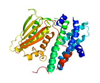
Pyruvate kinase is the enzyme involved in the last step of glycolysis. It catalyzes the transfer of a phosphate group from phosphoenolpyruvate (PEP) to adenosine diphosphate (ADP), yielding one molecule of pyruvate and one molecule of ATP. Pyruvate kinase was inappropriately named before it was recognized that it did not directly catalyze phosphorylation of pyruvate, which does not occur under physiological conditions. Pyruvate kinase is present in four distinct, tissue-specific isozymes in animals, each consisting of particular kinetic properties necessary to accommodate the variations in metabolic requirements of diverse tissues.

Fatty acid metabolism consists of various metabolic processes involving or closely related to fatty acids, a family of molecules classified within the lipid macronutrient category. These processes can mainly be divided into (1) catabolic processes that generate energy and (2) anabolic processes where they serve as building blocks for other compounds.

In oncology, the Warburg effect is the observation that most cancer cells release energy predominantly not through the 'usual' citric acid cycle and oxidative phosphorylation in the mitochondria as observed in normal cells, but through a less efficient process of 'aerobic glycolysis' consisting of a high level of glucose uptake and glycolysis followed by lactic acid fermentation taking place in the cytosol, not the mitochondria, even in the presence of abundant oxygen. This observation was first published by Otto Heinrich Warburg, who was awarded the 1931 Nobel Prize in Physiology for his "discovery of the nature and mode of action of the respiratory enzyme". The precise mechanism and therapeutic implications of the Warburg effect, however, remain unclear.

<span class="mw-page-title-main">Pyruvate dehydrogenase lipoamide kinase isozyme 1</span> Protein-coding gene in the species Homo sapiens

Pyruvate dehydrogenase lipoamide kinase isozyme 1, mitochondrial is an enzyme that in humans is encoded by the PDK1 gene. It codes for an isozyme of pyruvate dehydrogenase kinase (PDK).

In biochemistry, lipogenesis is the conversion of fatty acids and glycerol into fats, or a metabolic process through which acetyl-CoA is converted to triglyceride for storage in fat. Lipogenesis encompasses both fatty acid and triglyceride synthesis, with the latter being the process by which fatty acids are esterified to glycerol before being packaged into very-low-density lipoprotein (VLDL). Fatty acids are produced in the cytoplasm of cells by repeatedly adding two-carbon units to acetyl-CoA. Triacylglycerol synthesis, on the other hand, occurs in the endoplasmic reticulum membrane of cells by bonding three fatty acid molecules to a glycerol molecule. Both processes take place mainly in liver and adipose tissue. Nevertheless, it also occurs to some extent in other tissues such as the gut and kidney. A review on lipogenesis in the brain was published in 2008 by Lopez and Vidal-Puig. After being packaged into VLDL in the liver, the resulting lipoprotein is then secreted directly into the blood for delivery to peripheral tissues.

Tumor M2-PK is a synonym for the dimeric form of the pyruvate kinase isoenzyme type M2 (PKM2), a key enzyme within tumor metabolism. Tumor M2-PK can be elevated in many tumor types, rather than being an organ-specific tumor marker such as PSA. Increased stool (fecal) levels are being investigated as a method of screening for colorectal tumors, and EDTA plasma levels are undergoing testing for possible application in the follow-up of various cancers.

<span class="mw-page-title-main">Ribose 5-phosphate</span> Chemical compound

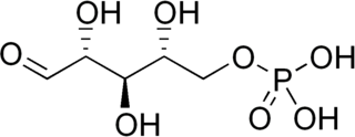
Ribose 5-phosphate (R5P) is both a product and an intermediate of the pentose phosphate pathway. The last step of the oxidative reactions in the pentose phosphate pathway is the production of ribulose 5-phosphate. Depending on the body's state, ribulose 5-phosphate can reversibly isomerize to ribose 5-phosphate. Ribulose 5-phosphate can alternatively undergo a series of isomerizations as well as transaldolations and transketolations that result in the production of other pentose phosphates as well as fructose 6-phosphate and glyceraldehyde 3-phosphate.

The Randle cycle, also known as the glucose fatty-acid cycle, is a metabolic process involving the competition of glucose and fatty acids for substrates. It is theorized to play a role in explaining type 2 diabetes and insulin resistance.

Glutaminolysis (glutamine + -lysis) is a series of biochemical reactions by which the amino acid glutamine is lysed to glutamate, aspartate, CO2, pyruvate, lactate, alanine and citrate.

<span class="mw-page-title-main">PKM2</span> Protein-coding gene in the species Homo sapiens

Pyruvate kinase isozymes M1/M2 (PKM1/M2), also known as pyruvate kinase muscle isozyme (PKM), pyruvate kinase type K, cytosolic thyroid hormone-binding protein (CTHBP), thyroid hormone-binding protein 1 (THBP1), or opa-interacting protein 3 (OIP3), is an enzyme that in humans is encoded by the PKM2 gene.

<span class="mw-page-title-main">TP53-inducible glycolysis and apoptosis regulator</span> Protein-coding gene in the species Homo sapiens

The TP53-inducible glycolysis and apoptosis regulator (TIGAR) also known as fructose-2,6-bisphosphatase TIGAR is an enzyme that in humans is encoded by the C12orf5 gene.

The lactate shuttle hypothesis describes the movement of lactate intracellularly and intercellularly. The hypothesis is based on the observation that lactate is formed and utilized continuously in diverse cells under both anaerobic and aerobic conditions. Further, lactate produced at sites with high rates of glycolysis and glycogenolysis can be shuttled to adjacent or remote sites including heart or skeletal muscles where the lactate can be used as a gluconeogenic precursor or substrate for oxidation. The hypothesis was proposed by professor George Brooks of the University of California at Berkeley.

Pseudohypoxia refers to increased cytosolic ratio of free NADH/NAD+ ratio in cells, where NADH is overly increased and NAD+ is overly decreased. It can be caused by diabetic hyperglycemia and by excessive alcohol consumption. The insufficiency of available NAD+ produces symptoms similar to hypoxia (lack of oxygen), because NAD+ is primarily needed by the Krebs cycle for oxidative phosphorylation, and to a lesser extent is needed in anaerobic glycolysis. Oxidative phosphorylation and glyocolysis are vital as these metabolic pathways produce ATP, which is the molecule that releases energy necessary for cells to function.

Jacques Pouysségur is a French engineer and researcher. He was born on November 10, 1943, in Toulouse, Haute-Garonne.

Immunometabolism is a branch of biology that studies the interplay between metabolism and immunology in all organisms. In particular, immunometabolism is the study of the molecular and biochemical underpinninngs for i) the metabolic regulation of immune function, and ii) the regulation of metabolism by molecules and cells of the immune system. Further categorization includes i) systemic immunometabolism and ii) cellular immunometabolism.

<span class="mw-page-title-main">Citrate–malate shuttle</span>

The citrate–malate shuttle is a series of chemical reactions – commonly referred to as a biochemical cycle or system – that transports acetyl-CoA in the mitochondrial matrix across the inner and outer mitochondrial membrane for fatty acid synthesis. Mitochondria is enclosed in a double membrane. As the inner mitochondrial membrane is impermeable to acetyl-CoA, the shuttle system is essential to fatty acid synthesis in the cytosol. It plays an important role in the generation of lipids in the liver.

References

  1. Vermeersch KA, Styczynski MP (2013). "Applications of metabolomics in cancer research". Journal of Carcinogenesis. 12 (9): 9. doi: 10.4103/1477-3163.113622 . PMC   3709411 . PMID   23858297.
  2. Mazurek S, Eigenbrodt E (March–April 2003). "The tumor metabolome". Anticancer Research. 23 (2A): 1149–54. PMID   12820363.
  3. Warburg O (February 1956). "On the origin of cancer cells". Science. 123 (3191): 309–14. Bibcode:1956Sci...123..309W. doi:10.1126/science.123.3191.309. PMID   13298683.
  4. Hanahan D, Weinberg RA (March 2011). "Hallmarks of cancer: the next generation". Cell. 144 (5): 646–74. doi: 10.1016/j.cell.2011.02.013 . PMID   21376230.
  5. 1 2 3 4 5 6 7 Cairns RA, Harris IS, Mak TW (February 2011). "Regulation of cancer cell metabolism". Nature Reviews. Cancer. 11 (2): 85–95. doi:10.1038/nrc2981. PMID   21258394. S2CID   8891526.
  6. Carmeliet P, Dor Y, Herbert JM, Fukumura D, Brusselmans K, Dewerchin M, et al. (July 1998). "Role of HIF-1alpha in hypoxia-mediated apoptosis, cell proliferation and tumour angiogenesis". Nature. 394 (6692): 485–90. Bibcode:1998Natur.394..485C. doi:10.1038/28867. PMID   9697772. S2CID   4419118.
  7. Semenza GL (February 2012). "Hypoxia-inducible factors in physiology and medicine". Cell. 148 (3): 399–408. doi:10.1016/j.cell.2012.01.021. PMC   3437543 . PMID   22304911.
  8. Semenza GL (February 2010). "HIF-1: upstream and downstream of cancer metabolism". Current Opinion in Genetics & Development. 20 (1): 51–6. doi:10.1016/j.gde.2009.10.009. PMC   2822127 . PMID   19942427.
  9. 1 2 3 4 Dang CV (May 2012). "Links between metabolism and cancer". Genes & Development. 26 (9): 877–90. doi:10.1101/gad.189365.112. PMC   3347786 . PMID   22549953.
  10. Khandekar MJ, Cohen P, Spiegelman BM (November 2011). "Molecular mechanisms of cancer development in obesity". Nature Reviews. Cancer. 11 (12): 886–95. doi:10.1038/nrc3174. PMID   22113164. S2CID   1978204.
  11. Guertin DA, Sabatini DM (July 2007). "Defining the role of mTOR in cancer". Cancer Cell. 12 (1): 9–22. doi: 10.1016/j.ccr.2007.05.008 . PMID   17613433.
  12. Mathupala SP, Heese C, Pedersen PL (September 1997). "Glucose catabolism in cancer cells. The type II hexokinase promoter contains functionally active response elements for the tumor suppressor p53". The Journal of Biological Chemistry. 272 (36): 22776–80. doi: 10.1074/jbc.272.36.22776 . PMID   9278438.
  13. Papandreou I, Cairns RA, Fontana L, Lim AL, Denko NC (March 2006). "HIF-1 mediates adaptation to hypoxia by actively downregulating mitochondrial oxygen consumption". Cell Metabolism. 3 (3): 187–97. doi: 10.1016/j.cmet.2006.01.012 . PMID   16517406.
  14. Kim JW, Tchernyshyov I, Semenza GL, Dang CV (March 2006). "HIF-1-mediated expression of pyruvate dehydrogenase kinase: a metabolic switch required for cellular adaptation to hypoxia". Cell Metabolism. 3 (3): 177–85. doi: 10.1016/j.cmet.2006.02.002 . PMID   16517405.
  15. Mazurek S, Boschek CB, Hugo F, Eigenbrodt E (August 2005). "Pyruvate kinase type M2 and its role in tumor growth and spreading". Seminars in Cancer Biology. 15 (4): 300–8. doi:10.1016/j.semcancer.2005.04.009. PMID   15908230.
  16. Vander Heiden MG, Cantley LC, Thompson CB (May 2009). "Understanding the Warburg effect: the metabolic requirements of cell proliferation". Science. 324 (5930): 1029–33. Bibcode:2009Sci...324.1029V. doi:10.1126/science.1160809. PMC   2849637 . PMID   19460998.
  17. Fang M, Shen Z, Huang S, Zhao L, Chen S, Mak TW, Wang X (November 2010). "The ER UDPase ENTPD5 promotes protein N-glycosylation, the Warburg effect, and proliferation in the PTEN pathway". Cell. 143 (5): 711–24. doi: 10.1016/j.cell.2010.10.010 . PMID   21074248. S2CID   11891493.
  18. Parsons DW, Jones S, Zhang X, Lin JC, Leary RJ, Angenendt P, et al. (September 2008). "An integrated genomic analysis of human glioblastoma multiforme". Science. 321 (5897): 1807–12. Bibcode:2008Sci...321.1807P. doi:10.1126/science.1164382. PMC   2820389 . PMID   18772396.
  19. Mardis ER, Ding L, Dooling DJ, Larson DE, McLellan MD, Chen K, et al. (September 2009). "Recurring mutations found by sequencing an acute myeloid leukemia genome". The New England Journal of Medicine. 361 (11): 1058–66. doi:10.1056/NEJMoa0903840. PMC   3201812 . PMID   19657110.
  20. Goldman A, Khiste S, Freinkman E, Dhawan A, Majumder B, Mondal J, et al. (August 2019). "Targeting tumor phenotypic plasticity and metabolic remodeling in adaptive cross-drug tolerance". Science Signaling. 12 (595). doi:10.1126/scisignal.aas8779. PMC   7261372 . PMID   31431543.
  21. Zhao Y, Butler EB, Tan M (March 2013). "Targeting cellular metabolism to improve cancer therapeutics". Cell Death & Disease. 4 (3): e532. doi:10.1038/cddis.2013.60. PMC   3613838 . PMID   23470539.
  22. Budihardjo II, Walker DL, Svingen PA, Buckwalter CA, Desnoyers S, Eckdahl S, et al. (January 1998). "6-Aminonicotinamide sensitizes human tumor cell lines to cisplatin". Clinical Cancer Research. 4 (1): 117–30. PMID   9516960.
  23. Griffin JL, Shockcor JP (July 2004). "Metabolic profiles of cancer cells". Nature Reviews. Cancer. 4 (7): 551–61. doi:10.1038/nrc1390. PMID   15229480. S2CID   527894.
  24. Chen JQ, Russo J (December 2012). "Dysregulation of glucose transport, glycolysis, TCA cycle and glutaminolysis by oncogenes and tumor suppressors in cancer cells". Biochimica et Biophysica Acta (BBA) - Reviews on Cancer. 1826 (2): 370–84. doi: 10.1016/j.bbcan.2012.06.004 . PMID   22750268.