Cardiopulmonary bypass

Last updated
Cardiopulmonary bypass
Coronary artery bypass surgery Image 657C-PH.jpg
A heart–lung machine (upper right) in a coronary artery bypass surgery.
ICD-10-PCS 12
ICD-9-CM 39.61
MeSH D002318
OPS-301 code 14
Other codes 22570829

Cardiopulmonary Bypass (CPB) is a machine that temporarily takes over the function of the heart and lungs during a cardiac surgery by maintaining the circulation of blood and oxygen throughout the body. [1]

Contents

CPB is used during many heart surgeries to improve the safety of the patient during a heart procedure. The machine mechanically circulates and oxygenates blood throughout the patient's body while bypassing the heart and lungs. Additionally, it maintains perfusion to other body organs and tissues of the patient while the surgeon works in a bloodless surgical field.

The CPB is often referred to as a heart-lung machine, "the pump," or CPB pumps. CPB is operated by perfusionists, and is a form of extracorporeal circulation.

Uses

Illustration of one typical way that a heart-lung machine may be connected to the veins and arteries near the heart. The three implements on the left represent (from top to bottom) the pump, the oxygenator, and the reservoir. Blausen 0468 Heart-Lung Machine.png
Illustration of one typical way that a heart-lung machine may be connected to the veins and arteries near the heart. The three implements on the left represent (from top to bottom) the pump, the oxygenator, and the reservoir.

CPB is commonly used in operations or surgical procedures involving the heart. The technique allows the surgical team to oxygenate and circulate the patient's blood, thus allowing the surgeon to operate safely on the heart. In many operations, such as coronary artery bypass grafting (CABG), the heart is arrested, due to the degree of the difficulty of operating on a beating heart.

Operations requiring the opening of the chambers of the heart, for example mitral valve repair or replacement, requires the use of CPB. This is to avoid engulfing air systemically, and to provide a bloodless field to increase visibility for the surgeon. The machine pumps the blood and, using an oxygenator, allows red blood cells to pick up oxygen, as well as allowing carbon dioxide levels to decrease. This mimics the function of the heart and the lungs, respectively.

Hypothermia

CPB can be used for the induction of total body hypothermia, a state in which the body can be maintained for up to 45 minutes without perfusion (blood flow). If blood flow is stopped at normal body temperature, permanent brain damage can occur in three to four minutes – death may follow. Similarly, CPB can be used to rewarm individuals who have hypothermia. This rewarming method of using CPB is successful if the core temperature of the patient is above 16 °C.

Cooled Blood

The blood is cooled during CPB and is returned to the body. The cooled blood slows the body's basal metabolic rate, decreasing its demand for oxygen. Cooled blood usually has a higher viscosity, but the various crystalloid or colloidal solutions that are used to prime the bypass tubing serve to dilute the blood. Maintaining appropriate blood pressure for organs is a challenge, but it is monitored carefully during the procedure. Hypothermia is also maintained (if necessary), and the body temperature is usually kept at 28 °C to 32 °C (82.4–89.6 °F).

Extracorporeal Membrane Oxygenation (ECMO)

Extracorporeal membrane oxygenation (ECMO) is a simplified version of the heart lung machine that includes a centrifugal pump and an oxygenator to temporarily take over the function of heart and/or the lungs. ECMO is useful for post-cardiac surgery patients with cardiac or pulmonary dysfunction, patients with acute pulmonary failure, massive pulmonary embolisms, lung trauma from infections, and a range of other problems that impair cardiac or pulmonary function.

ECMO gives the heart and/or lungs time to repair and recover, but is only a temporary solution. Patients with terminal conditions, cancer, severe nervous system damage, uncontrolled sepsis, and other conditions may not be candidates for ECMO.

When is Cardiopulmonary Bypass Used?

Contraindications and special considerations

There are no absolute contraindications to cardiopulmonary bypass. [6] However, there are several factors that need to be considered by the care team when planning an operation.

Heparin-induced thrombocytopenia (HIT) and heparin-induced thrombocytopenia and thrombosis (HITT) are potentially life-threatening conditions associated with the administration of heparin. In both of these conditions, antibodies against heparin are formed which causes platelet activation and the formation of blood clots. Because heparin is typically used in CPB, patients who are known to have the antibodies responsible for heparin-induced thrombocytopenia and heparin-induced thrombocytopenia and thrombosis require alternative forms of anticoagulation. Bivalirudin is the most studied heparin-alternative in people with heparin-induced thrombocytopenia and heparin-induced thrombocytopenia and thrombosis requiring CPB. [7]

A small percentage of patients, such as those with an antithrombin III deficiency, may exhibit resistance to heparin. In these patients, patients may need additional heparin, fresh frozen plasma, or other blood products such as recombinant anti-thrombin III to achieve adequate anticoagulation. [8]

A persistent left superior vena cava (PLSVC) is thoracic system variation in which the left-sided vena cava fails to involute during normal development. It is the most common variation of the thoracic venous system, occurring in approximately 0.3% of the population. [9] The abnormality is often detected on pre-operative imaging studies, but may also be discovered intra-operatively. A persistent left superior vena cava may make it difficult to achieve proper venous drainage or deliver of retrograde cardioplegia. Management of a persistent left superior vena cava during CPB depends on factors such as the size and drainage site of the vena cava variation. [10]

Risks and complications

Potential complications of cardiopulmonary bypass
ComplicationIncidence
(events/1000)
Death or serious
injury (%)
Protamine reaction [10] 1.310.5
Thrombosis [10] 0.3–0.42.6–5.2
Aortic dissection [10] 0.4–0.814.3–33.1
Gas embolism 0.2–1.30.2–8.7
Massive systemic gas embolism [10] 0.03–0.0750–52
Dislodging of cannula (causing massive bleeding) [10] 0.2–1.64.2–7.1
Acute respiratory distress syndrome [10]
Arrythmias [10]
Capillary leak syndrome [11]
Hemolysis [11]
Postperfusion syndrome ("pumphead") [11]

CPB is not benign and there are a number of associated problems. As a consequence, CPB is only used during the several hours a cardiac surgery may take. CPB is known to activate the coagulation cascade and stimulate inflammatory mediators, leading to hemolysis and coagulopathies. This problem worsens as complement proteins build on the membrane oxygenators. [12] For this reason, most oxygenators come with a manufacturer's recommendation that they are only used for a maximum of six hours, although they are sometimes used for up to ten hours, with care being taken to ensure they do not clot off and stop working. For longer periods than this, an ECMO (extracorporeal membrane oxygenation) is used, which can be in operation for up to 31 days – such as in a Taiwanese case, for 16 days, after which the patient received a heart transplant. [13]

The most common complication associated with CPB is a protamine reaction during anticoagulation reversal. [10] There are three types of protamine reactions, and each may cause life-threatening hypotension (type I), anaphylaxis (type II), or pulmonary hypertension (type III). [14] [12] Patients with prior exposure to protamine, such as those who have had a previous vasectomy (protamine is contained in sperm) or diabetics (protamine is contained in neutral protamine hagedorn (NPH) insulin formulations), are at an increased risk of type II protamine reactions due to cross-sensitivity. [12] Because protamine is a fast-acting drug, it is typically given slowly to allow for monitoring of possible reactions. [11] The first step in management of a protamine reaction is to immediately stop the protamine infusion. Corticosteroids are used for all types of protamine reactions. Chlorphenamine is used for type II (anaphylactic) reactions. For type III reactions, heparin is redosed and the patient may need to go back on bypass. [12]

CPB may contribute to immediate cognitive decline. The heart-lung blood circulation system and the connection surgery itself release a variety of debris into the bloodstream, including bits of blood cells, tubing, and plaque. For example, when surgeons clamp and connect the aorta to tubing, resulting emboli may block blood flow and cause mini strokes. Other heart surgery factors related to mental damage may be events of hypoxia, high or low body temperature, abnormal blood pressure, irregular heart rhythms, and fever after surgery. [15]

Components

Cardiopulmonary bypass devices consist of two main functional units: the pump and the oxygenator. These units remove oxygen-depleted blood from a patient's body and replace it with oxygen-rich blood through a series of tubes, also known as hoses. Additionally, a heat exchanger is used to control body temperature by heating or cooling the blood in the circuit. Note: It is important that all components of the circuit are coated internally by heparin or another anticoagulant to prevent clotting within the circuit. [10]

Perfusionist operating a modern heart lung machine Perfusionist opearting heart lung machine.jpg
Perfusionist operating a modern heart lung machine

Tubing

The components of the CPB circuit are interconnected by a series of tubes made of silicone rubber or PVC. [16]

Pumps

Centrifugal pump

Many CPB circuits now employ a centrifugal pump for the maintenance and control of blood flow during CPB. By altering the speed of revolution (RPM) of the pump head, blood flow is produced by centrifugal force. This type of pumping action is considered to be superior to the roller pump because it is thought to prevent over-pressurization, clamping, or kinking of lines, and makes less damage to blood products (hemolysis, etc.). [17]

Roller pump

The pump console usually comprises several rotating, motor-driven pumps that peristaltically "massage" the tubing. This action gently propels the blood through the tubing. This is commonly referred to as a roller pump, or peristaltic pump. The pumps are more affordable than their centrifugal counterparts but are susceptible to over-pressurization if the lines become clamped or kinked. [17] They are also more likely to cause a massive air embolism and require constant, close supervision by the perfusionist. [10]

Oxygenator

The oxygenator is designed to add oxygen to infused blood and remove some carbon dioxide from the venous blood.

Heat exchangers

Because hypothermia is frequently used in CPB (to reduce metabolic demands), heat exchangers are implemented to warm and cool blood within the circuit. Heating and cooling is accomplished by passing the line through a warm or ice water bath, and a separate heat exchanger is required for the cardioplegia line. [10]

Cannulae

Multiple cannulae are sewn into the patient's body in a variety of locations, depending on the type of surgery. A venous cannula removes oxygen depleted venous blood from a patient's body, and an arterial cannula infuses oxygen-rich blood into the arterial system. The main determinants of cannula size selection is determined by the patient's size and weight, anticipated flow rate, and the size of the vessel being cannulated. [10] A Cardioplegia cannula delivers a Cardioplegia solution to cause the heart to stop beating.

Some commonly used cannulation sites:

VenousArterialCardioplegia
Right atrium Proximal aorta, distal to the cross-clamp Proximal aorta, proximal to the cross-clamp
Vena cavae Femoral artery Coronary sinus (retrograde delivery)
Femoral vein Axillary artery Coronary ostia
Distal aorta Bypass grafts (during CABG)
Apex of the heart

Cardioplegia

Cardioplegia is a fluid solution used to protect the heart during CPB. It is delivered via a cannula to the opening of the coronary arteries (usually by way of the aortic root) and/or to the cardiac veins (by way of the coronary sinus). [17] These delivery methods are referred to antegrade or retrograde, respectively. Cardioplegia solution protects the heart by arresting, i.e. stopping. the heart. This then decreases the heart's metabolic demand. There are multiple types of cardioplegia solutions, but most work by inhibiting fast sodium currents in the heart, which prevent conduction of the action potential. Other types of solutions act by inhibiting calcium's actions on myocytes. [18]

Technique

Pre-operative planning

CPB requires significant forethought before surgery. In particular, the cannulation, cooling, and cardio-protective strategies must be coordinated between the surgeon, anesthesiologist, perfusionist, and nursing staff. [17]

Cannulation Strategy

The cannulation strategy varies on several operation-specific and patient-specific details. Nonetheless, a surgeon will place a cannula in the right atrium, vena cava, or femoral vein to withdraw blood from the body. The cannula used to return oxygenated blood is usually inserted in the ascending aorta, but there is a possibility that it is inserted in the femoral artery, axillary artery, or brachiocephalic artery according to the demand of the surgery. [19] [20] After the cannula is inserted, venous blood is drained from the body by the cannula into a reservoir. This blood is then filtered, cooled, or warmed, and oxygenated before it returns to the body through a mechanical pump.

Intra-Operative Technique

A CPB circuit must be primed with fluid and all air expunged from the arterial line/cannula before connection to the patient. The circuit is primed with a crystalloid solution and sometimes blood products are also added. Prior to cannulation (typically after opening the pericardium when using central cannulation), heparin or another anticoagulant is administered until the activated clotting time is above 480 seconds. [11]

The arterial cannulation site is inspected for calcification or other disease. Preoperative imaging or an ultrasound probe may be used to help identify aortic calcifications that could potentially become dislodged and cause an occlusion or stroke. Once the cannulation site has been deemed safe, two concentric, diamond-shaped pursestring sutures are placed in the distal ascending aorta. A stab incision with a scalpel is made within the pursestrings and the arterial cannula is passed through the incision. It is important the cannula is passed perpendicular to the aorta to avoid creating an aortic dissection. [11] The pursestrings sutures are cinched around the cannula using a tourniquet and secured to the cannula. [17] At this point, the perfusionist advances the arterial line of the CPB circuit and the surgeon connects the arterial line coming from the patient to the arterial line coming from the CPB machine. Care must be taken to ensure no air is in the circuit when the two are connected, or else the patient could develop an air embolism. [18] [11] Other sites for arterial cannulation include the axillary artery, brachiocephalic artery, or femoral artery.

Aside from the differences in location, venous cannulation is performed similarly to arterial cannulation. Since calcification of the venous system is less common, the inspection or use of an ultrasound for calcification at the cannulation sites is unnecessary. Also, because the venous system is under much less pressure than the arterial system, only a single suture is required to hold the cannula in place. [11] If only a single cannula is to be used (dual-stage cannulation), it is passed through the right atrial appendage, through the tricuspid valve, and into the inferior vena cava. [18] If two cannula are required (single-stage cannulation), the first one is typically passed through the superior vena cava and the second through the inferior vena cava. [18] The femoral vein may also be cannulated in select patients.

If the heart must be stopped for the operation, cardioplegia cannulas are also required. Antegrade cardioplegia (forward flowing, through the heart's arteries), retrograde cardioplegia (backwards flowing, through the heart's veins), or both types may be used depending on the operation and surgeon preference. For antegrade cardioplegia, a small incision is made in the aorta proximal to the arterial cannulation site (between the heart and arterial cannulation site) and the cannula is placed through this to deliver cardioplegia to the coronary arteries. For retrograde cardioplegia, an incision is made on the posterior (back) surface of the heart through the right ventricle. The cannula is placed in this incision, passed through the tricuspid valve, and into the coronary sinus. [17] [18] The cardioplegia lines are connected to the CPB machine.

At this point, the patient is ready to go on bypass. Blood from the venous cannula(s) enters the CPB machine by gravity where it is oxygenated and cooled (if necessary) before returning to the body through the arterial cannula. Cardioplegia can now be administered to stop the heart, and a cross-clamp is placed across the aorta between the arterial cannula and cardioplegia cannula to prevent the arterial blood from flowing backwards into the heart. Setting appropriate blood pressure targets to maintain the health and function of the organs including the brain and kidney are important considerations. [21]

Once the patient is ready to come off of bypass support, the cross-clamp and cannulas are removed and protamine sulfate is administered to reverse the anticoagulative effects of heparin.

History

A heart lung machine used in London's Middlesex Hospital in 1958. Science Museum, London (2008) 1955 heart lung machine.jpg
A heart lung machine used in London's Middlesex Hospital in 1958. Science Museum, London (2008)
Cardiopulmonary bypass machine used at the University of Michigan in the 1960s. Cardiopulmonary bypass machine.jpg
Cardiopulmonary bypass machine used at the University of Michigan in the 1960s.

The Austrian-German physiologist Maximilian von Frey constructed an early prototype of a heart-lung machine in 1885. This was conducted at Carl Ludwig's Physiological Institute of the University of Leipzig. [22]

The Soviet scientist Sergei Brukhonenko developed a heart-lung machine for total body perfusion in 1926 named the Autojektor , which was used in experiments with canines.

Soon after on April 5, 1951, Dr. Clarence Dennis led the team at the University of Minnesota Medical Center, conducted the first human operation involving Cardiotomy. This was a temporary mechanical takeover of both heart and lung functions.

The first successful mechanical support of left ventricular function was performed on July 3, 1952, by Forest Dewey Dodrill using a machine co-developed with General Motors, the Dodrill-GMR. The machine was later used to support the right ventricular function. [23]

The first successful open heart procedure on a human utilizing the heart lung machine was performed by John Gibbon and Frank F. Allbritten, Jr. [24] on May 6, 1953, at Thomas Jefferson University Hospital in Philadelphia.

In 1983, Ken Litzie patented a closed emergency heart bypass system which reduced circuit and component complexity. [25] This device improved patient survival after cardiac arrest because it could be rapidly deployed in non-surgical settings. [26]

See also

Membrane oxygenator

Related Research Articles

<span class="mw-page-title-main">Vein</span> Blood vessels that carry blood towards the heart

Veins are blood vessels in the circulatory system of humans and most other animals that carry blood towards the heart. Most veins carry deoxygenated blood from the tissues back to the heart; exceptions are those of the pulmonary and fetal circulations which carry oxygenated blood to the heart. In the systemic circulation, arteries carry oxygenated blood away from the heart, and veins return deoxygenated blood to the heart, in the deep veins.

<span class="mw-page-title-main">Circulatory system</span> Organ system for circulating blood in animals

The circulatory system is a system of organs that includes the heart, blood vessels, and blood which is circulated throughout the entire body of a human or other vertebrate. It includes the cardiovascular system, or vascular system, that consists of the heart and blood vessels. The circulatory system has two divisions, a systemic circulation or circuit, and a pulmonary circulation or circuit. Some sources use the terms cardiovascular system and vascular system interchangeably with circulatory system.

<span class="mw-page-title-main">Coronary artery bypass surgery</span> Surgical procedure to restore normal blood flow to an obstructed coronary artery

Coronary artery bypass surgery, also known as coronary artery bypass graft, is a surgical procedure to treat coronary artery disease (CAD), the buildup of plaques in the arteries of the heart. It can relieve chest pain caused by CAD, slow the progression of CAD, and increase life expectancy. It aims to bypass narrowings in heart arteries by using arteries or veins harvested from other parts of the body, thus restoring adequate blood supply to the previously ischemic heart.

<span class="mw-page-title-main">Extracorporeal membrane oxygenation</span> Technique of providing both cardiac and respiratory support

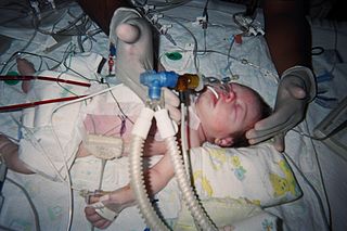
Extracorporeal membrane oxygenation (ECMO), is a form of extracorporeal life support, providing prolonged cardiac and respiratory support to persons whose heart and lungs are unable to provide an adequate amount of oxygen, gas exchange or blood supply (perfusion) to sustain life. The technology for ECMO is largely derived from cardiopulmonary bypass, which provides shorter-term support with arrested native circulation. The device used is a membrane oxygenator, also known as an artificial lung.

dextro-Transposition of the great arteries Medical condition

dextro-Transposition of the great arteries is a potentially life-threatening birth defect in the large arteries of the heart. The primary arteries are transposed.

A cyanotic heart defect is any congenital heart defect (CHD) that occurs due to deoxygenated blood bypassing the lungs and entering the systemic circulation, or a mixture of oxygenated and unoxygenated blood entering the systemic circulation. It is caused by structural defects of the heart such as right-to-left or bidirectional shunting, malposition of the great arteries, or any condition which increases pulmonary vascular resistance. The result may be the development of collateral circulation.

<span class="mw-page-title-main">Fontan procedure</span> Surgical procedure used in children with univentricular hearts

The Fontan procedure or Fontan–Kreutzer procedure is a palliative surgical procedure used in children with univentricular hearts. It involves diverting the venous blood from the inferior vena cava (IVC) and superior vena cava (SVC) to the pulmonary arteries. The procedure varies for differing congenital heart pathologies. For example in tricuspid atresia, the procedure can be done where the blood does not pass through the morphologic right ventricle; i.e., the systemic and pulmonary circulations are placed in series with the functional single ventricle. Whereas in hypoplastic left heart syndrome, the heart is more reliant on the more functional right ventricle to provide blood flow to the systemic circulation. The procedure was initially performed in 1968 by Francis Fontan and Eugene Baudet from Bordeaux, France, published in 1971, simultaneously described in July 1971 by Guillermo Kreutzer from Buenos Aires, Argentina, presented at the Argentinean National Cardilogy meeting of that year and finally published in 1973.

<span class="mw-page-title-main">Perfusionist</span> Healthcare professional who uses the cardiopulmonary bypass machine

A cardiovascular perfusionist, clinical perfusionist or perfusiologist, and occasionally a cardiopulmonary bypass doctor or clinical perfusion scientist, is a healthcare professional who operates the cardiopulmonary bypass machine during cardiac surgery and other surgeries that require cardiopulmonary bypass to manage the patient's physiological status. As a member of the cardiovascular surgical team, the perfusionist also known as the clinical perfusionist helps maintain blood flow to the body's tissues as well as regulate levels of oxygen and carbon dioxide in the blood, using a heart–lung machine.

<span class="mw-page-title-main">Atrium (heart)</span> Part of the human heart

The atrium is one of the two upper chambers in the heart that receives blood from the circulatory system. The blood in the atria is pumped into the heart ventricles through the atrioventricular mitral and tricuspid heart valves.

<span class="mw-page-title-main">Arterial switch operation</span> Open heart surgical procedure

Arterial switch operation (ASO) or arterial switch, is an open heart surgical procedure used to correct dextro-transposition of the great arteries (d-TGA).

<span class="mw-page-title-main">Pulmonary thromboendarterectomy</span>

In thoracic surgery, a pulmonary thromboendarterectomy (PTE), also referred to as pulmonary endarterectomy (PEA), is an operation that removes organized clotted blood (thrombus) from the pulmonary arteries, which supply blood to the lungs.

Cardioplegia is a solution given to the heart during cardiac surgery, to minimize the damage caused by myocardial ischemia while the heart is paused.

A cardiac shunt is a pattern of blood flow in the heart that deviates from the normal circuit of the circulatory system. It may be described as right-left, left-right or bidirectional, or as systemic-to-pulmonary or pulmonary-to-systemic. The direction may be controlled by left and/or right heart pressure, a biological or artificial heart valve or both. The presence of a shunt may also affect left and/or right heart pressure either beneficially or detrimentally.

<span class="mw-page-title-main">Oxygenator</span> Medical equipment

An oxygenator is a medical device that is capable of exchanging oxygen and carbon dioxide in the blood of human patients during surgical procedures that may necessitate the interruption or cessation of blood flow in the body, a critical organ or great blood vessel. These organs can be the heart, lungs or liver, while the great vessels can be the aorta, pulmonary artery, pulmonary veins or vena cava.

<span class="mw-page-title-main">Bidirectional Glenn procedure</span>

The bidirectional Glenn (BDG) shunt, or bidirectional cavopulmonary anastomosis, is a surgical technique used in pediatric cardiac surgery procedure used to temporarily improve blood oxygenation for patients with a congenital cardiac defect resulting in a single functional ventricle. Creation of a bidirectional shunt reduces the amount of blood volume that the heart needs to pump at the time of surgical repair with the Fontan procedure.

Cardiothoracic anesthesiology is a subspeciality of the medical practice of anesthesiology, devoted to the preoperative, intraoperative, and postoperative care of adult and pediatric patients undergoing cardiothoracic surgery and related invasive procedures.

The Senning procedure is an atrial switch heart operation performed to treat transposition of the great arteries. It is named after its inventor, the Swedish cardiac surgeon Åke Senning (1915–2000), also known for implanting the first permanent cardiac pacemaker in 1958.

Extracorporeal cardiopulmonary resuscitation is a method of cardiopulmonary resuscitation (CPR) that passes the patient's blood through a machine in a process to oxygenate the blood supply. A portable extracorporeal membrane oxygenation (ECMO) device is used as an adjunct to standard CPR. A patient who is deemed to be in cardiac arrest refractory to CPR has percutaneous catheters inserted into the femoral vein and artery. Theoretically, the application of ECPR allows for the return of cerebral perfusion in a more sustainable manner than with external compressions alone. By attaching an ECMO device to a person who has acutely undergone cardiovascular collapse, practitioners can maintain end-organ perfusion whilst assessing the potential reversal of causal pathology, with the goal of improving long-term survival and neurological outcomes.

Retrograde autologous priming (RAP) is a means to effectively and safely restrict the hemodilution caused by the direct homologous blood transfusion and reduce the blood transfusion requirements during cardiac surgery. It is also generally considered a blood conservation method used in most patients during the cardiopulmonary bypass (CPB). The processing of RAP includes three main steps, and the entire procedure of RAP could be completed within 5 to 8 minutes. This technique is proposed by Panico in 1960 for the first time and restated by Rosengart in 1998 to eliminate or reduce the risk of hemodilution during CPB. Moreover, to precisely determine the clinical efficacy of RAP, many related studies were conducted. Most results of researches indicate that RAP is available to provide some benefits to reducing the requirements for red blood cell transfusion. However, there are still some studies showing a failure of RAP to limit the hemodilution after the open heart operation.

Raghib syndrome is rare a congenital heart defect where the left superior vena cava (LSVC) is draining into the left atrium in addition to an absent coronary sinus and an atrial septal defect. This can be considered a dangerous heart condition because it puts the individual at a high risk of stroke. Other defects that are often associated with Raghib syndrome can include ventricular septal defects, enlargement of the tricuspid annulus, and pulmonary stenosis. While this is considered an extremely rare developmental complex, cases regarding a persistent left superior vena cava (PLSVC) are relatively common among congenital heart defects. It is also important to note that the PLSVC often drains into the right atrium, and only drains into the left atrium in approximately 10 to 20% of individuals with the defect.

References

  1. Stefanou D, Dimarakis I (2020). "9. Adult cardiopulmonary bypass". In Raja SG (ed.). Cardiac Surgery: A Complete Guide. Switzerland: Springer. pp. 93–99. ISBN   978-3-030-24176-6.
  2. Al-Atassi, Talal; Toeg, Hadi D.; Chan, Vincent; Ruel, Marc (2016). "Coronary Artery Bypass Grafting". In Frank Sellke; Pedro J. del Nido (eds.). Sabiston and Spencer Surgery of the Chest. p. 1553–1554. ISBN   978-0-323-24126-7.
  3. Madani, Michael M. (2016). "50. Pulmonary Thromboendarterectomy". In Peacock, Andrew J.; Naeije, Robert; Rubin, Lewis J. (eds.). Pulmonary Circulation: Diseases and Their Treatment, Fourth Edition. CRC Press. p. 541. ISBN   978-1-4987-1991-9.
  4. Casazza F, Roncon L, Greco F (Oct 2005). "Pulmonary embolism: treatment of the acute episode". Ital Heart J. 6 (10): 818–23. PMID   16270473.
  5. Lich B, Brown M (2004). The Manual of Clinical Perfusion (2nd ed.). Fort Myers, Florida: perfusion.com. ISBN   978-0-9753396-0-2.
  6. Ismail A, Miskolczi SY (2019), "Cardiopulmonary Bypass", StatPearls, StatPearls Publishing, PMID   29489210 , retrieved 2020-01-21
  7. Shore-Lesserson L, Baker RA, Ferraris VA, Greilich PE, Fitzgerald D, Roman P, Hammon JW (February 2018). "The Society of Thoracic Surgeons, The Society of Cardiovascular Anesthesiologists, and The American Society of ExtraCorporeal Technology: Clinical Practice Guidelines-Anticoagulation During Cardiopulmonary Bypass". The Annals of Thoracic Surgery. 105 (2): 650–662. doi: 10.1016/j.athoracsur.2017.09.061 . PMID   29362176.
  8. Finley A, Greenberg C (June 2013). "Review article: heparin sensitivity and resistance: management during cardiopulmonary bypass". Anesthesia and Analgesia. 116 (6): 1210–1222. doi: 10.1213/ANE.0b013e31827e4e62 . PMID   23408671. S2CID   22500786.
  9. Berg C, Knüppel M, Geipel A, Kohl T, Krapp M, Knöpfle G, et al. (March 2006). "Prenatal diagnosis of persistent left superior vena cava and its associated congenital anomalies". Ultrasound in Obstetrics & Gynecology. 27 (3): 274–280. doi: 10.1002/uog.2704 . PMID   16456841. S2CID   26364072.
  10. 1 2 3 4 5 6 7 8 9 10 11 12 13 Cohn LH (2017-08-28). Cardiac surgery in the adult (Fifth ed.). New York. ISBN   978-0-07-184487-1. OCLC   930496902.{{cite book}}: CS1 maint: location missing publisher (link)
  11. 1 2 3 4 5 6 7 8 Kouchoukos NT, Kirklin JW (2013). Kirklin/Barratt-Boyes cardiac surgery : morphology, diagnostic criteria, natural history, techniques, results, and indications (4th ed.). Philadelphia: Elsevier/Saunders. ISBN   978-1-4557-4605-7. OCLC   812289395.
  12. 1 2 3 4 Lapar DJ (2016). Tsra review of cardiothoracic surgery. [Place of publication not identified]: Createspace. ISBN   978-1-5232-1716-8. OCLC   953497320.
  13. Man survives 16 days without a heart United Press International. April 3, 2008.
  14. Nybo M, Madsen JS (August 2008). "Serious anaphylactic reactions due to protamine sulfate: a systematic literature review". Basic & Clinical Pharmacology & Toxicology. 103 (2): 192–196. doi: 10.1111/j.1742-7843.2008.00274.x . PMID   18816305.
  15. Stutz B (9 January 2009). "Pumphead: Does the heart-lung machine have a dark side?". Scientific American.
  16. Davies H. "Cardiopulmonary bypass machine - CPB". www.ebme.co.uk. Retrieved 2019-11-21.
  17. 1 2 3 4 5 6 Mokadam NA (2015). Cardiopulmonary bypass : a primer. University of Washington. OCLC   922073684.
  18. 1 2 3 4 5 Youssef SJ, Williams JA (2013). TSRA Primer of Cardiothoracic Surgery. Chicago, IL: TSRA/TSDA. ISBN   978-0-9894023-0-9.
  19. Cohn LH (2017-08-28). Cardiac surgery in the adult (Fifth ed.). New York. ISBN   978-0-07-184487-1. OCLC   930496902.{{cite book}}: CS1 maint: location missing publisher (link)
  20. Kouchoukos NT, Kirklin JW (2013). Kirklin/Barratt-Boyes cardiac surgery : morphology, diagnostic criteria, natural history, techniques, results, and indications (4th ed.). Philadelphia: Elsevier/Saunders. ISBN   978-1-4557-4605-7. OCLC   812289395.
  21. Kotani, Yuki; Kataoka, Yuki; Izawa, Junichi; Fujioka, Shoko; Yoshida, Takuo; Kumasawa, Junji; Kwong, Joey SW (2022-11-30). Cochrane Heart Group (ed.). "High versus low blood pressure targets for cardiac surgery while on cardiopulmonary bypass". Cochrane Database of Systematic Reviews. 2022 (11). doi:10.1002/14651858.CD013494.pub2. PMC   9709767 . PMID   36448514.
  22. Zimmer HG (September 2003). "The heart-lung machine was invented twice--the first time by Max von Frey". Clinical Cardiology. 26 (9): 443–445. doi:10.1002/clc.4960260914. PMC   6654655 . PMID   14524605.
  23. Norton J (2008). Surgery: Basic science and clinical evidence . NY: springer. pp.  1473. ISBN   978-0-387-30800-5.
  24. Hedlund KD (2001). "A Tribute to Frank F Alibritten, Jr. Origin of the left ventricular vent during the early years of open-heart surgery with the Gibbon heart-lung machine". Texas Heart Institute Journal. 28 (4): 292–296. PMC   101205 . PMID   11777154.
  25. US 4540399,Litzie K, Roberts CP,"US Patent for Emergency bypass system",issued 10 September 1985, assigned to C.R. Bard, Incand Lifestream International, LLC.
  26. Reichman RT, Joyo CI, Dembitsky WP, Griffith LD, Adamson RM, Daily PO, et al. (January 1990). "Improved patient survival after cardiac arrest using a cardiopulmonary support system". The Annals of Thoracic Surgery. 49 (1): 101–105. doi: 10.1016/0003-4975(90)90363-B . PMID   2297254.

Further reading