
A computed tomography scan is a medical imaging technique used to obtain detailed internal images of the body. The personnel that perform CT scans are called radiographers or radiology technologists.

Radiology is the medical discipline that uses medical imaging to diagnose diseases and guide their treatment, within the bodies of humans and other animals. It began with radiography, but today it includes all imaging modalities, including those that use no electromagnetic radiation, as well as others that do, such as computed tomography (CT), fluoroscopy, and nuclear medicine including positron emission tomography (PET). Interventional radiology is the performance of usually minimally invasive medical procedures with the guidance of imaging technologies such as those mentioned above.

Tomography is imaging by sections or sectioning that uses any kind of penetrating wave. The method is used in radiology, archaeology, biology, atmospheric science, geophysics, oceanography, plasma physics, materials science, astrophysics, quantum information, and other areas of science. The word tomography is derived from Ancient Greek τόμος tomos, "slice, section" and γράφω graphō, "to write" or, in this context as well, "to describe." A device used in tomography is called a tomograph, while the image produced is a tomogram.
The Hounsfield scale, named after Sir Godfrey Hounsfield, is a quantitative scale for describing radiodensity. It is frequently used in CT scans, where its value is also termed CT number.

Treacher Collins syndrome (TCS) is a genetic disorder characterized by deformities of the ears, eyes, cheekbones, and chin. The degree to which a person is affected, however, may vary from mild to severe. Complications may include breathing problems, problems seeing, cleft palate, and hearing loss. Those affected generally have normal intelligence.

Virtual colonoscopy is the use of CT scanning or magnetic resonance imaging (MRI) to produce two- and three-dimensional images of the colon, from the lowest part, the rectum, to the lower end of the small intestine, and to display the images on an electronic display device. The procedure is used to screen for colon cancer and polyps, and may detect diverticulosis. A virtual colonoscopy can provide 3D reconstructed endoluminal views of the bowel. VC provides a secondary benefit of revealing diseases or abnormalities outside the colon.

Forensic facial reconstruction is the process of recreating the face of an individual from their skeletal remains through an amalgamation of artistry, anthropology, osteology, and anatomy. It is easily the most subjective—as well as one of the most controversial—techniques in the field of forensic anthropology. Despite this controversy, facial reconstruction has proved successful frequently enough that research and methodological developments continue to be advanced.
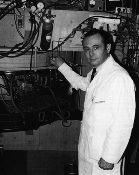
Michel Matthew Ter-Pogossian was an American medical physicist. He was professor of radiology at the Washington University School of Medicine for over 30 years. A pioneer in nuclear medicine, he is best known for his research on the positron emission tomography (PET). He is considered one of its creators and often referred to as the "father of PET."
Willi A. Kalender is a German medical physicist and professor and former chairman of the Institute of Medical Physics of the University of Erlangen-Nuremberg. Kalender has produced several new technologies in the field of diagnostic radiology imaging.
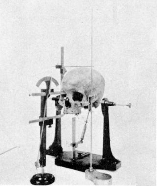
Cephalometry is the study and measurement of the head, usually the human head, especially by medical imaging such as radiography. Craniometry, the measurement of the cranium (skull), is a large subset of cephalometry. Cephalometry also has a history in phrenology, which is the study of personality and character as well as physiognomy, which is the study of facial features. Cephalometry as applied in a comparative anatomy context informs biological anthropology. In clinical contexts such as dentistry and oral and maxillofacial surgery, cephalometric analysis helps in treatment and research; cephalometric landmarks guide surgeons in planning and operating.

ITK-SNAP is an interactive software application that allows users to navigate three-dimensional medical images, manually delineate anatomical regions of interest, and perform automatic image segmentation. The software was designed with the audience of clinical and basic science researchers in mind, and emphasis has been placed on having a user-friendly interface and maintaining a limited feature set to prevent feature creep. ITK-SNAP is most frequently used to work with magnetic resonance imaging (MRI), cone-beam computed tomography (CBCT) and computed tomography (CT) data sets.
Virtopsy is a virtual alternative to a traditional autopsy, conducted with scanning and imaging technology. The name is a portmanteau of "virtual" and "autopsy" and is a trademark registered to Richard Dirnhofer (de), the former head of the Institute of Forensic Medicine of the University of Bern, Switzerland.
Bone segment navigation is a surgical method used to find the anatomical position of displaced bone fragments in fractures, or to position surgically created fragments in craniofacial surgery. Such fragments are later fixed in position by osteosynthesis. It has been developed for use in craniofacial and oral and maxillofacial surgery.

Surgical planning is the preoperative method of pre-visualising a surgical intervention, in order to predefine the surgical steps and furthermore the bone segment navigation in the context of computer-assisted surgery. The surgical planning is most important in neurosurgery and oral and maxillofacial surgery. The transfer of the surgical planning to the patient is generally made using a medical navigation system.

Meresamun was an ancient Egyptian singer-priestess in the inner sanctum at the temple in Karnak. Her mummy, ca. 800 BC, was on exhibit at the Oriental Institute of Chicago Museum of the University of Chicago from February 10 to December 6, 2009. A special exhibition, “The Life of Meresamun: A Temple Singer in Ancient Egypt,” opened in February 2009 and provides a personal look into Meresamun's life.
Allan George Farman is a Professor of Radiology and Imaging Science, Department of Surgical and Hospital Dentistry, The University of Louisville School of Dentistry, and also serves both as Adjunct Professor of Anatomical Sciences and Neurobiology and as Clinical Professor of Diagnostic Radiology of the School of Medicine in the same institution.

Cone beam computed tomography is a medical imaging technique consisting of X-ray computed tomography where the X-rays are divergent, forming a cone.

Rotational angiography is a medical imaging technique based on x-ray, that allows to acquire CT-like 3D volumes during hybrid surgery or during a catheter intervention using a fixed C-Arm. The fixed C-Arm thereby rotates around the patient and acquires a series of x-ray images that are then reconstructed through software algorithms into a 3D image. Synonyms for rotational angiography include flat-panel volume CT and cone-beam CT.
The Mallinckrodt Institute of Radiology (MIR), established 1931, is an academic radiology center associated with the Washington University School of Medicine, located within the Washington University Medical Center in St. Louis, Missouri. In addition to providing diagnostic and therapeutic patient-care services, the institute is a top research and education center. It employs over 140 academic staff and is among the top recipients of National Institutes of Health funding of radiology departments. The center provides radiology services to Barnes-Jewish and St. Louis Children's hospitals, as well as multiple other hospitals and outpatient centers in the St. Louis area. The center performs 700,000 examinations and procedures annually.
Pamela K. Woodard is an American cardiovascular physician who is the Hugh Monroe Wilson Professor of Radiology at the Mallinckrodt Institute of Radiology. She was elected a Fellow of the American Association for the Advancement of Science in 2022.