Nipah virus infection

Last updated

Nipah virus infection
Henipavirus structure.svg
Structure of a Henipavirus
Specialty Infectious diseases   OOjs UI icon edit-ltr-progressive.svg
Symptoms None, fever, cough, headache, diarrhea, confusion [1]
Complications Inflammation of the brain, seizures [2]
Usual onset5 to 14 days after exposure [1]
CausesNipah virus (spread by direct contact) [1]
Diagnostic method Based on symptoms, confirmed by laboratory testing [1]
PreventionAvoiding exposure to bats and to sick pigs and people.
Treatment Supportive care only [2]
Prognosis The case fatality rate of Nipah virus infection is estimated at 40–75% but can vary by outbreak depending on surveillance and clinical management in affected areas. [3]
Frequency~701 human cases (1998 to May 2018) [4] [5]
Deaths~50 to 75% risk of death [4] [6]

A Nipah virus infection is a viral infection caused by the Nipah virus. Symptoms from infection vary from none to fever, cough, headache, shortness of breath, and confusion. This may worsen into a coma over a day or two, and 50 to 75% of those infected die. Complications can include inflammation of the brain and seizures following recovery. [2] [1]

Contents

The Nipah virus (NiV) is a type of RNA virus in the genus Henipavirus, which normally circulates among fruit bats of the genus Pteropus. [7] Spread typically requires direct contact with an infected source; it can both spread between people and from other animals to people. Diagnosis is based on symptoms and confirmed by laboratory testing. [2] [1]

Management is restricted to supportive care; as of 2021 there is neither vaccine nor specific treatment. [2] Preventive measures include avoiding exposure to bats and infected animals such as pigs, and not drinking raw date palm sap. [1] As of May 2018 about 700 human cases of Nipah virus were estimated to have occurred, and 50 to 75 percent of those infected died. [4] [6] [5]

The disease was first identified in 1998 by a team of researchers at the Faculty of Medicine, University of Malaya during an outbreak in Malaysia. [8] The majority of the patients in Malaysia diagnosed with the disease were referred to and treated at the University of Malaya Medical Centre. The virus was isolated and identified in 1999. [2] The disease is named after a village in Malaysia, Sungai Nipah. Pigs may also be infected, and millions were killed by Malaysian authorities in 1999 to stop the spread of disease, a measure which proved to be successful. [2] [9]

Signs and symptoms

Human infections range from asymptomatic infection to acute respiratory infection, seizures and fatal encephalitis. This illness typically initially presents as 3-14 days of fever and headache, often accompanied by a cough, sore throat, difficulty breathing, and other signs of respiratory illness. [1] Infected people initially develop symptoms that include fever, headaches, myalgia, vomiting and sore throat. This can be followed by dizziness, drowsiness, altered consciousness, and neurological signs that indicate acute encephalitis. Some people can also experience atypical pneumonia and severe respiratory problems, including acute respiratory distress. Encephalitis and seizures occur in severe cases, progressing to coma within 24 to 48 hours. [1]

The incubation period is from 4 to 14 days but an incubation period as long as 45 days has been reported. [1]

Death occurs in 40-75% of cases, and some long-term side effects of infection include persistent convulsions and personality changes. Most survivors make a full recovery, although some are left with residual neurological conditions after acute encephalitis. Some cases of relapse have been reported. [1]

Transmission

The initial case in human outbreaks of Nipah virus has always been zoonotic [8] from exposure to contaminated secretions or tissues of infected bats or pigs. Subsequent human-to-human transmission of Nipah virus occurs via close contact with NiV-infected persons or exposure to NiV-infected body fluids (e.g., blood, urine, nasal secretions). [1]

Most experts do not classify Nipah virus as airborne, though there is consensus that transmission can—and does—occur from short-range exposure to NiV-infected respiratory droplets in close contact settings. [10]

Indirect transmission of Nipah virus via contaminated fomites is likely responsible for many cases in which there was no known direct contact with a NiV-infected person or animal. [1]

Risk factors

The risk of exposure is high for hospital workers and caretakers of those infected with the virus. In Malaysia and Singapore, Nipah virus infected people with close contact to infected pigs. In Bangladesh and India, the disease has been linked to consumption of raw date palm sap (toddy), eating of fruits partially consumed by bats, and using water from wells inhabited by bats. [11] [12]

Diagnosis

Transmission electron micrograph (TEM) depicted a number of Nipah virus virions from a person's cerebrospinal fluid (CSF). Nipah.jpg
Transmission electron micrograph (TEM) depicted a number of Nipah virus virions from a person's cerebrospinal fluid (CSF).

During acute and convalescent stages of the disease, RNA can be detected using reverse transcriptase polymerase chain reaction (RT-PCR) from throat swabs, cerebrospinal fluid, urine and blood analysis. [1]

After recovery, IgG and IgM antibody detection can confirm a prior Nipah virus infection. Immunohistochemistry on tissues collected during autopsy also confirms the disease. [1]

Prevention

Prevention through sanitary practices is the best protection. The likelihood of infection through animal transmission can be reduced by avoiding exposure to sick pigs, and to bats where the disease is endemic. Bats harbor a significantly higher proportion of zoonotic viruses than all other mammalian orders, [14] and are known not to be affected by the many viruses they carry, apparently due to their developing special immune systems to deal with the stress of flying. [15] Infection via bats can be caused by drinking raw palm sap (palm toddy) contaminated by bat excreta, [16] eating fruits partially consumed by bats, and using water from wells infested by bats. [12] Bats are known to drink toddy that is collected in open containers, and occasionally urinate in it, which contaminates it with the virus. [16]

Standard infection control practices can protect against human-to-human hospital-acquired infections. [1]

In January 2024 a candidate vaccine, ChAdOx1 NipahB, commenced Phase I clinical trials after completing laboratory and animal testing. [17] [18]

Treatment

As of 2020, there is no specific treatment for Nipah virus infection. [19] The mainstay of treatment is supportive care. While tentative evidence supports the use of ribavirin, it has not yet been studied in people with the disease. [1] Specific antibodies have also been studied in an animal model with potential benefit. [1] Acyclovir, favipiravir, [19] and remdesivir [20] have been assessed as potential antivirals against Nipah virus.

M 102.4 is a nonpatented monoclonal antibody developed by Christopher C. Broder, a professor of immunology and microbiology at Uniformed Services University of the Health Sciences in Maryland. It proved highly effective in animal models. 50 doses were sent to Kerala in 2018, but it is not clear if they were used in humans.[ citation needed ]

Prognosis

Nipah virus infection is fatal in 40 to 70 percent of cases, [1] though some outbreaks have had 100% case fatality rate. [21]

Sequelae

Those who survive the initial infection often struggle with debilitating long-term neurological sequelae, including memory loss, impaired cognition, seizures, convulsions, and personality changes. [1]

Moreover, Nipah virus is known to be able to persist and lie dormant in survivors and to re-activate many months or years after the initial infection. [1] Deaths from re-activation of latent Nipah virus have been reported. [22]

Outbreaks

Map showing locations of outbreaks of Nipah and Hendra virus as well as the range of Pteropus bats as of 2014 Hendra-distribution-map.jpg
Map showing locations of outbreaks of Nipah and Hendra virus as well as the range of Pteropus bats as of 2014

Nipah virus outbreaks have been reported in Malaysia, Singapore, Bangladesh and India. The area is known as the Nipah Belt. The highest mortality due to Nipah virus infection was found in Bangladesh,[ citation needed ] where outbreaks are typically seen in winter. [23] Nipah virus was first seen in 1998 in peninsular Malaysia in pigs and pig farmers. By mid-1999, more than 265 human cases of encephalitis, including 105 deaths, had been reported in Malaysia, and 11 cases of either encephalitis or respiratory illness with one fatality were reported in Singapore. [24] In 2001, Nipah virus was reported from Meherpur District, Bangladesh [25] [26] and Siliguri, India. [25] The outbreak again appeared in 2003, 2004 and 2005 in Naogaon District, Manikganj District, Rajbari District, Faridpur District and Tangail District. [26] In Bangladesh there were further outbreaks in subsequent years. [27] [6]

Research

Ribavirin, m102.4 monoclonal antibody, and favipiravir were being studied as treatments as of 2019. [51]

Medication

Ribavirin has been studied in a small number of people. As of 2011, it was unclear whether it was useful, although a few people had returned to normal life after treatment. [52] In vitro studies and animal studies have shown conflicting results in the efficacy of ribavirin against NiV and Hendra, with some studies showing effective inhibition of viral replication in cell lines, [53] [54] whereas some studies in animal models showed that ribavirin treatment only delayed but did not prevent death after NiV or Hendra virus infection. [55] [56]

In 2013, the anti-malarial drug chloroquine was shown to block the critical functions needed for maturation of Nipah virus, although no clinical benefit was observed. [4]

Immunization

Passive immunization using a human monoclonal antibody, m102.4, that targets the ephrin-B2 and ephrin-B3 receptor-binding domain of the henipavirus Nipah G glycoprotein was evaluated in the ferret model as post-exposure prophylaxis. [4] [1] m102.4 has been used in people on a compassionate use basis in Australia, and was in pre-clinical development in 2013. [4]

Society and culture

The fictional MEV-1 virus featured in the 2011 film Contagion was based on a combination of Nipah and measles virus. [57]

A Malayalam movie, Virus , was released in 2019, based on the 2018 outbreak of Nipah virus in Kerala, India. [58] [59]

In series 4 episode 1 of The Good Karma Hospital , a patient is admitted with Nipah.[ citation needed ]

Episode 7 of the Korean drama Doctor John also features a Nipah virus outbreak.[ citation needed ]

See also

Related Research Articles

<span class="mw-page-title-main">Lassa fever</span> Viral disease spread by a type of mouse

Lassa fever, also known as Lassa hemorrhagic fever, is a type of viral hemorrhagic fever caused by the Lassa virus. Many of those infected by the virus do not develop symptoms. When symptoms occur they typically include fever, weakness, headaches, vomiting, and muscle pains. Less commonly there may be bleeding from the mouth or gastrointestinal tract. The risk of death once infected is about one percent and frequently occurs within two weeks of the onset of symptoms. Of those who survive, about a quarter have hearing loss, which improves within three months in about half of these cases.

<span class="mw-page-title-main">Hand, foot, and mouth disease</span> Common human disease caused by a group of viruses

Hand, foot, and mouth disease (HFMD) is a common infection caused by a group of enteroviruses. It typically begins with a fever and feeling generally unwell. This is followed a day or two later by flat discolored spots or bumps that may blister, on the hands, feet and mouth and occasionally buttocks and groin. Signs and symptoms normally appear 3–6 days after exposure to the virus. The rash generally resolves on its own in about a week. Fingernail and toenail loss may occur a few weeks later, but they will regrow with time.

<i>Henipavirus</i> Genus of RNA viruses

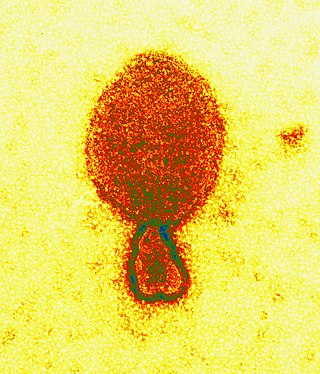
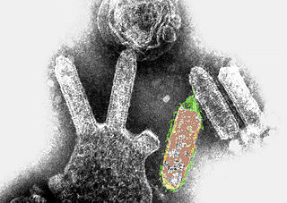
Henipavirus is a genus of negative-strand RNA viruses in the family Paramyxoviridae, order Mononegavirales containing six established species, and numerous others still under study. Henipaviruses are naturally harboured by several species of small mammals, notably pteropid fruit bats, microbats of several species, and shrews. Henipaviruses are characterised by long genomes and a wide host range. Their recent emergence as zoonotic pathogens capable of causing illness and death in domestic animals and humans is a cause of concern.

<span class="mw-page-title-main">Japanese encephalitis</span> Infection of the brain caused by the Japanese encephalitis virus

Japanese encephalitis (JE) is an infection of the brain caused by the Japanese encephalitis virus (JEV). While most infections result in little or no symptoms, occasional inflammation of the brain occurs. In these cases, symptoms may include headache, vomiting, fever, confusion and seizures. This occurs about 5 to 15 days after infection.

<span class="mw-page-title-main">Hendra virus</span> Species of virus

Hendra virus is a zoonotic virus found solely in Australia. First isolated in 1994, the virus has since been connected to numerous outbreaks of disease in domestic horses and seven human cases. Hendra virus belongs to the genus Henipavirus, which also contains the zoonotic Nipah virus. The reservoir species of Hendra virus are four species of bat within the genus Pteropus native to Australia.

<i>Nipah virus</i> Species of virus

Nipah virus is a bat-borne, zoonotic virus that causes Nipah virus infection in humans and other animals, a disease with a very high mortality rate (40-75%). Numerous disease outbreaks caused by Nipah virus have occurred in South East Africa and Southeast Asia. Nipah virus belongs to the genus Henipavirus along with the Hendra virus, which has also caused disease outbreaks.

<span class="mw-page-title-main">Viral hemorrhagic fever</span> Type of illnesses

Viral hemorrhagic fevers (VHFs) are a diverse group of animal and human illnesses. VHFs may be caused by five distinct families of RNA viruses: the families Filoviridae, Flaviviridae, Rhabdoviridae, and several member families of the Bunyavirales order such as Arenaviridae, and Hantaviridae. All types of VHF are characterized by fever and bleeding disorders and all can progress to high fever, shock and death in many cases. Some of the VHF agents cause relatively mild illnesses, such as the Scandinavian nephropathia epidemica, while others, such as Ebola virus, can cause severe, life-threatening disease.

<span class="mw-page-title-main">Kyasanur Forest disease</span> Human disease

Kyasanur forest disease (KFD) is a tick-borne viral haemorrhagic fever endemic to South-western part of India. The disease is caused by a virus belonging to the family Flaviviridae. KFDV is transmitted to humans through the bite of infected hard ticks which act as a reservoir of KFDV.

<i>Australian bat lyssavirus</i> Species of virus

Australian bat lyssavirus (ABLV), originally named Pteropid lyssavirus (PLV), is a enzootic virus closely related to the rabies virus. It was first identified in a 5-month-old juvenile black flying fox collected near Ballina in northern New South Wales, Australia, in January 1995 during a national surveillance program for the recently identified Hendra virus. ABLV is the seventh member of the genus Lyssavirus and the only Lyssavirus member present in Australia. ABLV has been categorized to the Phylogroup I of the Lyssaviruses.

<span class="mw-page-title-main">Crimean–Congo hemorrhagic fever</span> Disease of humans and other animals

Crimean–Congo hemorrhagic fever (CCHF) is a viral disease. Symptoms of CCHF may include fever, muscle pains, headache, vomiting, diarrhea, and bleeding into the skin. Onset of symptoms is less than two weeks following exposure. Complications may include liver failure. Survivors generally recover around two weeks after onset.

The central nervous system (CNS) controls most of the functions of the body and mind. It comprises the brain, spinal cord and the nerve fibers that branch off to all parts of the body. The CNS viral diseases are caused by viruses that attack the CNS. Existing and emerging viral CNS infections are major sources of human morbidity and mortality.

Lujo is a bisegmented RNA virus—a member of the family Arenaviridae—and a known cause of viral hemorrhagic fever (VHF) in humans. Its name was suggested by the Special Pathogens Unit of the National Institute for Communicable Diseases of the National Health Laboratory Service (NICD-NHLS) by using the first two letters of the names of the cities involved in the 2008 outbreak of the disease, Lusaka (Zambia) and Johannesburg. It is the second pathogenic Arenavirus to be described from the African continent—the first being Lassa virus—and since 2012 has been classed as a "Select Agent" under U.S. law.

<span class="mw-page-title-main">Favipiravir</span> Experimental antiviral drug with potential activity against RNA viruses

Favipiravir, sold under the brand name Avigan among others, is an antiviral medication used to treat influenza in Japan. It is also being studied to treat a number of other viral infections, including SARS-CoV-2. Like the experimental antiviral drugs T-1105 and T-1106, it is a pyrazinecarboxamide derivative.

<span class="mw-page-title-main">Bat virome</span> Group of viruses associated with bats

The bat virome is the group of viruses associated with bats. Bats host a diverse array of viruses, including all seven types described by the Baltimore classification system: (I) double-stranded DNA viruses; (II) single-stranded DNA viruses; (III) double-stranded RNA viruses; (IV) positive-sense single-stranded RNA viruses; (V) negative-sense single-stranded RNA viruses; (VI) positive-sense single-stranded RNA viruses that replicate through a DNA intermediate; and (VII) double-stranded DNA viruses that replicate through a single-stranded RNA intermediate. The greatest share of bat-associated viruses identified as of 2020 are of type IV, in the family Coronaviridae.

<span class="mw-page-title-main">Nipah virus outbreaks in Kerala</span> Nipah virus outbreaks occurring in India

There have been several outbreaks of Nipah virus in Kerala, some of which have been traced to fruit bats. The National Institute of Virology in Pune confirmed the first case of Nipah virus in Kerala in May 2018.

<i>Virus</i> (2019 film) 2019 film directed by Aashiq Abu

Virus is a 2019 Indian Malayalam-language medical thriller film co-produced and directed by Aashiq Abu set against the backdrop of the 2018 Nipah virus outbreak in Kerala. The film was written by Muhsin Parari and Suhas-Sharfu. It stars an ensemble cast that includes Kunchacko Boban, Indrajith Sukumaran, Asif Ali, Tovino Thomas, Parvathy Thiruvothu, Rahman, Indrans, Jinu Joseph, Sreenath Bhasi, Dileesh Pothan, Rima Kallingal, Madonna Sebastian, Ramya Nambeeshan, Joju George, Soubin Shahir and Revathi.

Daniel R. Lucey is an American physician, researcher, senior scholar and adjunct professor of infectious diseases at Georgetown University, and a research associate in anthropology at the Smithsonian National Museum of Natural History, where he has co-organised an exhibition on eight viral outbreaks.

<span class="mw-page-title-main">1998–1999 Malaysia Nipah virus outbreak</span> Disease outbreak in Malaysia

The 1998–1999 Malaysia Nipah virus outbreak was a Nipah virus outbreak occurring from September 1998 to May 1999 in the states of Perak, Negeri Sembilan and Selangor in Malaysia. A total of 265 cases of acute encephalitis with 105 deaths caused by the virus were reported in the three states throughout the outbreak. The Malaysian health authorities at first thought that Japanese encephalitis (JE) was the cause of the infection. This misunderstanding hampered the deployment of effective measures to prevent the spread, before the disease was identified by a local virologist from the Faculty of Medicine, University of Malaya as a newly discovered agent. It was named Nipah virus (NiV). The disease was as deadly as the Ebola virus disease (EVD), but attacked the brain system instead of the blood vessels. University of Malaya's Faculty of Medicine and the University of Malaya Medical Centre played a major role in serving as a major referral centre for the outbreak, treating majority of the Nipah patients and was instrumental in isolating the novel virus and researched on its features.

Ghanaian bat henipavirus (also known Kumasi virus belongs to the genus Henipavirus in the family Paramyxoviridae. Human infections are caused by zoonotic events where the virus crosses over from another animal species. Therefore, humans are not the innate host for this virus family but instead become infected by peripheral viral reservoirs such as bats and other carriers of the virus. When these virus are spread to humans through zoonotic events they have been found to be one of the most deadly viruses with the capability to infect humans, with mortality rates between 50 and 100%. Therefore, these viruses have been classified as a biosafety level four virus with regards to its pathogenesis when it infects humans.

References

  1. 1 2 3 4 5 6 7 8 9 10 11 12 13 14 15 16 17 18 19 20 21 22 "Nipah Virus (NiV)". Centers for Disease Control and Prevention, National Center for Emerging and Zoonotic Infectious Diseases (NCEZID), Division of High-Consequence Pathogens and Pathology (DHCPP), Viral Special Pathogens Branch (VSPB). 19 October 2022. Retrieved 17 September 2023.
  2. 1 2 3 4 5 6 7 "Nipah Virus (NiV) Infection". World Health Organization (WHO). Archived from the original on 26 September 2020. Retrieved 14 October 2020.
  3. "Nipah Virus Infection". who.int. World Health Organization. Retrieved 16 February 2023.
  4. 1 2 3 4 5 6 Broder CC, Xu K, Nikolov DB, Zhu Z, Dimitrov DS, Middleton D, et al. (October 2013). "A treatment for and vaccine against the deadly Hendra and Nipah viruses". Antiviral Research. 100 (1): 8–13. doi:10.1016/j.antiviral.2013.06.012. PMC   4418552 . PMID   23838047.
  5. 1 2 "Morbidity and mortality due to Nipah or Nipah-like virus encephalitis in WHO South-East Asia Region, 2001-2018" (PDF). SEAR. Archived from the original (PDF) on 13 June 2018. Retrieved 2 June 2018. 112 cases since Oct 2013
  6. 1 2 3 "Nipah virus outbreaks in the WHO South-East Asia Region". South-East Asia Regional Office. WHO. Archived from the original on 23 May 2018. Retrieved 23 May 2018.
  7. Singh RK, Dhama K, Chakraborty S, Tiwari R, Natesan S, Khandia R, et al. (1 January 2019). "Nipah virus: epidemiology, pathology, immunobiology and advances in diagnosis, vaccine designing and control strategies – a comprehensive review". Veterinary Quarterly. 39 (1): 26–55. doi:10.1080/01652176.2019.1580827. ISSN   0165-2176. PMC   6830995 . PMID   31006350.
  8. 1 2 "Nipah virus factsheet". World Health Organization. 30 May 2018. Retrieved 17 September 2023.
  9. "Seven things you need to know about Nipah virus | Gavi, the Vaccine Alliance". www.gavi.org. 15 September 2023. Retrieved 17 September 2023.
  10. "Nipah virus airborne". Australian Veterinary Journal. 77 (9): 558. 1999. doi:10.1111/j.1751-0813.1999.tb13182.x.
  11. Luby SP, Gurley ES, Hossain MJ (2012). Transmission of Human Infection with Nipah Virus. National Academies Press (US). Archived from the original on 22 May 2018. Retrieved 21 May 2018.
  12. 1 2 Balan S (21 May 2018). "6 Nipah virus deaths in Kerala: Bat-infested house well of first victims sealed". The News Minute. Archived from the original on 22 May 2018. Retrieved 21 May 2018.
  13. "Nipah Virus Infection symptoms, causes, treatment, medicine, prevention, diagnosis". myUpchar. Retrieved 27 January 2020.
  14. Olival KJ, Hosseini PR, Zambrana-Torrelio C, Ross N, Bogich TL, Daszak P (2017). "Host and viral traits predict zoonotic spillover from mammals". Nature. 546 (7660): 646–650. Bibcode:2017Natur.546..646O. doi:10.1038/nature22975. ISSN   0028-0836. PMC   5570460 . PMID   28636590.
  15. Huang P (9 February 2020). "Bats Carry Many Viruses. So Why Don't They Get Sick?". National Public Radio (npr).
  16. 1 2 Islam MS, Sazzad HM, Satter SM, Sultana S, Hossain MJ, Hasan M, et al. (April 2016). "Nipah Virus Transmission from Bats to Humans Associated with Drinking Traditional Liquor Made from Date Palm Sap, Bangladesh, 2011-2014". Emerging Infectious Diseases. 22 (4): 664–70. doi:10.3201/eid2204.151747. PMC   4806957 . PMID   26981928.
  17. "First in-human vaccine trial for Nipah virus". Oxford Vaccine Group. 11 January 2024. Retrieved 13 January 2024.
  18. Doremalen Nv, Avanzato VA, Feldmann F, Schulz JE, Haddock E, Okumura A, et al. (20 July 2021). "ChAdOx1 NiV vaccination protects against lethal Nipah Bangladesh virus infection in African green monkeys". bioRxiv: 2021.07.20.452991. doi:10.1101/2021.07.20.452991. S2CID   236179976.
  19. 1 2 Sharma V, Kaushik S, Kumar R, Yadav JP, Kaushik S (January 2019). "Emerging trends of Nipah virus: A review". Reviews in Medical Virology. 29 (1): e2010. doi: 10.1002/rmv.2010 . PMC   7169151 . PMID   30251294.
  20. Lo MK, Feldmann F, Gary JM, Jordan R, Bannister R, Cronin J, et al. (29 May 2019). "Remdesivir (GS-5734) protects African green monkeys from Nipah virus challenge". Science Translational Medicine. 11 (494): eaau9242. doi:10.1126/scitranslmed.aau9242. ISSN   1946-6234. PMC   6732787 . PMID   31142680.
  21. Shoemaker T, Choi MJ (24 June 2019). "Henipaviruses - Chapter 4 - 2020 Yellow Book | Travelers' Health | CDC". Centers for Disease Control and Prevention. Retrieved 12 December 2021.
  22. "Nipah virus: epidemiology, outbreaks and guidance". Public Health England. 30 May 2018. Retrieved 12 December 2021.
  23. Chadha MS, Comer JA, Lowe L, Rota PA, Rollin PE, Bellini WJ, et al. (February 2006). "Nipah virus-associated encephalitis outbreak, Siliguri, India". Emerging Infectious Diseases. 12 (2): 235–40. doi:10.3201/eid1202.051247. PMC   3373078 . PMID   16494748.
  24. Eaton BT, Broder CC, Middleton D, Wang LF (January 2006). "Hendra and Nipah viruses: different and dangerous". Nature Reviews. Microbiology. 4 (1): 23–35. doi:10.1038/nrmicro1323. PMC   7097447 . PMID   16357858. S2CID   24764543.
  25. 1 2 Chadha MS, Comer JA, Lowe L, Rota PA, Rollin PE, Bellini WJ, et al. (February 2006). "Nipah virus-associated encephalitis outbreak, Siliguri, India". Emerging Infectious Diseases. 12 (2): 235–40. doi:10.3201/eid1202.051247. PMC   3373078 . PMID   16494748.
  26. 1 2 Hsu VP, Hossain MJ, Parashar UD, Ali MM, Ksiazek TG, Kuzmin I, et al. (December 2004). "Nipah virus encephalitis reemergence, Bangladesh". Emerging Infectious Diseases. 10 (12): 2082–7. doi:10.3201/eid1012.040701. PMC   3323384 . PMID   15663842.
  27. "Basic preventive measures can reduce Nipah virus attack". The Daily Star (Bangladesh). 8 March 2008.
  28. 1 2 Looi L, Chua K (2007). "Lessons from the Nipah virus outbreak in Malaysia" (PDF). The Malaysian Journal of Pathology. Department of Pathology, University of Malaya and National Public Health Laboratory of the Ministry of Health, Malaysia. 29 (2): 63–7. PMID   19108397. Archived (PDF) from the original on 30 August 2019.
  29. Chadha MS, Comer JA, Lowe L (2006). "Nipah virus-associated encephalitis outbreak, Siliguri, India". Emerging Infectious Diseases. 12 (2): 235–40. doi:10.3201/eid1202.051247. PMC   3373078 . PMID   16494748.
  30. 1 2 Hsu VP, Hossain MJ, Parashar UD (2004). "Nipah virus encephalitis reemergence, Bangladesh". Emerging Infectious Diseases. 10 (12): 2082–7. doi:10.3201/eid1012.040701. PMC   3323384 . PMID   15663842.
  31. ICDDR,B (December 2005). "Nipah virus outbreak from date palm juice" (PDF). Centre for Population Research - Health and Science Bulletin. 3 (4): 1–5. ISSN   1729-343X. Archived from the original (PDF) on 22 July 2020. Retrieved 14 October 2020.
  32. "Person-to-person transmission of Nipah infection in Bangladesh" (PDF). Health and Science Bulletin. 5 (4): 1–6. December 2007. ISSN   1729-343X. Archived from the original on 3 February 2009.
  33. ICDDR,B (2008). "Outbreaks of Nipah virus in Rajbari and Manikgonj". Health and Science Bulletin. 6 (1): 12–3. Archived from the original on 3 February 2009.
  34. ICDDR,B (2010). "Nipah outbreak in Faridpur District, Bangladesh, 2010". Health and Science Bulletin. 8 (2): 6–11. Archived from the original on 28 September 2011.
  35. "Arguments in Bahodderhat murder case begin". The Daily Star. 18 March 2008. Retrieved 21 May 2014.
  36. তাহেরকে ফাঁসি দেওয়ার সিদ্ধান্ত নেন জিয়া. prothom-alo.com. 4 February 2011
  37. "Nipah virus outbreak: Death toll rises in Kerala, two more cases confirmed". indianexpress.com. 27 May 2018. Retrieved 29 May 2018.
  38. "Kozhikode on high alert as three deaths attributed to Nipah virus". The Indian Express. 20 May 2018. Retrieved 20 May 2018.
  39. "Deadly Nipah virus claims victims in India". BBC News. 21 May 2018. Retrieved 11 December 2018.
  40. 1 2 "Kerala Govt Confirms Nipah Virus, 86 Under Observation". New Delhi. 4 June 2019. Archived from the original on 14 July 2019. Retrieved 15 July 2019.
  41. Sharma NC (4 June 2019). "Centre gears up to contain re-emergence of Nipah virus in Kerala". Mint. Retrieved 7 June 2019.
  42. "Ernakulam district declared Nipah virus free, says Kerala health minister". India Today. Press Trust of India. 23 July 2019.
  43. "Nipah in Kozhikode: Health Minister Veena George says next one week crucial". OnManorama. Retrieved 5 September 2021.
  44. "Nipah returns to Kerala, 12-year-old dead in Kozhikode". The Indian Express. 5 September 2021. Retrieved 5 September 2021.
  45. "Nipah Back to Haunt Kerala as 12-Yr-Old Succumbs to Virus. Symptoms, Treatment & All You Must Know". News18. 5 September 2021. Retrieved 5 September 2021.
  46. Unnithan PS (5 September 2021). "12-year-old boy dies of Nipah virus in Kerala's Kozhikode". India Today. Retrieved 5 September 2021.
  47. "Authorities race to contain deadly Nipah virus outbreak in India". www.cbsnews.com. 7 September 2021. Retrieved 8 September 2021.
  48. "Nipah virus infection - Bangladesh".
  49. Bureau TH (13 September 2023). "Nipah outbreak | Number of cases rises to 5 in Kerala; 789 contacts kept under watch". The Hindu. ISSN   0971-751X . Retrieved 14 September 2023.
  50. Millson A (14 September 2023). "What Is Nipah and Why Is the Deadly Virus Flaring Up Again". Bloomberg.com. Retrieved 14 September 2023.
  51. Banerjee S, Gupta N, Kodan P, Mittal A, Ray Y, Nischal N, et al. (February 2019). "Nipah virus disease: A rare and intractable disease". Intractable & Rare Diseases Research. 8 (1): 1–8. doi:10.5582/irdr.2018.01130. PMC   6409114 . PMID   30881850.
  52. Vigant F, Lee B (June 2011). "Hendra and nipah infection: pathology, models and potential therapies". Infectious Disorders Drug Targets. 11 (3): 315–36. doi:10.2174/187152611795768097. PMC   3253017 . PMID   21488828.
  53. Wright PJ, Crameri G, Eaton BT (March 2005). "RNA synthesis during infection by Hendra virus: an examination by quantitative real-time PCR of RNA accumulation, the effect of ribavirin and the attenuation of transcription". Archives of Virology. 150 (3): 521–32. doi:10.1007/s00705-004-0417-5. PMID   15526144. S2CID   23885676.
  54. Aljofan M, Saubern S, Meyer AG, Marsh G, Meers J, Mungall BA (June 2009). "Characteristics of Nipah virus and Hendra virus replication in different cell lines and their suitability for antiviral screening". Virus Research. 142 (1–2): 92–9. doi:10.1016/j.virusres.2009.01.014. PMC   2744099 . PMID   19428741.
  55. Georges-Courbot MC, Contamin H, Faure C, Loth P, Baize S, Leyssen P, et al. (May 2006). "Poly(I)-poly(C12U) but not ribavirin prevents death in a hamster model of Nipah virus infection". Antimicrobial Agents and Chemotherapy. 50 (5): 1768–72. doi:10.1128/AAC.50.5.1768-1772.2006. PMC   1472238 . PMID   16641448.
  56. Freiberg AN, Worthy MN, Lee B, Holbrook MR (March 2010). "Combined chloroquine and ribavirin treatment does not prevent death in a hamster model of Nipah and Hendra virus infection". The Journal of General Virology. 91 (Pt 3): 765–72. doi:10.1099/vir.0.017269-0. PMC   2888097 . PMID   19889926.
  57. "Nipah virus inspired "Contagion." We're testing a vaccine". www.path.org. Retrieved 17 May 2020.
  58. Virus , retrieved 1 September 2019
  59. "Virus Movie Review {3.5/5}: A well-crafted multi-starrer, fictional documentation on the Nipah virus attack", The Times of India, retrieved 1 September 2019