Related Research Articles
A magneto-optic effect is any one of a number of phenomena in which an electromagnetic wave propagates through a medium that has been altered by the presence of a quasistatic magnetic field. In such a medium, which is also called gyrotropic or gyromagnetic, left- and right-rotating elliptical polarizations can propagate at different speeds, leading to a number of important phenomena. When light is transmitted through a layer of magneto-optic material, the result is called the Faraday effect: the plane of polarization can be rotated, forming a Faraday rotator. The results of reflection from a magneto-optic material are known as the magneto-optic Kerr effect.

Raman spectroscopy is a spectroscopic technique typically used to determine vibrational modes of molecules, although rotational and other low-frequency modes of systems may also be observed. Raman spectroscopy is commonly used in chemistry to provide a structural fingerprint by which molecules can be identified.

In physics, Raman scattering or the Raman effect is the inelastic scattering of photons by matter, meaning that there is both an exchange of energy and a change in the light's direction. Typically this effect involves vibrational energy being gained by a molecule as incident photons from a visible laser are shifted to lower energy. This is called normal Stokes-Raman scattering.
Coherent anti-Stokes Raman spectroscopy, also called Coherent anti-Stokes Raman scattering spectroscopy (CARS), is a form of spectroscopy used primarily in chemistry, physics and related fields. It is sensitive to the same vibrational signatures of molecules as seen in Raman spectroscopy, typically the nuclear vibrations of chemical bonds. Unlike Raman spectroscopy, CARS employs multiple photons to address the molecular vibrations, and produces a coherent signal. As a result, CARS is orders of magnitude stronger than spontaneous Raman emission. CARS is a third-order nonlinear optical process involving three laser beams: a pump beam of frequency ωp, a Stokes beam of frequency ωS and a probe beam at frequency ωpr. These beams interact with the sample and generate a coherent optical signal at the anti-Stokes frequency (ωpr+ωp-ωS). The latter is resonantly enhanced when the frequency difference between the pump and the Stokes beams (ωp-ωS) coincides with the frequency of a Raman resonance, which is the basis of the technique's intrinsic vibrational contrast mechanism.
A fiber laser is a laser in which the active gain medium is an optical fiber doped with rare-earth elements such as erbium, ytterbium, neodymium, dysprosium, praseodymium, thulium and holmium. They are related to doped fiber amplifiers, which provide light amplification without lasing.
Dual-polarization interferometry (DPI) is an analytical technique that probes molecular layers adsorbed to the surface of a waveguide using the evanescent wave of a laser beam. It is used to measure the conformational change in proteins, or other biomolecules, as they function.
Fluorescence anisotropy or fluorescence polarization is the phenomenon where the light emitted by a fluorophore has unequal intensities along different axes of polarization. Early pioneers in the field include Aleksander Jablonski, Gregorio Weber, and Andreas Albrecht. The principles of fluorescence polarization and some applications of the method are presented in Lakowicz's book.

In optics, a supercontinuum is formed when a collection of nonlinear processes act together upon a pump beam in order to cause severe spectral broadening of the original pump beam, for example using a microstructured optical fiber. The result is a smooth spectral continuum. There is no consensus on how much broadening constitutes a supercontinuum; however researchers have published work claiming as little as 60 nm of broadening as a supercontinuum. There is also no agreement on the spectral flatness required to define the bandwidth of the source, with authors using anything from 5 dB to 40 dB or more. In addition the term supercontinuum itself did not gain widespread acceptance until this century, with many authors using alternative phrases to describe their continua during the 1970s, 1980s and 1990s.

Second-harmonic imaging microscopy (SHIM) is based on a nonlinear optical effect known as second-harmonic generation (SHG). SHIM has been established as a viable microscope imaging contrast mechanism for visualization of cell and tissue structure and function. A second-harmonic microscope obtains contrasts from variations in a specimen's ability to generate second-harmonic light from the incident light while a conventional optical microscope obtains its contrast by detecting variations in optical density, path length, or refractive index of the specimen. SHG requires intense laser light passing through a material with a noncentrosymmetric molecular structure, either inherent or induced externally, for example by an electric field.

A beam of light has radial polarization if at every position in the beam the polarization vector points towards the center of the beam. In practice, an array of waveplates may be used to provide an approximation to a radially polarized beam. In this case the beam is divided into segments, and the average polarization vector of each segment is directed towards the beam centre.

Cross-polarized wave (XPW) generation is a nonlinear optical process that can be classified in the group of frequency degenerate processes. It can take place only in media with anisotropy of third-order nonlinearity. As a result of such nonlinear optical interaction at the output of the nonlinear crystal, it is generated a new linearly polarized wave at the same frequency, but with polarization oriented perpendicularly to the polarization of input wave

The Raman microscope is a laser-based microscopic device used to perform Raman spectroscopy. The term MOLE is used to refer to the Raman-based microprobe. The technique used is named after C. V. Raman, who discovered the scattering properties in liquids.
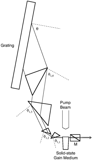
Multiple-prism grating laser oscillators, or MPG laser oscillators, use multiple-prism beam expansion to illuminate a diffraction grating mounted either in Littrow configuration or grazing-incidence configuration. Originally, these narrow-linewidth tunable dispersive oscillators were introduced as multiple-prism Littrow (MPL) grating oscillators, or hybrid multiple-prism near-grazing-incidence (HMPGI) grating cavities, in organic dye lasers. However, these designs were quickly adopted for other types of lasers such as gas lasers, diode lasers, and more recently fiber lasers.
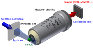
Light sheet fluorescence microscopy (LSFM) is a fluorescence microscopy technique with an intermediate-to-high optical resolution, but good optical sectioning capabilities and high speed. In contrast to epifluorescence microscopy only a thin slice of the sample is illuminated perpendicularly to the direction of observation. For illumination, a laser light-sheet is used, i.e. a laser beam which is focused only in one direction. A second method uses a circular beam scanned in one direction to create the lightsheet. As only the actually observed section is illuminated, this method reduces the photodamage and stress induced on a living sample. Also the good optical sectioning capability reduces the background signal and thus creates images with higher contrast, comparable to confocal microscopy. Because light sheet fluorescence microscopy scans samples by using a plane of light instead of a point, it can acquire images at speeds 100 to 1,000 times faster than those offered by point-scanning methods.
Brillouin spectroscopy is an empirical spectroscopy technique which allows the determination of elastic moduli of materials. The technique uses inelastic scattering of light when it encounters acoustic phonons in a crystal, a process known as Brillouin scattering, to determine phonon energies and therefore interatomic potentials of a material. The scattering occurs when an electromagnetic wave interacts with a density wave, photon-phonon scattering.
Stimulated Raman spectroscopy, also referred to as stimulated Raman scattering (SRS) is a form of spectroscopy employed in physics, chemistry, biology, and other fields. The basic mechanism resembles that of spontaneous Raman spectroscopy: a pump photon, of the angular frequency , which is scattered by a molecule has some small probability of inducing some vibrational transition, as opposed to inducing a simple Rayleigh transition. This makes the molecule emit a photon at a shifted frequency. However, SRS, as opposed to spontaneous Raman spectroscopy, is a third-order non-linear phenomenon involving a second photon—the Stokes photon of angular frequency —which stimulates a specific transition. When the difference in frequency between both photons resembles that of a specific vibrational transition the occurrence of this transition is resonantly enhanced. In SRS, the signal is equivalent to changes in the intensity of the pump and Stokes beams. The signals are typically rather low, of the order of a part in 10^5, thus calling for modulation-transfer techniques: one beam is modulated in amplitude and the signal is detected on the other beam via a lock-in amplifier. Employing a pump laser beam of a constant frequency and a Stokes laser beam of a scanned frequency allows for the unraveling of the spectral fingerprint of the molecule. This spectral fingerprint differs from those obtained by other spectroscopy methods such as Rayleigh scattering as the Raman transitions confer to different exclusion rules than those that apply for Rayleigh transitions.
Micro-spatially offset Raman spectroscopy (micro-SORS) is an analytical technique developed in 2014 that combines SORS with microscopy. The technique derives its sublayer‐resolving properties from its parent technique SORS. The main difference between SORS and micro-SORS is the spatial resolution: while SORS is suited to the analysis of millimetric layers, micro-SORS is able to resolve thin, micrometric-scale layers. Similarly to SORS technique, micro-SORS is able to preferentially collect the Raman photons generated under the surface in turbid media. In this way, it is possible to reconstruct the chemical makeup of micrometric multi-layered turbid system in a non destructive way. Micro-SORS is particularly useful when dealing with precious or unique objects as for Cultural Heritage field and Forensic Science or in biomedical applications, where a non-destructive molecular characterization constitute a great advantage.
Coherent Raman scattering (CRS) microscopy is a multi-photon microscopy technique based on Raman-active vibrational modes of molecules. The two major techniques in CRS microscopy are stimulated Raman scattering (SRS) and coherent anti-Stokes Raman scattering (CARS). SRS and CARS were theoretically predicted and experimentally realized in the 1960s. In 1982 the first CARS microscope was demonstrated. In 1999, CARS microscopy using a collinear geometry and high numerical aperture objective were developed in Xiaoliang Sunney Xie's lab at Harvard University. This advancement made the technique more compatible with modern laser scanning microscopes. Since then, CRS's popularity in biomedical research started to grow. CRS is mainly used to image lipid, protein, and other bio-molecules in live or fixed cells or tissues without labeling or staining. CRS can also be used to image samples labeled with Raman tags, which can avoid interference from other molecules and normally allows for stronger CRS signals than would normally be obtained for common biomolecules. CRS also finds application in other fields, such as material science and environmental science.

Anisotropic terahertz microspectroscopy (ATM) is a spectroscopic technique in which molecular vibrations in an anisotropic material are probed with short pulses of terahertz radiation whose electric field is linearly polarized parallel to the surface of the material. The technique has been demonstrated in studies involving single crystal sucrose, fructose, oxalic acid, and molecular protein crystals in which the spatial orientation of molecular vibrations are of interest.
References
- ↑ de Vito, Giuseppe; Bifone, Angelo; Vincenzo, Piazza (2012). "Rotating-polarization CARS microscopy: combining chemical and molecular orientation sensitivity". Optics Express. OSA Publishing. 20 (28): 29369–29377. doi: 10.1364/OE.20.029369 . hdl: 2158/1243645 .
- ↑ Bélanger, E.; Bégin, S.; Laffray, S.; De Koninck, Y.; Vallée, R.; Côté, D. (2009). "Quantitative myelin imaging with coherent anti-Stokes Raman scattering microscopy: alleviating the excitation polarization dependence with circularly polarized laser beams". Optics Express. 17 (21): 18419. Bibcode:2009OExpr..1718419B. doi: 10.1364/OE.17.018419 . ISSN 1094-4087.
- ↑ de Vito, Giuseppe; Bifone, Angelo; Piazza, Vincenzo (2012). "Rotating-polarization CARS microscopy: combining chemical and molecular orientation sensitivity". Optics Express. 20 (28): 29369. Bibcode:2012OExpr..2029369D. doi: 10.1364/OE.20.029369 . hdl: 2158/1243645 . ISSN 1094-4087.
- 1 2 de Vito, Giuseppe; Piazza, Vincenzo (2014). "Fast signal analysis in Rotating-Polarization CARS microscopy". Optical Data Processing and Storage. 1 (1). doi: 10.2478/odps-2014-0001 . hdl: 2158/1243643 . ISSN 2084-8862.