Thoracic aorta injury

Last updated
Thoracic aorta injury
Aorta Anatomy.jpg
Anatomy of the thoracic aorta

Injury of the thoracic aorta refers to any injury which affects the portion of the aorta which lies within the chest cavity. Injuries of the thoracic aorta are usually the result of physical trauma; however, they can also be the result of a pathological process. The main causes of this injury are deceleration (such as a car accident) and crush injuries. There are different grades to injuries to the aorta depending on the extent of injury, and the treatment whether surgical or medical depends on that grade. [1] It is difficult to determine if a patient has a thoracic injury just by their symptoms, but through imaging and a physical exam the extent of injury can be determined. [2] All patients with a thoracic aortic injury need to be treated either surgically with endovascular repair or open surgical repair or with medicine to keep their blood pressure and heart rate in the appropriate range. [3] However, most patients that have a thoracic aortic injury do not live for 24 hours. [4]

Contents

Mechanism

Injuries to the aorta are usually the result of trauma, such as deceleration and crush injuries. Deceleration injuries almost always occur during high speed impacts, such as those in motor vehicle crashes and falls from a substantial height. Several mechanical processes can occur and are reflected in the injury itself. A more recently proposed mechanism is that the aorta can be compressed between bony structures (such as the manubrium, clavicle, and first rib) and the spine. In the ascending aorta (the portion of the aorta which is almost vertical), one mechanism of injury is torsion (a two-way twisting). [5] There are clinical predictors of an aortic injury. [6] The predictors include if a patient is older than 50, was an unrestrained patient, has hypotension, has a thoracic injury requiring thoracotomy, has a spinal injury, or has a head injury. [6] If four of these criteria are met their likelihood for an aortic injury is 30% [6]

The aortic wall is made up of three different components the inner layer (intima), the muscle layer (media), and the outer layer (adventitia). A traumatic injury to the thoracic aorta can cause disruption of any of these parts. Therefore, aortic injury is on a scale from injury to a part of the inner layer to a complete tear of all three layers. [7]

There are 4 grades of aortic injury. [1]

In addition to the 4 grades of aortic injury, the risk of rupture can also be categorized. If both the inner layer and the muscle layer of the aortic wall are both involved in the injury then the injury is categorized as significant aortic injury. [8] If just the inner layer and a portion of the muscle layer are involved in the injury then the injury is characterized as minimal aortic injury. [8] Radiographically this would be seen as an intimal flap less than 1 cm in size. [8]

Between the mobile ascending aorta and the relatively fixed descending thoracic aorta is the aortic isthmus. When there is a sudden deceleration the mobile ascending aorta pushes forward creating a whiplash effect on the aortic isthmus. [9] However, a different mechanism is involved when the ascending aorta proximal to the isthmus is torn. When there is a rapid deceleration the heart is pushed to the left posterior chest. This causes a sudden increase in intra-aortic pressure and can cause aortic rupture. This is known as the water hammer effect. [9]

Based on the location of the injury in the thorax subsequent injuries can take place. If the injury is in the descending thoracic aorta this could lead to a hemothorax. [4] Where as an injury to the ascending aorta could lead to hemoperricardium and subsequent tamponade or could compress the SVC. [4]

Symptoms

It is difficult to rely on symptoms to diagnose a thoracic aortic injury. However some symptoms do include severe chest pain, cough, shortness of breath, difficulty swallowing due to compression of the esophagus, back pain, and hoarseness due to involvement of the recurrent laryngeal nerve. [4] There might be external signs such as bruising on the anterior chest wall due to a traumatic injury. [9] Clinical signs are uncommon and nonspecific but can include generalized hypertension due to the injury involving the sympathetic afferent nerves in the aortic isthmus. [9] A murmur can also be audible as turbulent blood flow goes over the tear. [9]

Diagnosis

Classification

There are inconsistencies in the terminology of aortic injury. There are several terms which are interchangeably used to describe injury to the aorta such as tear, laceration, transection, and rupture. Laceration is used as a term for the consequence of a tear, whereas a transection is a section across an axis or cross section. For all intents and purposes, the latter is used when a tear occurs across all or nearly all of the circumference of the aorta. Rupture is defined as a forcible disruption of tissue. Some disagree with the usage of rupture as they believe it implies that a tear is incompatible with life; however, the term accurately gauges the severity of tears in the aorta. A rupture can be either complete or partial, and can be classified further by the position of the tear. [5]

Imaging

The gold standard for diagnosis of thoracic aortic injury is aortography. This method involves inserting a catheter into the aorta and directly injecting contrast material. The primary benefit of aortography is the ability to precisely determine the location of injury for surgical planning. [4] Another imaging modality is CT angiogram which has a sensitivity of 100%. [4] A CT angiogram relies on timing the CT scan after a bolus of IV contrast is administered from a peripheral IV site. Since a CT angiogram has a sensitivity of 100% and less invasive due to the peripheral placement of the IV line than aortagraphy it is the primary imaging choice. [4] This allows visualization of the aorta and provides precise locations of traumatic injury. [9] A CT angiogram does show both direct and indirect signs of aortic injury. The indirect sign that you can see is effacement of fat due to a hematoma. [2] This sign should clue in a radiologist that there is an underlying injury. Some direct signs from a CT include having an intimal flap, irregularity of the shape of the aorta, filling defects secondary to a thrombus, or out pouching of the aorta. [2]

However, non contrasted CT scans, chest X-rays, and transesophageal echos can also be used. Chest X-rays most sensitive finding is a widened mediastinum of greater than 8 cm. [4] An apical cap and displacement of the trachea to either side of the chest from midline can also be seen. [9] A normal chest X-ray, however, does not exclude a diagnosis of thoracic aortic injury. [4] A chest X-ray can also be useful to diagnose subsequent problems caused by aortic rupture such as pneumothorax or hemothorax. [9] Non contrasted CT scans might show an intimal flap, periaortic hematoma, luminal filling defect, aortic contour abnormality, pseudoaneurysm, contained rupture, vessel wall disruption, active extravasation of intravenous contrast from the aorta and is therefore useful to assess for minimal aortic injury. [9] Trans esophageal echos are useful in patients that are hemodynamically unstable, but the sensitivity and specificity of this study varies based on clinical user. [4] The trans esophageal echo relies on placement an ultrasound probe into the patient's esophagus in order to get an ultrasound of the heart. If esophageal injury is expected, the patient has a facial injury, or if the patient has difficulty maintaining their away then the trans esophageal echo is contraindicated. [8]

Treatment

The first line treatment for patients with thoracic aortic injury is maintaining the patient's airway with intubation and treating secondary injuries such as a hemothorax. [4] After ensuring the patient has a patent airway and other life-threatening injuries are treated then treatment for the aortic injury can be started.

Due to the constant risk of sudden rupture or exsanguination urgent treatment is necessary. A patient can either undergo endovascular repair or surgical repair. [9] Endovascular repair is the current gold standard due to increased success rates and lower complications. [9] [1] Patients that are able to undergo endovascular repair without contraindications should proceed with it. [1] Repair should be delayed if there is life-threatening intra-abdominal or intracranial bleeding or if the patient is at risk for infection. [9]

Endovascular Repair

Endovascular repair is done by first gaining vascular access usually through the femoral artery. [8] A catheter is inserted to the point of injury and a luminal stent is deployed. [1] Blood is then able to be pumped through the stent and prevent the aortic wall from rupturing. [1]

Open Surgical Repair

Surgical repair is done by way of a thoracotomy or opening of the chest wall. [8] From this point multiple methods can be used, but the most successful methods enable distal perfusion to prevent ischemia. [8] When the surgery is performed a constant check of blood flow to the parts of the body away from the injury should be monitored to know if oxygenation is occurring. [8]

Medical Management

While waiting for surgery careful regulation of blood pressure and heart rate is necessary. [3] Systolic blood pressure should be maintained between 100 and 120 mmHg allowing for perfusion distal to the injury but decreasing the risk of rupture while the heart rate should be kept under 100 beats per minute. Esmolol is first choice to maintain blood pressure and heart rate due to its short time of action, but if the blood pressure is not within range adding nitroprusside sodium can be added as a second agent. [9] The treatment is similar to what is done for aortic dissections. [7]

If the patient has minimal aortic injury then the patient can be managed non surgically. [8] Rather the patient can be followed with serial images. If the patient does develop a more severe injury including a full thickness injury through the media layer then the patient should be treated with surgery. [8]

Outcomes

Thoracic aortic injury is the 2nd leading cause of death involving both blunt trauma. 80% of patients that have a thoracic aortic injury will die immediately. [4] Of the patients that do make it to be evaluated only 50% will survive 24 hours. [1] Of the patients that do survive the first 24 hours 14% develop paraplegia. [6]

Epidemiology

Thoracic aortic injury is most commonly caused by a penetrating trauma in up to 90% of cases. [10] Of these cases around 28% are confined to the thoracic portion of the aorta including the ascending aorta, aorta arch, and the descending aorta. [10] Of the thoracic aortic injuries the ligament arteriosum is the most common location followed by the portion of the aorta after the origin of the left subclavian artery. [10] The most common mechanism leading to thoracic aortic injury is a motor vehicle collision. Other mechanisms include airplane crashes, falling from a large height and landing on a hard surface, or any injury that causes substantial pressure to the sternum. [10] The incidence of thoracic aortic injuries is approximately 1 in 100,000. [6]

Related Research Articles

<span class="mw-page-title-main">Aorta</span> Largest artery in the human body

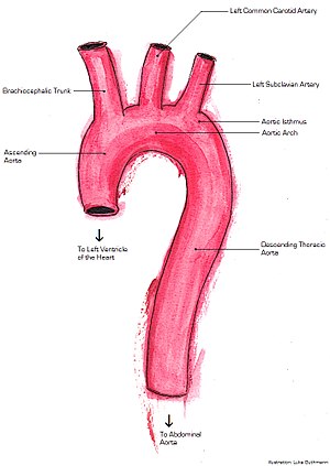
The aorta is the main and largest artery in the human body, originating from the left ventricle of the heart, branching upwards immediately after, and extending down to the abdomen, where it splits at the aortic bifurcation into two smaller arteries. The aorta distributes oxygenated blood to all parts of the body through the systemic circulation.

<span class="mw-page-title-main">Aortic dissection</span> Injury to the innermost layer of the aorta

Aortic dissection (AD) occurs when an injury to the innermost layer of the aorta allows blood to flow between the layers of the aortic wall, forcing the layers apart. In most cases, this is associated with a sudden onset of severe chest or back pain, often described as "tearing" in character. Also, vomiting, sweating, and lightheadedness may occur. Other symptoms may result from decreased blood supply to other organs, such as stroke, lower extremity ischemia, or mesenteric ischemia. Aortic dissection can quickly lead to death from insufficient blood flow to the heart or complete rupture of the aorta.

<span class="mw-page-title-main">Aneurysm</span> Bulge in the wall of a blood vessel

An aneurysm is an outward bulging, likened to a bubble or balloon, caused by a localized, abnormal, weak spot on a blood vessel wall. Aneurysms may be a result of a hereditary condition or an acquired disease. Aneurysms can also be a nidus for clot formation (thrombosis) and embolization. As an aneurysm increases in size, the risk of rupture, which leads to uncontrolled bleeding, increases. Although they may occur in any blood vessel, particularly lethal examples include aneurysms of the Circle of Willis in the brain, aortic aneurysms affecting the thoracic aorta, and abdominal aortic aneurysms. Aneurysms can arise in the heart itself following a heart attack, including both ventricular and atrial septal aneurysms. There are congenital atrial septal aneurysms, a rare heart defect.

<span class="mw-page-title-main">Interventional radiology</span> Medical subspecialty

Interventional radiology (IR) is a medical specialty that performs various minimally-invasive procedures using medical imaging guidance, such as x-ray fluoroscopy, computed tomography, magnetic resonance imaging, or ultrasound. IR performs both diagnostic and therapeutic procedures through very small incisions or body orifices. Diagnostic IR procedures are those intended to help make a diagnosis or guide further medical treatment, and include image-guided biopsy of a tumor or injection of an imaging contrast agent into a hollow structure, such as a blood vessel or a duct. By contrast, therapeutic IR procedures provide direct treatment—they include catheter-based medicine delivery, medical device placement, and angioplasty of narrowed structures.

<span class="mw-page-title-main">Vascular surgery</span> Medical specialty, operative procedures for the treatment of vascular disorders

Vascular surgery is a surgical subspecialty in which vascular diseases involving the arteries, veins, or lymphatic vessels, are managed by medical therapy, minimally-invasive catheter procedures and surgical reconstruction. The specialty evolved from general and cardiovascular surgery where it refined the management of just the vessels, no longer treating the heart or other organs. Modern vascular surgery includes open surgery techniques, endovascular techniques and medical management of vascular diseases - unlike the parent specialities. The vascular surgeon is trained in the diagnosis and management of diseases affecting all parts of the vascular system excluding the coronaries and intracranial vasculature. Vascular surgeons also are called to assist other physicians to carry out surgery near vessels, or to salvage vascular injuries that include hemorrhage control, dissection, occlusion or simply for safe exposure of vascular structures.

<span class="mw-page-title-main">Aortic aneurysm</span> Excessive enlargement of the human aorta

An aortic aneurysm is an enlargement (dilatation) of the aorta to greater than 1.5 times normal size. They usually cause no symptoms except when ruptured. Occasionally, there may be abdominal, back, or leg pain. The prevalence of abdominal aortic aneurysm ("AAA") has been reported to range from 2 to 12% and is found in about 8% of men more than 65 years of age. The mortality rate attributable to AAA is about 15,000 per year in the United States and 6,000 to 8,000 per year in the United Kingdom and Ireland. Between 2001 and 2006, there were approximately 230,000 AAA surgical repairs performed on Medicare patients in the United States.

<span class="mw-page-title-main">Abdominal aortic aneurysm</span> Medical condition

Abdominal aortic aneurysm (AAA) is a localized enlargement of the abdominal aorta such that the diameter is greater than 3 cm or more than 50% larger than normal. An AAA usually causes no symptoms, except during rupture. Occasionally, abdominal, back, or leg pain may occur. Large aneurysms can sometimes be felt by pushing on the abdomen. Rupture may result in pain in the abdomen or back, low blood pressure, or loss of consciousness, and often results in death.

<span class="mw-page-title-main">Thoracic aortic aneurysm</span> Medical condition

A thoracic aortic aneurysm is an aortic aneurysm that presents primarily in the thorax.

<span class="mw-page-title-main">Pseudoaneurysm</span> Collection of blood between outer artery layers

A pseudoaneurysm, also known as a false aneurysm, is a locally contained hematoma outside an artery or the heart due to damage to the vessel wall. The injury passes through all three layers of the arterial wall, causing a leak, which is contained by a new, weak "wall" formed by the products of the clotting cascade. A pseudoaneurysm does not contain any layer of the vessel wall.

<span class="mw-page-title-main">Blunt trauma</span> Physical trauma caused to a body part, either by impact, injury or physical attack

Blunt trauma, also known as blunt force trauma or non-penetrating trauma, is physical trauma or impactful force to a body part, often occurring with road traffic collisions, direct blows, assaults, injuries during sports, and particularly in the elderly who fall. It is contrasted with penetrating trauma which occurs when an object pierces the skin and enters a tissue of the body, creating an open wound and bruise.

<span class="mw-page-title-main">Chest injury</span> Medical condition

A chest injury, also known as chest trauma, is any form of physical injury to the chest including the ribs, heart and lungs. Chest injuries account for 25% of all deaths from traumatic injury. Typically chest injuries are caused by blunt mechanisms such as direct, indirect, compression, contusion, deceleration, or blasts caused by motor vehicle collisions or penetrating mechanisms such as stabbings.

Hemoperitoneum is the presence of blood in the peritoneal cavity. The blood accumulates in the space between the inner lining of the abdominal wall and the internal abdominal organs. Hemoperitoneum is generally classified as a surgical emergency; in most cases, urgent laparotomy is needed to identify and control the source of the bleeding. In selected cases, careful observation may be permissible. The abdominal cavity is highly distensible and may easily hold greater than five liters of blood, or more than the entire circulating blood volume for an average-sized individual. Therefore, large-scale or rapid blood loss into the abdomen will reliably induce hemorrhagic shock and, if untreated, may rapidly lead to death.

<span class="mw-page-title-main">Computed tomography angiography</span>

Computed tomography angiography is a computed tomography technique used for angiography—the visualization of arteries and veins—throughout the human body. Using contrast injected into the blood vessels, images are created to look for blockages, aneurysms, dissections, and stenosis. CTA can be used to visualize the vessels of the heart, the aorta and other large blood vessels, the lungs, the kidneys, the head and neck, and the arms and legs. CTA can also be used to localise arterial or venous bleed of the gastrointestinal system.

<span class="mw-page-title-main">Traumatic aortic rupture</span> Medical condition

Traumatic aortic rupture, also called traumatic aortic disruption or transection, is a condition in which the aorta, the largest artery in the body, is torn or ruptured as a result of trauma to the body. The condition is frequently fatal due to the profuse bleeding that results from the rupture. Since the aorta branches directly from the heart to supply blood to the rest of the body, the pressure within it is very great, and blood may be pumped out of a tear in the blood vessel very rapidly. This can quickly result in shock and death. Thus traumatic aortic rupture is a common killer in automotive accidents and other traumas, with up to 18% of deaths that occur in automobile collisions being related to the injury. In fact, aortic disruption due to blunt chest trauma is the second leading cause of injury death behind traumatic brain injury.

<span class="mw-page-title-main">Endovascular aneurysm repair</span> Surgery used to treat abdominal aortic aneurysm

Endovascular aneurysm repair (EVAR) is a type of minimally-invasive endovascular surgery used to treat pathology of the aorta, most commonly an abdominal aortic aneurysm (AAA). When used to treat thoracic aortic disease, the procedure is then specifically termed TEVAR for "thoracic endovascular aortic/aneurysm repair." EVAR involves the placement of an expandable stent graft within the aorta to treat aortic disease without operating directly on the aorta. In 2003, EVAR surpassed open aortic surgery as the most common technique for repair of AAA, and in 2010, EVAR accounted for 78% of all intact AAA repair in the United States.

<span class="mw-page-title-main">Aortic rupture</span> Rupture or breakage of the aorta, the largest artery in the body

Aortic rupture is the rupture or breakage of the aorta, the largest artery in the body. Aortic rupture is a rare, extremely dangerous condition. The most common cause is an abdominal aortic aneurysm that has ruptured spontaneously. Aortic rupture is distinct from aortic dissection, which is a tear through the inner wall of the aorta that can block the flow of blood through the aorta to the heart or abdominal organs.

<span class="mw-page-title-main">Familial aortic dissection</span> Medical condition

Familial aortic dissection or FAD refers to the splitting of the wall of the aorta in either the arch, ascending or descending portions. FAD is thought to be passed down as an autosomal dominant disease and once inherited will result in dissection of the aorta, and dissecting aneurysm of the aorta, or rarely aortic or arterial dilation at a young age. Dissection refers to the actual tearing open of the aorta. However, the exact gene(s) involved has not yet been identified. It can occur in the absence of clinical features of Marfan syndrome and of systemic hypertension. Over time this weakness, along with systolic pressure, results in a tear in the aortic intima layer thus allowing blood to enter between the layers of tissue and cause further tearing. Eventually complete rupture of the aorta occurs and the pleural cavity fills with blood. Warning signs include chest pain, ischemia, and hemorrhaging in the chest cavity. This condition, unless found and treated early, usually results in death. Immediate surgery is the best treatment in most cases. FAD is not to be confused with PAU and IMH, both of which present in ways similar to that of familial aortic dissection.

<span class="mw-page-title-main">Hybrid cardiac surgery</span>

A hybrid cardiac surgical procedure in a narrow sense is defined as a procedure that combines a conventional, more invasive surgical part with an interventional part, using some sort of catheter-based procedure guided by fluoroscopy imaging in a hybrid operating room (OR) without interruption. The hybrid technique has a reduced risk of surgical complications and has shown decreased recovery time. It can be used to treat numerous heart diseases and conditions and with the increasing complexity of each case, the hybrid surgical technique is becoming more common.

<span class="mw-page-title-main">Open aortic surgery</span> Surgical technique

Open aortic surgery (OAS), also known as open aortic repair (OAR), describes a technique whereby an abdominal, thoracic or retroperitoneal surgical incision is used to visualize and control the aorta for purposes of treatment, usually by the replacement of the affected segment with a prosthetic graft. OAS is used to treat aneurysms of the abdominal and thoracic aorta, aortic dissection, acute aortic syndrome, and aortic ruptures. Aortobifemoral bypass is also used to treat atherosclerotic disease of the abdominal aorta below the level of the renal arteries. In 2003, OAS was surpassed by endovascular aneurysm repair (EVAR) as the most common technique for repairing abdominal aortic aneurysms in the United States.

<span class="mw-page-title-main">Inflammatory aortic aneurysm</span> Medical condition

Inflammatory aortic aneurysm (IAA), also known as Inflammatory abdominal aortic aneurysm (IAAA), is a type of abdominal aortic aneurysm (AAA) where the walls of the aneurysm become thick and inflamed. Similar to AAA, IAA occurs in the abdominal region. IAA is closely associated and believed to be a response to and extensive peri-aneurysmal fibrosis, which is the formation of excess fibrous connective tissue in an organ or tissue in a reparative or reactive process IAA accounts for 5-10% of aortic aneurysms. IAA occurs mainly in a population that is on average younger by 10 years than most AAA patients. Some common symptoms of IAA may include back pain, abdominal tenderness, fevers, weight loss or elevated Erythrocyte sedimentation rate (ESR) levels. Corticosteroids and other immunosuppressive drugs have been found to decrease symptoms and the degree of peri-aortic inflammation and fibrosis

References

  1. 1 2 3 4 5 6 7 Lee, W. Anthony; Matsumura, Jon; Mitchell, R. Scott; Farber, Mark; Greenburg, Roy; Murad, Mohammad; Fairman, Ronald (2011). "Endovascular repair of traumatic thoracic aortic injury: Clinical practice guidelines of the Society for Vascular Surgery". Journal of Vascular Surgery. 53 (1): 187–192. doi: 10.1016/j.jvs.2010.08.027 . PMID   20974523.
  2. 1 2 3 Problem solving in chest imaging. Digumarthy, Subba R. (Subba Rao),, Abbara, Suhny,, Chung, Jonathan H. Philadelphia, PA. March 2019. ISBN   978-0-323-04132-4. OCLC   1126790420.{{cite book}}: CS1 maint: location missing publisher (link) CS1 maint: others (link)
  3. 1 2 Emergency medicine secrets. Markovchick, Vincent J.,, Pons, Peter T.,, Bakes, Katherine M.,, Buchanan, Jennie A. (Sixth ed.). Philadelphia, PA. 2015-11-10. ISBN   978-0-323-37483-5. OCLC   932082432.{{cite book}}: CS1 maint: location missing publisher (link) CS1 maint: others (link)
  4. 1 2 3 4 5 6 7 8 9 10 11 12 Emergency medicine : clinical essentials. Adams, James, 1962- (2nd ed.). Philadelphia, Pa: Elsevier/ Saunders. 2013. ISBN   978-1-4377-3548-2. OCLC   820203833.{{cite book}}: CS1 maint: others (link)
  5. 1 2 Creasy JD, Chiles C, Routh WD, Dyer RB (1997). "Overview of traumatic injury of the thoracic aorta". Radiographics. 17 (1): 27–45. doi:10.1148/radiographics.17.1.9017797. PMID   9017797.
  6. 1 2 3 4 5 Müller's imaging of the chest. Walker, Christopher M.,, Chung, Jonathan H. (2nd ed.). Philadelphia, PA. 2018-08-17. ISBN   978-0-323-53179-5. OCLC   1051135278.{{cite book}}: CS1 maint: location missing publisher (link) CS1 maint: others (link)
  7. 1 2 Miller's anesthesia. Gropper, Michael A., 1958-, Miller, Ronald D., 1939- (Ninth ed.). Philadelphia, PA. 2019-10-07. ISBN   978-0-323-61264-7. OCLC   1124935549.{{cite book}}: CS1 maint: location missing publisher (link) CS1 maint: others (link)
  8. 1 2 3 4 5 6 7 8 9 10 Oh's intensive care manual. Bersten, Andrew D.,, Handy, Jonathan M. (Eighth ed.). [Oxford, U.K.] 2018-08-15. ISBN   978-0-7020-7606-0. OCLC   1053859479.{{cite book}}: CS1 maint: location missing publisher (link) CS1 maint: others (link)
  9. 1 2 3 4 5 6 7 8 9 10 11 12 13 Rosen's emergency medicine : concepts and clinical practice. Walls, Ron M.,, Hockberger, Robert S.,, Gausche-Hill, Marianne (Ninth ed.). Philadelphia, PA. 2017-03-09. ISBN   978-0-323-39016-3. OCLC   989157341.{{cite book}}: CS1 maint: location missing publisher (link) CS1 maint: others (link)
  10. 1 2 3 4 Rutherford's vascular surgery and endovascular therapy. Sidawy, Anton N.,, Perler, Bruce A. (9th ed.). Philadelphia, PA. 2018-04-03. ISBN   978-0-323-58130-1. OCLC   1037557259.{{cite book}}: CS1 maint: location missing publisher (link) CS1 maint: others (link)