
Conjunctivitis, also known as pink eye, is inflammation of the outermost layer of the white part of the eye and the inner surface of the eyelid. It makes the eye appear pink or reddish. Pain, burning, scratchiness, or itchiness may occur. The affected eye may have increased tears or be "stuck shut" in the morning. Swelling of the white part of the eye may also occur. Itching is more common in cases due to allergies. Conjunctivitis can affect one or both eyes.

Adenoviruses are medium-sized, nonenveloped viruses with an icosahedral nucleocapsid containing a double-stranded DNA genome. Their name derives from their initial isolation from human adenoids in 1953.

Dry eye syndrome, also known as keratoconjunctivitis sicca, is the condition of having dry eyes. Symptoms include dryness in the eye, irritation, redness, discharge, blurred vision, and easily fatigued eyes. Symptoms range from mild and occasional to severe and continuous. Dry eye syndrome can lead to blurred vision, instability of the tear film, increased risk of damage to the ocular surface such as scarring of the cornea, and changes in the eye including the neurosensory system.

A red eye is an eye that appears red due to illness or injury. It is usually injection and prominence of the superficial blood vessels of the conjunctiva, which may be caused by disorders of these or adjacent structures. Conjunctivitis and subconjunctival hemorrhage are two of the less serious but more common causes.

Allergic conjunctivitis (AC) is inflammation of the conjunctiva due to allergy. Although allergens differ among patients, the most common cause is hay fever. Symptoms consist of redness, edema (swelling) of the conjunctiva, itching, and increased lacrimation. If this is combined with rhinitis, the condition is termed allergic rhinoconjunctivitis (ARC).
Keratoconjunctivitis is a term used to describe inflammation of both the cornea and the conjunctiva. This condition can have various causes, and its presentation may vary depending on the underlying factors.
Adenovirus infection is a contagious viral disease, caused by adenoviruses, commonly resulting in a respiratory tract infection. Typical symptoms range from those of a common cold, such as nasal congestion, coryza and cough, to difficulty breathing as in pneumonia. Other general symptoms include fever, fatigue, muscle aches, headache, abdominal pain and swollen neck glands. Onset is usually two to fourteen days after exposure to the virus. A mild eye infection may occur on its own, combined with a sore throat and fever, or as a more severe adenoviral keratoconjunctivitis with a painful red eye, intolerance to light and discharge. Very young children may just have an earache. Adenovirus infection can present as a gastroenteritis with vomiting, diarrhoea and abdominal pain, with or without respiratory symptoms. However, some people have no symptoms.

Phlyctenular keratoconjunctivitis is an inflammatory syndrome caused by a delayed hypersensitivity reaction to one or more antigens. The triggering antigen is usually a bacterial protein, but may also be a virus, fungus, or nematode.

A corneal ulcer, or ulcerative keratitis, is an inflammatory condition of the cornea involving loss of its outer layer. It is very common in dogs and is sometimes seen in cats. In veterinary medicine, the term corneal ulcer is a generic name for any condition involving the loss of the outer layer of the cornea, and as such is used to describe conditions with both inflammatory and traumatic causes.

Vernal keratoconjunctivitis is a recurrent, bilateral, and self-limiting type of conjunctivitis having a periodic seasonal incidence.

Feline viral rhinotracheitis (FVR) is an upper respiratory or pulmonary infection of cats caused by Felid alphaherpesvirus 1 (FeHV-1), of the family Herpesviridae. It is also commonly referred to as feline influenza, feline coryza, and feline pneumonia but, as these terms describe other very distinct collections of respiratory symptoms, they are misnomers for the condition. Viral respiratory diseases in cats can be serious, especially in catteries and kennels. Causing one-half of the respiratory diseases in cats, FVR is the most important of these diseases and is found worldwide. The other important cause of feline respiratory disease is feline calicivirus.

Idoxuridine is an anti-herpesvirus antiviral drug.
Infectious bovine keratoconjunctivitis (IBK), also known as pinkeye, New Forest eye or blight, is a veterinary infection of cattle caused by Moraxella bovis, a Gram-negative, β-haemolytic, aerobic, rod-shaped bacterium. It is spread by direct contact or by flies serving as vectors. It is the most common ocular disease of cattle. IBK is similar to human pink eye and causes severe infection of the conjunctiva, edema, corneal opacity, and ulceration. This disease is highly contagious and occurs worldwide. Younger animals are more susceptible, but recovery with minimal damage is usual, if they are treated early.

Corneal neovascularization (CNV) is the in-growth of new blood vessels from the pericorneal plexus into avascular corneal tissue as a result of oxygen deprivation. Maintaining avascularity of the corneal stroma is an important aspect of healthy corneal physiology as it is required for corneal transparency and optimal vision. A decrease in corneal transparency causes visual acuity deterioration. Corneal tissue is avascular in nature and the presence of vascularization, which can be deep or superficial, is always pathologically related.

Corneal ulcer, also called keratitis, is an inflammatory or, more seriously, infective condition of the cornea involving disruption of its epithelial layer with involvement of the corneal stroma. It is a common condition in humans particularly in the tropics and in farming. In developing countries, children afflicted by vitamin A deficiency are at high risk for corneal ulcer and may become blind in both eyes persisting throughout life. In ophthalmology, a corneal ulcer usually refers to having an infection, while the term corneal abrasion refers more to a scratch injury.

A cold sore is a type of herpes infection caused by the herpes simplex virus that affects primarily the lip. Symptoms typically include a burning pain followed by small blisters or sores. The first attack may also be accompanied by fever, sore throat, and enlarged lymph nodes. The rash usually heals within ten days, but the virus remains dormant in the trigeminal ganglion. The virus may periodically reactivate to create another outbreak of sores in the mouth or lip.
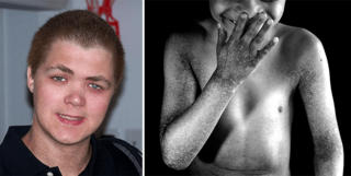
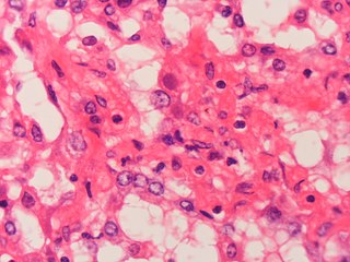
X-linked reticulate pigmentary disorder is a rare X-linked genetic condition in which males manifest multiple systemic symptoms and a reticulated mottled brown pigmentation of the skin, which, on biopsy, demonstrated dermal deposits of amyloid. Females usually only have linear streaks of hyperpigmentation.
Acute Haemmorrhagic Conjunctivitis is the inflammation of the conjunctiva of sudden onset. It presents as a reddening of the eye due to the infection of the conjunctiva. The conjunctiva is the thin transparent tissue that covers the eye from the Corneal limbus to the lid margin. Many conditions can lead to the inflammation of the conjunctiva. They include allergies, bacterial infection, viral infection etc. A common form of the condition that occurs every rainy season is the seasonal conjunctivitis popularly referred to as "Apollo" by West Africans because the reports of its first epidemic in Accra coincided with the Apollo 11 Moon landing. Every year prior to the rainy season in the country, various health warnings are given to remind citizens of the condition.

Herpetic simplex keratitis is a form of keratitis caused by recurrent herpes simplex virus (HSV) infection in the cornea.
