Acanthamoeba

Last updated

Acanthamoeba
Acanthamoeba polyphaga cyst.jpg
Phase contrast micrograph of an Acanthamoeba polyphaga cyst.
Scientific classification OOjs UI icon edit-ltr.svg
Domain: Eukaryota
Phylum: Amoebozoa
Class: Discosea
Order: Centramoebida
Family: Acanthamoebidae
Genus: Acanthamoeba
Volkonsky 1931
Type species
Acanthamoeba castellanii
Volkonsky 1931

Acanthamoeba is a genus of amoebae that are commonly recovered from soil, fresh water, and other habitats. The genus Acanthamoeba has two stages in its life cycle, the metabolically active trophozoite stage and a dormant, stress-resistant cyst stage. In nature, Acanthamoeba species are generally free-living bacterivores. However, they are also opportunistic pathogens able to cause serious and sometimes fatal infections in humans and other animals. [1]

Contents

Distribution

Acanthamoeba spp. are among the most prevalent protozoa found in the environment. [1] They are distributed worldwide, and have been isolated from soil, air, sewage, seawater, chlorinated swimming pools, domestic tap water, bottled water, dental treatment units, hospitals, air-conditioning units, and contact lens cases. Additionally, they have been isolated from human skin, nasal cavities, throats, and intestines, as well as plants and other mammals. [2]

Role in disease

Acanthamoeba encephalitis microscopy and molecular diagnostics: scale bar: 10 mm Acanthamoeba pathology.jpg
Acanthamoeba encephalitis microscopy and molecular diagnostics: scale bar: 10 μm

Diseases caused by Acanthamoeba include keratitis and granulomatous amoebic encephalitis (GAE). [3] The latter is often but not always seen in immunosuppressed patients. [4] GAE is caused by the amoebae entering the body through an open wound and then spreading to the brain. [5] The combination of host immune responses and secreted amoebal proteases causes massive brain swelling [6] resulting in death in about 95% of those infected.

Granulomatous amoebic encephalitis (GAE)

Granulomatous amoebic encephalitis (GAE) is caused by amoebic infection of the central nervous system (CNS). It is characterized by neurological symptoms including headache, seizures, and mental-status abnormalities. [1] These worsen progressively over weeks to months, leading to death in most patients. [1] Infection is generally associated with underlying conditions such as immunodeficiency, diabetes, malignancies, malnutrition, systemic lupus erythematosus, and alcoholism. [1] The parasite enters the body through cuts in the skin or by being inhaled into the upper respiratory tract. [1] The parasite then spreads through the blood into the CNS. Acanthamoeba crosses the blood–brain barrier by means that are not yet understood. Subsequent invasion of the connective tissue and induction of pro-inflammatory responses leads to neuronal damage that can be fatal within days. Pure granulomatous lesions are rare in patients with AIDS and other related immunodeficiency states, as the patients do not have adequate numbers of CD+ve T-cells to mount a granulomatous response to Acanthamoeba infection in CNS and other organs and tissues. [4] A perivascular cuffing with amoebae in necrotic tissue is usual finding in the AIDS and related T-cell immunodeficiency conditions.

Brain biopsy normally reveals severe oedema and hemorrhagic necrosis. [7] A patient who has contracted this illness usually displays subacute symptoms, including altered mental status, headaches, fever, neck stiffness, seizures, and focal neurological signs (such as cranial nerve palsies and coma), all leading to death within one week to several months. [8] Due to the rarity of this parasite and a lack of knowledge, no good diagnoses or treatments for Acanthamoeba infection are now known. Acanthamoeba keratitis cases in the past have resolved from a therapy consisting of atropine and some other drugs with no antimicrobial effects. Recent publications show atropine to interfere with the protist's CHRM1 receptor, causing cell death. [9]

Infection usually mimics that of bacterial leptomeningitis, tuberculous meningitis, or viral encephalitis. The misdiagnosis often leads to erroneous, ineffective treatment. In the case that the Acanthamoeba is diagnosed correctly, the current treatments, such as amphotericin B, rifampicin, trimethoprim-sulfamethoxazole, ketoconazole, fluconazole, sulfadiazine, or albendazole, are only tentatively successful. Correct and timely diagnosis, as well as improved treatment methods and an understanding of the parasite, are important factors in improving the outcome of infection by Acanthamoeba. A paper published in 2013 has shown substantial effects of some FDA-approved drugs with an in vitro kill rate above 90%. [4] These results were in vitro effects, but as the drugs are already approved, human infections can be targeted after dose calculations in clinical trials done with these diverse groups of drugs.

Acanthamoebic keratitis

Pathogenic Acanthamoeba as seen under a light microscope Parasite140120-fig3 Acanthamoeba keratitis Figure 3B.png
Pathogenic Acanthamoeba as seen under a light microscope

When present in the eye, Acanthamoeba strains can cause acanthamoebic keratitis, which may lead to corneal ulcers or even blindness. [10] This condition occurs most often among contact lens wearers who do not properly disinfect their lenses, exacerbated by a failure to wash hands prior to handling the lenses. Multipurpose contact lens solutions are largely ineffective against Acanthamoeba, whereas hydrogen peroxide-based solutions have good disinfection characteristics. [11] [12]

The first cure of a corneal infection was achieved in 1985 at Moorfields Eye Hospital. [13]

In May 2007, Advanced Medical Optics, manufacturer of Complete Moisture Plus Contact Lens Solution products, issued a voluntary recall of their Complete Moisture Plus solutions. The fear was that contact lens wearers who used their solution were at higher risk of acanthamoebic keratitis than contact lens wearers who used other solutions. The manufacturer recalled the product after the Centers for Disease Control in the United States found that 21 people had possibly received an Acanthamoeba infection after using Complete Moisture Plus in the month prior to diagnosis. [14]

As a bacterial reservoir

Several species of bacteria that can cause human disease are also able to infect and replicate within Acanthamoeba species. [1] These include Legionella pneumophila , Pseudomonas aeruginosa , and some strains of Escherichia coli and Staphylococcus aureus . [1] [15] For some of these bacteria, replication inside Acanthamoeba has been associated with enhanced growth in macrophages, and increased resistance to some antibiotics. [1] Furthermore, due to the high prevalence of Acanthamoeba in the environment, these amoebae have been proposed to serve as an environmental reservoir for some human pathogens. [1]

Ecology

A. castellanii can be found at high densities in various soil ecosystems. It preys on bacteria, but also fungi and other protozoa.

This species is able to lyse bacteria and produce a wide range of enzymes, such as cellulases or chitinases, [16] and probably contributes to the breakdown of organic matter in soil, contributing to the microbial loop.

Physiology

Role as a model organism

Because Acanthamoeba does not differ greatly at the ultrastructural level from a mammalian cell, it is an attractive model for cell-biology studies; it is important in cellular microbiology, environmental biology, physiology, cellular interactions, molecular biology, biochemistry, and evolutionary studies, due to the organisms' versatile roles in the ecosystem and ability to capture prey by phagocytosis, act as vectors and reservoirs for microbial pathogens, and to produce serious human infections. In addition, Acanthamoeba has been used extensively to understand the molecular biology of cell motility [17] and cancer cell dormancy by in-depth exploration of the process of encystation. [18]

The recently available Acanthamoeba genome sequence revealed several orthologs of genes employed in meiosis of sexual eukaryotes. These genes included Spo11, Mre11, Rad50, Rad51, Rad52, Mnd1, Dmc1, Msh , and Mlh . [19] This finding suggests that Acanthamoeba is capable of some form of meiosis and may be able to undergo sexual reproduction. Furthermore, since Acanthamoeba diverged early from the eukaryotic family tree, these results suggest that meiosis was present early in eukaryotic evolution.

Owing to its ease and economy of cultivation, the Neff strain of A. castellanii, discovered in a pond in Golden Gate Park in the 1960s, has been effectively used as a classic model organism in the field of cell biology. From just 30 L of simple medium inoculated with A. castellanii, about 1 kg of cells can be obtained after several days of aerated culture at room temperature. Pioneered in the laboratory of Edward D. Korn at the National Institutes of Health (NIH), many important biological molecules have been discovered and their pathways elucidated using the Acanthamoeba model. Thomas Dean Pollard applied this model at the NIH, Harvard Medical School, Johns Hopkins University School of Medicine, and the Salk Institute for Biological Studies to discover and characterize many proteins that are essential for cell motility, not only in amoebae, but also in many other eukaryotic cells, especially those of the human nervous and immune systems, the developing embryo, and cancer cells. Acanthamoeba also has served as a model to study the evolution of certain G-proteins. This unicellular eukaryote expresses few GPCRs over its cell membrane that serve vital role for the microorganism, structural homology bioinformatics tools have been used to show the presence of a homolog of human M1-muscarinic receptor in A. castellanii. [20] Blocking these muscarinic receptors in past studies has proven to be amoebicidal in Acanthamoeba spp. [5] More recently, voltage-gated calcium channels in Acanthamoeba spp. (CavAc) have been reported to have similarities with human voltage-gated calcium channels such as TPC-1 and L-type calcium channels and respond to Ca-channel blockers such as loperamide. [21] This model microbe has been studied to understand complex neurodegenerative states including Alzheimer's disease. Scientists have isolated a neurotransmitter acetylcholine in Acanthamoeba and the enzymatic machinery needed for its synthesis. [22]

Endosymbionts

Acanthamoeba spp. contain diverse bacterial endosymbionts that are similar to human pathogens, so they are considered to be potential emerging human pathogens. [23] The exact nature of these symbionts and the benefit they represent for the amoebic host still have to be clarified. These include Legionella and Legionella-like pathogens. [24]

Giant viruses

The giant viruses Mimivirus, Megavirus , and Pandoravirus infect Acanthamoeba. [25]

Members of the genus Acanthamoeba are unusual in serving as hosts for a variety of giant viruses (that have more than 1000 protein-coding genes; for instance, Pandoravirus, which has about 2500 protein-coding genes in its genome).

Diversity

Acanthamoeba can be distinguished from other genera of amoebae based on morphological characteristics. [26] However, differentiating one species of Acanthamoeba from another by morphology has proven difficult. Based on 18S rDNA sequencing, known Acanthamoeba strains can be organized into 12 groups, denoted T1-T12. [26] Most disease-causing isolates belong to type T4. [26]

Below is a list of described species of Acanthamoeba, with sequence types noted where known. Species that have been identified in diseased patients are marked with *.

Etymology

From the Greek akantha (spike/thorn), which was added before "amoeba" (change) to describe this organism as having a spine-like structure (acanthopodia). This organism is now well known as Acanthamoeba, an amphizoic, opportunistic, and nonopportunistic protozoan protist widely distributed in the environment. [27]

See also

Related Research Articles

<span class="mw-page-title-main">Keratitis</span> Medical condition

Keratitis is a condition in which the eye's cornea, the clear dome on the front surface of the eye, becomes inflamed. The condition is often marked by moderate to intense pain and usually involves any of the following symptoms: pain, impaired eyesight, photophobia, red eye and a 'gritty' sensation. Diagnosis of infectious keratitis is usually made clinically based on the signs and symptoms as well as eye examination, but corneal scrapings may be obtained and evaluated using microbiological culture or other testing to identify the causative pathogen.

Free-living amoebae in the Amoebozoa group are important causes of disease in humans and animals.

<span class="mw-page-title-main">Amoebozoa</span> Phylum of protozoans

Amoebozoa is a major taxonomic group containing about 2,400 described species of amoeboid protists, often possessing blunt, fingerlike, lobose pseudopods and tubular mitochondrial cristae. In traditional classification schemes, Amoebozoa is usually ranked as a phylum within either the kingdom Protista or the kingdom Protozoa. In the classification favored by the International Society of Protistologists, it is retained as an unranked "supergroup" within Eukaryota. Molecular genetic analysis supports Amoebozoa as a monophyletic clade. Modern studies of eukaryotic phylogenetic trees identify it as the sister group to Opisthokonta, another major clade which contains both fungi and animals as well as several other clades comprising some 300 species of unicellular eukaryotes. Amoebozoa and Opisthokonta are sometimes grouped together in a high-level taxon, variously named Unikonta, Amorphea or Opimoda.

<span class="mw-page-title-main">Naegleriasis</span> Rare and usually fatal brain infection by a protist

Naegleriasis is an almost invariably fatal infection of the brain by the free-living unicellular eukaryote Naegleria fowleri. Symptoms are meningitis-like and include headache, fever, nausea, vomiting, a stiff neck, confusion, hallucinations and seizures. Symptoms progress rapidly over around five days, and death usually results within one to two weeks of symptoms.

<i>Naegleria</i> Genus of protists

Naegleria is a free living amoebae protist genus consisting of 47 described species often found in warm aquatic environments as well as soil habitats worldwide. It has three life cycle forms: the amoeboid stage, the cyst stage, and the flagellated stage, and has been routinely studied for its ease in change from amoeboid to flagellated stages. The Naegleria genera became famous when Naegleria fowleri, a human pathogenic strain and the causative agent of primary amoebic meningoencephalitis (PAM), was discovered in 1965. Most species in the genus, however, are nonpathogenic, meaning they do not cause disease.

<i>Legionella pneumophila</i> Species of bacterium

Legionella pneumophila is an aerobic, pleomorphic, flagellated, non-spore-forming, Gram-negative bacterium of the genus Legionella. L. pneumophila is the primary human pathogenic bacterium in this group. In nature, L. pneumophila infects freshwater and soil amoebae of the genera Acanthamoeba and Naegleria. This pathogen is found commonly near freshwater environments and will then invade the amoebae found in these environments, using them to carry out metabolic functions.

<i>Acanthamoeba</i> keratitis Eye infection caused by a protist

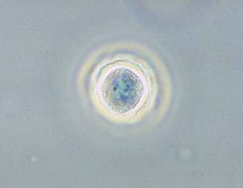
Acanthamoeba keratitis (AK) is a rare disease in which amoebae of the genus Acanthamoeba invade the clear portion of the front (cornea) of the eye. It affects roughly 100 people in the United States each year. Acanthamoeba are protozoa found nearly ubiquitously in soil and water and can cause infections of the skin, eyes, and central nervous system.

<i>Balamuthia mandrillaris</i> Species of pathogenic Amoebozoa

Balamuthia mandrillaris is a free-living amoeba that causes the rare but deadly neurological condition granulomatous amoebic encephalitis (GAE). B. mandrillaris is a soil-dwelling amoeba and was first discovered in 1986 in the brain of a mandrill that died in the San Diego Wild Animal Park.

<span class="mw-page-title-main">Granulomatous amoebic encephalitis</span> Rare and usually fatal brain infection by certain amoebae

.

<span class="mw-page-title-main">Miltefosine</span> Phospholipid drug

Miltefosine, sold under the trade name Impavido among others, is a medication mainly used to treat leishmaniasis and free-living amoeba infections such as Naegleria fowleri and Balamuthia mandrillaris. This includes the three forms of leishmaniasis: cutaneous, visceral and mucosal. It may be used with liposomal amphotericin B or paromomycin. It is taken by mouth.

Sappinia diploidea is a free-living amoeba species.

Sappinia is a genus of heterotrophic, lobose amoebae within the family Thecamoebidae. A defining feature of Sappinia, which separates it from its sister genus Thecamoeba, is the presence of two closely apposed nuclei with a central, flattened connection. Sappinia species have two life cycle stages: a trophozoite and a cyst. Up until 2015, only two species had been discovered, Sappinia pedata and Sappinia diploidea. Sequencing of the small subunit rRNA of a particular isolate from a sycamore tree revealed a new species, Sappinia platani.Sappinia species were once thought to be coprozoic, as the first strains were isolated from animal dung. More research has shown that they are typical free-living amoebae, and can be found worldwide in soil, plant litter, and standing decaying plants, as well as freshwater ponds. In 2001, the first and only case of human pathogenesis in Sappinia was confirmed. The patient was a non-immunocompromised 38-year-old male who presented signs of amoebic encephalitis and who patient made a full recovery after treatment with several antimicrobials. The CDC initially classified the causative agent as S. diploidea based on morphological characteristics, but in 2009, Qvarnstrom et al. used molecular data to confirm that the true causative agent was S. pedata.

Cutaneous amoebiasis, refers to a form of amoebiasis that presents primarily in the skin. It can be caused by Acanthamoeba or Entamoeba histolytica. When associated with Acanthamoeba, it is also known as "cutaneous acanthamoebiasis". Balamuthia mandrillaris can also cause cutaneous amoebiasis, but can prove fatal if the amoeba enters the bloodstream It is characterized by ulcers. Diagnosis of amebiasis cutis calls for high degree of clinical suspicion. This needs to be backed with demonstration of trophozoites from lesions. Unless an early diagnosis can be made such patients can develop significant morbidity.

<span class="mw-page-title-main">Acanthamoebidae</span> Family of protozoans

Acanthamoebidae is a family of single-celled eukaryotes within the group Amoebozoa.

Sappinia pedata is a species of Amoebozoa. Sappinia is a free-living amoeba, found in the environment. This organism can cause granulomatous amoebic encephalitis (GAE), however, only one case of GAE due to S. pedata infection has ever been reported, and the patient survived without any long-term consequences.

Sappinia amoebic encephalitis (SAE) is the name for amoebic encephalitis caused by species of Sappinia.

Parachlamydia acanthamoebae are bacterium that fall into the category of host-associated microorganisms. This bacterium lives within free-living amoebae that are an intricate part of their reproduction. Originally named Candidatus Parachlamydia acanthamoebae, its current scientific name was introduced shortly after. This species has shown to have over eighty percent 16S rRNA gene sequencing identity with the class Chlamydiia. Parachlamydia acanthamoebae has the same family as the genus Neochlamydia with which it shares many similarities.

<span class="mw-page-title-main">Amoeba</span> Polyphyletic group of unicellular eukaryotes with the ability to shapeshift

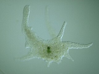
An amoeba, often called an amoeboid, is a type of cell or unicellular organism with the ability to alter its shape, primarily by extending and retracting pseudopods. Amoebae do not form a single taxonomic group; instead, they are found in every major lineage of eukaryotic organisms. Amoeboid cells occur not only among the protozoa, but also in fungi, algae, and animals.

<i>Naegleria fowleri</i> Species of free-living excavate form of protist

Naegleria fowleri, colloquially known as the "brain-eating amoeba", is a species of the genus Naegleria. It belongs to the phylum Percolozoa and is technically classified as an amoeboflagellate excavate, rather than a true amoeba. This free-living microorganism primarily feeds on bacteria but can become pathogenic in humans, causing an extremely rare, sudden, severe, and usually fatal brain infection known as naegleriasis or primary amoebic meningoencephalitis (PAM).

<span class="mw-page-title-main">Symbiosis in Amoebozoa</span>

Amoebozoa of the free living genus Acanthamoeba and the social amoeba genus Dictyostelium are single celled eukaryotic organisms that feed on bacteria, fungi, and algae through phagocytosis, with digestion occurring in phagolysosomes. Amoebozoa are present in most terrestrial ecosystems including soil and freshwater. Amoebozoa contain a vast array of symbionts that range from transient to permanent infections, confer a range of effects from mutualistic to pathogenic, and can act as environmental reservoirs for animal pathogenic bacteria. As single celled phagocytic organisms, amoebas simulate the function and environment of immune cells like macrophages, and as such their interactions with bacteria and other microbes are of great importance in understanding functions of the human immune system, as well as understanding how microbiomes can originate in eukaryotic organisms.

References

  1. 1 2 3 4 5 6 7 8 9 10 Marciano-Cabral F, Cabral G (April 2003). "Acanthamoeba spp. as agents of disease in humans". Clinical Microbiology Reviews. 16 (2): 273–307. doi:10.1128/CMR.16.2.273-307.2003. PMC   153146 . PMID   12692099.
  2. De Jonckheere JF (1991). "Ecology of Acanthamoeba". Reviews of Infectious Diseases. 13 Suppl 5: S385–7. doi:10.1093/clind/13.supplement_5.s385. PMID   2047667.
  3. Di Gregorio C, Rivasi F, Mongiardo N, De Rienzo B, Wallace S, Visvesvara GS (December 1992). "Acanthamoeba meningoencephalitis in a patient with acquired immunodeficiency syndrome". Archives of Pathology & Laboratory Medicine. 116 (12): 1363–5. PMID   1456885.
  4. 1 2 3 Baig AM (December 2014). "Granulomatous amoebic encephalitis: ghost response of an immunocompromised host?". Journal of Medical Microbiology. 63 (Pt 12): 1763–6. doi: 10.1099/jmm.0.081315-0 . PMID   25239626. S2CID   28069984.
  5. 1 2 Baig AM, Iqbal J, Khan NA (August 2013). "In vitro efficacies of clinically available drugs against growth and viability of an Acanthamoeba castellanii keratitis isolate belonging to the T4 genotype". Antimicrobial Agents and Chemotherapy. 57 (8): 3561–7. doi:10.1128/AAC.00299-13. PMC   3719691 . PMID   23669391.
  6. Baig AM (August 2015). "Pathogenesis of amoebic encephalitis: Are the amoebae being credited to an 'inside job' done by the host immune response?". Acta Tropica. 148: 72–6. doi:10.1016/j.actatropica.2015.04.022. PMID   25930186.
  7. Khan NA (February 2007). "Acanthamoeba invasion of the central nervous system". International Journal for Parasitology. 37 (2): 131–8. doi:10.1016/j.ijpara.2006.11.010. PMID   17207487.
  8. Kaushal V, Chhina DK, Kumar R, Pannu HS, Dhooria HP, Chhina RS (March 2007). "Acanthamoeba encephalitis". Indian Journal of Medical Microbiology. 26 (2): 182–4. doi: 10.1016/S0255-0857(21)01941-1 . hdl: 1807/53578 . PMID   18445961.
  9. Baig AM, Zuberi H, Khan NA (May 2014). "Recommendations for the management of Acanthamoeba keratitis". Journal of Medical Microbiology. 63 (Pt 5): 770–1. doi: 10.1099/jmm.0.069237-0 . PMID   24509420.
  10. Lorenzo-Morales J, Khan NA, Walochnik J (2015). "An update on Acanthamoeba keratitis: diagnosis, pathogenesis and treatment". Parasite. 22: 10. doi:10.1051/parasite/2015010. PMC   4330640 . PMID   25687209. Open Access logo PLoS transparent.svg
  11. Shoff ME, Joslin CE, Tu EY, Kubatko L, Fuerst PA (July 2008). "Efficacy of contact lens systems against recent clinical and tap water Acanthamoeba isolates". Cornea. 27 (6): 713–9. doi: 10.1097/QAI.0b013e31815e7251 . PMID   18580265. S2CID   54503941.
  12. Johnston SP, Sriram R, Qvarnstrom Y, Roy S, Verani J, Yoder J, Lorick S, Roberts J, Beach MJ, Visvesvara G (July 2009). "Resistance of Acanthamoeba cysts to disinfection in multiple contact lens solutions". Journal of Clinical Microbiology. 47 (7): 2040–5. doi:10.1128/JCM.00575-09. PMC   2708465 . PMID   19403771.
  13. Wright P, Warhurst D, Jones BR (October 1985). "Acanthamoeba keratitis successfully treated medically". The British Journal of Ophthalmology. 69 (10): 778–82. doi:10.1136/bjo.69.10.778. PMC   1040738 . PMID   4052364.
  14. "Abbott Medical Optics" (PDF). Archived from the original (PDF) on 7 July 2011. Retrieved 31 May 2009.
  15. Huws SA, Morley RJ, Jones MV, Brown MR, Smith AW (May 2008). "Interactions of some common pathogenic bacteria with Acanthamoeba polyphaga". FEMS Microbiology Letters. 282 (2): 258–65. doi: 10.1111/j.1574-6968.2008.01123.x . PMID   18399997.
  16. Anderson IJ, Watkins RF, Samuelson J, Spencer DF, Majoros WH, Gray MW, Loftus BJ (August 2005). "Gene discovery in the Acanthamoeba castellanii genome". Protist. 156 (2): 203–14. doi:10.1016/j.protis.2005.04.001. PMID   16171187.
  17. Khan N (2009). Acanthamoeba: Biology and Pathogenesis. Caister Academic Press. ISBN   978-1-904455-43-1.
  18. Baig AM, Khan NA, Abbas F. Eukaryotic cell encystation and cancer cell dormancy: is a greater devil veiled in the details of a lesser evil? Cancer Biol
  19. Khan NA, Siddiqui R (June 2015). "Is there evidence of sexual reproduction (meiosis) in Acanthamoeba?". Pathogens and Global Health. 109 (4): 193–5. doi:10.1179/2047773215Y.0000000009. PMC   4530557 . PMID   25800982.
  20. Baig AM, Ahmad HR (June 2017). "1-muscarinic GPCR homolog in unicellular eukaryotes: featuring Acanthamoeba spp bioinformatics 3D-modelling and experimentations". Journal of Receptor and Signal Transduction Research. 37 (3): 267–275. doi:10.1080/10799893.2016.1217884. PMID   27601178. S2CID   5234123.
  21. Baig AM, Rana Z, Mannan M, Tariq S, Ahmad HR (2017). "Antibiotic Effects of Loperamide: Homology of Human Targets of Loperamide with Targets in Acanthamoeba spp". Recent Patents on Anti-Infective Drug Discovery. 12 (1): 44–60. doi:10.2174/1574891X12666170425170544. PMID   28506204.
  22. Baig AM, Rana Z, Tariq S, Lalani S, Ahmad HR (March 2018). "Traced on the Timeline: Discovery of Acetylcholine and the Components of the Human Cholinergic System in a Primitive Unicellular Eukaryote Acanthamoeba spp". ACS Chemical Neuroscience. 9 (3): 494–504. doi:10.1021/acschemneuro.7b00254. PMID   29058403.
  23. Horn M, Wagner M (September–October 2004). "Bacterial endosymbionts of free-living amoebae". The Journal of Eukaryotic Microbiology. 51 (5): 509–14. doi:10.1111/j.1550-7408.2004.tb00278.x. PMID   15537084. S2CID   21052932.
  24. Schuster FL, Visvesvara GS (February 2004). "Opportunistic amoebae: challenges in prophylaxis and treatment". Drug Resistance Updates. 7 (1): 41–51. doi:10.1016/j.drup.2004.01.002. PMID   15072770.
  25. Philippe N, Legendre M, Doutre G, Couté Y, Poirot O, Lescot M, Arslan D, Seltzer V, Bertaux L, Bruley C, Garin J, Claverie JM, Abergel C (July 2013). "Pandoraviruses: amoeba viruses with genomes up to 2.5 Mb reaching that of parasitic eukaryotes" (PDF). Science. 341 (6143): 281–6. Bibcode:2013Sci...341..281P. doi:10.1126/science.1239181. PMID   23869018. S2CID   16877147.
  26. 1 2 3 Kong HH (October 2009). "Molecular phylogeny of acanthamoeba". The Korean Journal of Parasitology. 47 Suppl (Suppl): S21–8. doi:10.3347/kjp.2009.47.S.S21. PMC   2769217 . PMID   19885332.
  27. Pradhan, Nitika (August 2020). "Etymologia: Acanthamoeba". Emerg Infect Dis. 26 (8): 1855. doi: 10.3201/eid2608.et2608 . PMC   7392430 . citing public domain text from the CDC