Amoebozoa

Last updated

Amoebozoa
Temporal range:
Chaos carolinense.jpg
Chaos carolinensis
Scientific classification OOjs UI icon edit-ltr.svg
Domain: Eukaryota
(unranked): Amorphea
Phylum: Amoebozoa
Lühe, 1913 emend. Cavalier-Smith, 1998 [3]
Clades [4] [5]
Synonyms

Amoebozoa is a major taxonomic group containing about 2,400 described species of amoeboid protists, [7] often possessing blunt, fingerlike, lobose pseudopods and tubular mitochondrial cristae. [6] [8] In traditional classification schemes, Amoebozoa is usually ranked as a phylum within either the kingdom Protista [9] or the kingdom Protozoa. [10] [11] In the classification favored by the International Society of Protistologists, it is retained as an unranked "supergroup" within Eukaryota. [6] Molecular genetic analysis supports Amoebozoa as a monophyletic clade. Modern studies of eukaryotic phylogenetic trees identify it as the sister group to Opisthokonta, another major clade which contains both fungi and animals as well as several other clades comprising some 300 species of unicellular eukaryotes. [7] [8] Amoebozoa and Opisthokonta are sometimes grouped together in a high-level taxon, variously named Unikonta, [10] Amorphea [6] or Opimoda. [12]

Contents

Amoebozoa includes many of the best-known amoeboid organisms, such as Chaos , Entamoeba , Pelomyxa and the genus Amoeba itself. Species of Amoebozoa may be either shelled (testate) or naked, and cells may possess flagella. Free-living species are common in both salt and freshwater as well as soil, moss and leaf litter. Some live as parasites or symbionts of other organisms, and some are known to cause disease in humans and other organisms.

While the majority of amoebozoan species are unicellular, the group also includes several clades of slime molds, which have a macroscopic, multicellular stage of life during which individual amoeboid cells remain together after multiple cell division to form a macroscopic plasmodium or, in cellular slime molds, aggregate to form one.

Amoebozoa vary greatly in size. Some are only 10–20 μm in diameter, while others are among the largest protozoa. The well-known species Amoeba proteus, which may reach 800 μm in length, is often studied in schools and laboratories as a representative cell or model organism, partly because of its convenient size. Multinucleate amoebae like Chaos and Pelomyxa may be several millimetres in length, and some multicellular amoebozoa, such as the "dog vomit" slime mold Fuligo septica , can cover an area of several square meters. [13]

Morphology

An amoeba of the genus Mayorella (Amoebozoa, Discosea) Stroenie ameby Mayorella sp.jpg
An amoeba of the genus Mayorella (Amoebozoa, Discosea)

Amoebozoa is a large and diverse group, but certain features are common to many of its members. The amoebozoan cell is typically divided into a granular central mass, called endoplasm, and a clear outer layer, called ectoplasm. During locomotion, the endoplasm flows forwards and the ectoplasm runs backwards along the outside of the cell. In motion, many amoebozoans have a clearly defined anterior and posterior and may assume a "monopodial" form, with the entire cell functioning as a single pseudopod. Large pseudopods may produce numerous clear projections called subpseudopodia (or determinate pseudopodia), which are extended to a certain length and then retracted, either for the purpose of locomotion or food intake. A cell may also form multiple indeterminate pseudopodia, through which the entire contents of the cell flow in the direction of locomotion. These are more or less tubular and are mostly filled with granular endoplasm. The cell mass flows into a leading pseudopod, and the others ultimately retract, unless the organism changes direction. [14]

While most amoebozoans are "naked," like the familiar Amoeba and Chaos, or covered with a loose coat of minute scales, like Cochliopodium and Korotnevella, members of the order Arcellinida form rigid shells, or tests, equipped with a single aperture through which the pseudopods emerge. Arcellinid tests may be secreted from organic materials, as in Arcella , or built up from collected particles cemented together, as in Difflugia.

In all amoebozoa, the primary mode of nutrition is phagocytosis, in which the cell surrounds potential food particles with its pseudopods, sealing them into vacuoles within which they may be digested and absorbed. Some amoebozoans have a posterior bulb called a uroid, which may serve to accumulate waste, periodically detaching from the rest of the cell.[ citation needed ] When food is scarce, most species can form cysts, which may be carried aerially and introduce them to new environments.[ citation needed ] In slime moulds, these structures are called spores, and form on stalked structures called fruiting bodies or sporangia. Mixotrophic species living in a symbiotic relationship with microalgae of the genus Chlorella, which lives inside the cytoplasm of their host, have been found in Arcellinida and Mayorella. [15] [16]

The majority of Amoebozoa lack flagella and more generally do not form microtubule-supported structures except during mitosis. However, flagella do occur among the Archamoebae, and many slime moulds produce biflagellate gametes [ citation needed ]. The flagellum is generally anchored by a cone of microtubules, suggesting a close relationship to the opisthokonts. [ citation needed ] The mitochondria in amoebozoan cells characteristically have branching tubular cristae. However, among the Archamoebae, which are adapted to anoxic or microaerophilic habitats, mitochondria have been lost.

Classification

Place of Amoebozoa in the eukaryote tree

It appears (based on molecular genetics) that the members of Amoebozoa form a sister group to animals and fungi, diverging from this lineage after it had split from the other groups, [17] as illustrated below in a simplified diagram:

Opimoda

Loukozoa Giardia lamblia.jpg

Podiata

CRuMs Collodictyon anterior view, showing sulcus, nucleus, blepharoplast, rhizoplast, and four flagella..jpg

Amorphea

Amoebozoa

Obazoa

Breviata Mastigamoeba invertens.jpg

Apusomonadida Apusomonas.png

Opisthokonta

Fungi Asco1013.jpg

Animalia Comb jelly.jpg

Strong similarities between Amoebozoa and Opisthokonts lead to the hypothesis that they form a distinct clade. [18] Thomas Cavalier-Smith proposed the name "unikonts" (formally, Unikonta) for this branch, whose members were believed to have been descended from a common ancestor possessing a single emergent flagellum rooted in one basal body.[1][2] However, while the close relationship between Amoebozoa and Opisthokonta is robustly supported, recent work has shown that the hypothesis of a uniciliate ancestor is probably false. In their Revised Classification of Eukaryotes (2012), Adl et al. proposed Amorphea as a more suitable name for a clade of approximately the same composition, a sister group to the Diaphoretickes. [6] More recent work places the members of Amorphea together with the malawimonids and collodictyonids in a proposed clade called Opimoda, which comprises one of two major lineages diverging at the root of the eukaryote tree of life, the other being Diphoda. [12]

Subphyla within Amoebozoa: Lobosa and Conosa

Traditionally all amoebozoa with lobose pseudopods were grouped together in the class Lobosea, placed with other amoeboids in the phylum Sarcodina or Rhizopoda, but these were considered to be unnatural groups. Structural and genetic studies identified the percolozoans and several archamoebae as independent groups. In phylogenies based on rRNA their representatives were separate from other amoebae, and appeared to diverge near the base of eukaryotic evolution, as did most slime molds.

However, revised trees by Cavalier-Smith and Chao in 1996 [19] suggested that the remaining lobosans do form a monophyletic group, to which the Archamoebae and Mycetozoa were closely related, although the percolozoans were not. Subsequently, they emended the phylum Amoebozoa to include both the subphylum Lobosa and a new subphylum Conosa, comprising the Archamoebae and the Mycetozoa. [3]

Recent molecular genetic data appear to support this primary division of the Amoebozoa into Lobosa and Conosa. [8] The former, as defined by Cavalier-Smith and his collaborators, consists largely of the classic Lobosea: non-flagellated amoebae with blunt, lobose pseudopods (Amoeba, Acanthamoeba, Arcella, Difflugia etc.). The latter is made up of both amoeboid and flagellated cells, characteristically with more pointed or slightly branching subpseudopodia (Archamoebae and the Mycetozoan slime molds).

Phylogeny and taxonomy within Amoebozoa

From older studies by Cavalier-Smith, Chao & Lewis 2016 [20] and Silar 2016. [21] Also recent phylogeny indicates the Lobosa are paraphyletic: Conosa is sister of the Cutosea. [5] [22] [23]

Amoebozoa phylogeny
Discosea
Centramoebia

Centramoebida

Himatismenida

Himatismenida

Flabellinia

Thecamoebida

Dermamoebida

Vannellida

Dactylopodida

Tevosa
Tubulinea
Corycidia

Trichosida

Microcoryciidae

Echinamoebia

Echinamoebida

Elardia
Leptomyxia

Leptomyxida

Eulobosia

Euamoebida

Arcellinida

Evosida
Cutosea

Squamocutida

Conosa
Archamoebae
Archamoebea

Entamoebida

Pelobiontida

Semiconosia
Variosea

Phalansteriida

Flamellidae

Ramamoebida

Profiliida

Fractovitellida

Eumycetozoa
Dictyostelea

Acytosteliales

Dictyosteliida

Myxogastria
Ceratiomyxomycetes

Ceratiomyxida

Protosporangiida

Myxomycetes
Lucisporomycetidae
Cribrarianae

Cribrariales

Trichianae

Reticulariales

Liceida

Trichiida

Columellomycetidae
Echinostelianae

Echinosteliida

Stemonitanae

Clastodermatales

Meridermatales

Stemonitales

Physarales

Phylum Amoebozoa Lühe 1913 emend. Cavalier-Smith 1998 [Amoebobiota; Eumycetozoa Zopf 1884 emend Olive 1975]

Fossil record

Vase-shaped microfossils (VSMs) discovered around the world show that amoebozoans have existed since the Neoproterozoic Era. The fossil species Melanocyrillium hexodiadema , Palaeoarcella athanata , and Hemisphaeriella ornata come from rocks 750 million years old. All three VSMs share a hemispherical shape, invaginated aperture, and regular indentations, that strongly resemble modern arcellinids, which are shell-bearing amoebozoans belonging to the class Tubulinea. P. athanata in particular looks the same as the extant genus Arcella . [1] [25]

List of amoebozoan protozoa pathogenic to humans

Meiosis

The recently available Acanthamoeba genome sequence revealed several orthologs of genes employed in meiosis of sexual eukaryotes. These genes included Spo11, Mre11, Rad50, Rad51, Rad52, Mnd1, Dmc1, Msh and Mlh . [26] This finding suggests that Acanthamoeba is capable of some form of meiosis and may be able to undergo sexual reproduction.

In sexually reproducing eukaryotes, homologous recombination (HR) ordinarily occurs during meiosis. The meiosis-specific recombinase, Dmc1, is required for efficient meiotic HR, and Dmc1 is expressed in Entamoeba histolytica . [27] The purified Dmc1 from E. histolytica forms presynaptic filaments and catalyzes ATP-dependent homologous DNA pairing and DNA strand exchange over at least several thousand base pairs. [27] The DNA pairing and strand exchange reactions are enhanced by the eukaryotic meiosis-specific recombination accessory factor (heterodimer) Hop2-Mnd1. [27] These processes are central to meiotic recombination, suggesting that E. histolytica undergoes meiosis. [27]

Studies of Entamoeba invadens found that, during the conversion from the tetraploid uninucleate trophozoite to the tetranucleate cyst, homologous recombination is enhanced. [28] Expression of genes with functions related to the major steps of meiotic recombination also increased during encystations. [28] These findings in E. invadens, combined with evidence from studies of E. histolytica indicate the presence of meiosis in the Entamoeba. A comparative genetic analysis indicated that meiotic processes are present in all major amoebozoan lineages. [29]

Since Amoebozoa diverged early from the eukaryotic family tree, these results also suggest that meiosis was present early in eukaryotic evolution.

Human health

Amoebiasis, also known as amebiasis or entamoebiasis, [30] [31] is an infection caused by any of the amoebozoans of the Entamoeba group. Symptoms are most common upon infection by Entamoeba histolytica . Amoebiasis can present with no, mild, or severe symptoms. Symptoms may include abdominal pain, mild diarrhoea, bloody diarrhea or severe colitis with tissue death and perforation. This last complication may cause peritonitis. People affected may develop anemia due to loss of blood. [32]

Invasion of the intestinal lining causes amoebic bloody diarrhea or amoebic colitis. If the parasite reaches the bloodstream it can spread through the body, most frequently ending up in the liver where it causes amoebic liver abscesses.Liver abscesses can occur without previous diarrhea. Cysts of Entamoeba can survive for up to a month in soil or for up to 45 minutes under fingernails. It is important to differentiate between amoebiasis and bacterial colitis. The preferred diagnostic method is through faecal examination under microscope, but requires a skilled microscopist and may not be reliable when excluding infection. This method however may not be able to separate between specific types.Increased white blood cell count is present in severe cases, but not in mild ones. The most accurate test is for antibodies in the blood, but it may remain positive following treatment. [32]

Prevention of amoebiasis is by separating food and water from faeces and by proper sanitation measures. There is no vaccine. There are two treatment options depending on the location of the infection. Amoebiasis in tissues is treated with either metronidazole, tinidazole, nitazoxanide, dehydroemetine or chloroquine, while luminal infection is treated with diloxanide furoate or iodoquinoline. For treatment to be effective against all stages of the amoeba may require a combination of medications. Infections without symptoms do not require treatment but infected individuals can spread the parasite to others and treatment can be considered. Treatment of other Entamoeba infections apart from E. histolytica is not needed. [32]

Amoebiasis is present all over the world. [33] About 480 million people are infected with what appears to be E. histolytica and these result in the death of between 40,000–110,000 people every year. Most infections are now ascribed to E. dispar.E. dispar is more common in certain areas and symptomatic cases may be fewer than previously reported. The first case of amoebiasis was documented in 1875 and in 1891 the disease was described in detail, resulting in the terms amoebic dysentery and amoebic liver abscess. Further evidence from the Philippines in 1913 found that upon ingesting cysts of E. histolytica volunteers developed the disease. It has been known since 1897 that at least one non-disease-causing species of Entamoeba existed (Entamoeba coli), but it was first formally recognized by the WHO in 1997 that E. histolytica was two species, despite this having first been proposed in 1925. In addition to the now-recognized E. dispar evidence shows there are at least two other species of Entamoeba that look the same in humans - E. moshkovskii and Entamoeba bangladeshi. The reason these species haven't been differentiated until recently is because of the reliance on appearance. [32]

Notes

  1. The oldest fossils are dated at 800 Mya, [1] but molecular clocks estimate the divergence time of Amoebozoa between 1624 and 1384 Mya. [2]
  2. The term Eumycetozoa, before its redefinition in 2017, [5] was considered to be equal to Amoebozoa. [6]

Related Research Articles

<i>Entamoeba</i> Genus of internal parasites

Entamoeba is a genus of Amoebozoa found as internal parasites or commensals of animals. In 1875, Fedor Lösch described the first proven case of amoebic dysentery in St. Petersburg, Russia. He referred to the amoeba he observed microscopically as Amoeba coli; however, it is not clear whether he was using this as a descriptive term or intended it as a formal taxonomic name. The genus Entamoeba was defined by Casagrandi and Barbagallo for the species Entamoeba coli, which is known to be a commensal organism. Lösch's organism was renamed Entamoeba histolytica by Fritz Schaudinn in 1903; he later died, in 1906, from a self-inflicted infection when studying this amoeba. For a time during the first half of the 20th century the entire genus Entamoeba was transferred to Endamoeba, a genus of amoebas infecting invertebrates about which little is known. This move was reversed by the International Commission on Zoological Nomenclature in the late 1950s, and Entamoeba has stayed 'stable' ever since.

<i>Entamoeba histolytica</i> Anaerobic parasitic protist

Entamoeba histolytica is an anaerobic parasitic amoebozoan, part of the genus Entamoeba. Predominantly infecting humans and other primates causing amoebiasis, E. histolytica is estimated to infect about 35-50 million people worldwide. E. histolytica infection is estimated to kill more than 55,000 people each year. Previously, it was thought that 10% of the world population was infected, but these figures predate the recognition that at least 90% of these infections were due to a second species, E. dispar. Mammals such as dogs and cats can become infected transiently, but are not thought to contribute significantly to transmission.

<span class="mw-page-title-main">Opisthokont</span> Group of eukaryotes which includes animals and fungi, among other groups

The opisthokonts are a broad group of eukaryotes, including both the animal and fungus kingdoms. The opisthokonts, previously called the "Fungi/Metazoa group", are generally recognized as a clade. Opisthokonts together with Apusomonadida and Breviata comprise the larger clade Obazoa.

<i>Pelomyxa</i> Genus of flagellar amoeboids

Pelomyxa is a genus of giant flagellar amoebae, usually 500–800 μm but occasionally up to 5 mm in length, found in anaerobic or microaerobic bottom sediments of stagnant freshwater ponds or slow-moving streams.

<span class="mw-page-title-main">Lobosa</span> Phylum of protozoans

Lobosa is a taxonomic group of amoebae in the phylum Amoebozoa. Most lobosans possess broad, bluntly rounded pseudopods, although one genus in the group, the recently discovered Sapocribrum, has slender and threadlike (filose) pseudopodia. In current classification schemes, Lobosa is a subphylum, composed mainly of amoebae that have lobose pseudopods but lack cilia or flagella.

<span class="mw-page-title-main">Amorphea</span> Members of the Unikonta, a taxonomic group proposed by Thomas Cavalier-Smith

Amorphea is a taxonomic supergroup that includes the basal Amoebozoa and Obazoa. That latter contains the Opisthokonta, which includes the Fungi, Animals and the Choanomonada, or Choanoflagellates. The taxonomic affinities of the members of this clade were originally described and proposed by Thomas Cavalier-Smith in 2002.

<span class="mw-page-title-main">Tubulinea</span> Class of protozoans

The Tubulinea are a major grouping of Amoebozoa, including most of the more familiar amoebae genera like Amoeba, Arcella, Difflugia and Hartmannella.

<span class="mw-page-title-main">Discosea</span> Class of amoebae

Discosea is a class of Amoebozoa, consisting of naked amoebae with a flattened, discoid body shape. Members of the group do not produce tubular or subcylindrical pseudopodia, like amoebae of the class Tubulinea. When a discosean is in motion, a transparent layer called hyaloplasm forms at the leading edge of the cell. In some discoseans, short "subpseudopodia" may be extended from this hyaloplasm, but the granular contents of the cell do not flow into these, as in true pseudopodia. Discosean amoebae lack hard shells, but some, like Cochliopodium and Korotnevella secrete intricate organic scales which may cover the upper (dorsal) surface of the cell. No species have flagella or flagellated stages of life.

<span class="mw-page-title-main">Archamoebae</span> Phylum of protists

The Archamoebae are a group of protists originally thought to have evolved before the acquisition of mitochondria by eukaryotes. They include genera that are internal parasites or commensals of animals. A few species are human pathogens, causing diseases such as amoebic dysentery. The other genera of archamoebae live in freshwater habitats and are unusual among amoebae in possessing flagella. Most have a single nucleus and flagellum, but the giant amoeba Pelomyxa has many of each.

<span class="mw-page-title-main">Eumycetozoa</span> Taxonomic group of slime molds

Eumycetozoa, or true slime molds, is a diverse group of protists that behave as slime molds and develop fruiting bodies, either as sorocarps or as sporocarps. It is a monophyletic group or clade within the phylum Amoebozoa that contains the myxogastrids, dictyostelids and protosporangiids.

<span class="mw-page-title-main">Corticata</span> Type of plant

Corticata, in the classification of eukaryotes, is a clade suggested by Thomas Cavalier-Smith to encompass the eukaryote supergroups of the following two groups:

<span class="mw-page-title-main">Conosa</span> Phylum of protozoans

Conosa is a grouping of Amoebozoa. It is subdivided into three groups: Archamoeba, Variosea and Mycetozoa.

<i>Trichosphaerium</i> Genus of amoebae

Trichosphaerium is a genus of amoebozoan protists that present extraordinary morphological transformations, both in size and shape, during their life cycle. They can present a test that may or may not be covered in spicules. They are related to the family Microcoryciidae, which contains other amoebae with tests, within the clade Corycidia of the phylum Amoebozoa.

<span class="mw-page-title-main">Taxonomy of Protista</span> Classification of eukaryotes

A protist is any eukaryotic organism that is not an animal, plant, or fungus. The protists do not form a natural group, or clade, since they exclude certain eukaryotes with whom they share a common ancestor; but, like algae or invertebrates, the grouping is used for convenience. In some systems of biological classification, such as the popular five-kingdom scheme proposed by Robert Whittaker in 1969, the protists make up a kingdom called Protista, composed of "organisms which are unicellular or unicellular-colonial and which form no tissues". In the 21st century, the classification shifted toward a two-kingdom system of protists: Chromista and Protozoa.

<span class="mw-page-title-main">Amoeba</span> Polyphyletic group of unicellular eukaryotes with the ability to shapeshift

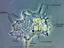
An amoeba, often called an amoeboid, is a type of cell or unicellular organism with the ability to alter its shape, primarily by extending and retracting pseudopods. Amoebae do not form a single taxonomic group; instead, they are found in every major lineage of eukaryotic organisms. Amoeboid cells occur not only among the protozoa, but also in fungi, algae, and animals.

Cutosea is a small group of marine amoeboid protists proposed in 2016. It is a monotypic class of Amoebozoa containing the order Squamocutida. Cutosean organisms are characterized by a cell coat of microscales separated from the cell membrane. Three genera, Armaparvus, Sapocribrum and Squamamoeba, belong to this group, distributed in two families.

<span class="mw-page-title-main">Orthokaryotes</span>

The Orthokaryotes are a proposed Eukaryote clade consisting of the Jakobea and the Neokaryotes. Together with its sister Discicristata it forms a basal Eukaryote clade. They are characterized by stacked Golgi, orthogonal centrioles, and two opposite posterior ciliary roots.

<span class="mw-page-title-main">Neokaryotes</span> Eukaryote clade consisting of most protists

The neokaryotes are a proposed eukaryote clade consisting of the unikonts and the bikonts as sister of for instance the Jakobea. It arises because the Euglenozoa, Percolozoa, Tsukubea, and Jakobea are seen in this view as more basal eukaryotes. These four groups, are traditionally grouped together in the Discoba. However, the Discoba may well be paraphyletic as the neokaryotes may have emerged in them.

The Scotokaryotes (Cavalier-Smith) is a proposed basal Neokaryote clade as sister of the Diaphoretickes. Basal Scotokaryote groupings are the Metamonads, the Malawimonas and the Podiata. In this phylogeny the Discoba are sometimes seen as paraphyletic and basal Eukaryotes.

<span class="mw-page-title-main">Evosea</span> Group of amoebae

Evosea is a diverse clade of amoeboid protists discovered through molecular analyses. Along with Tubulinea and Discosea, Evosea is one of the three major groups within Amoebozoa, an important clade of eukaryotic organisms. It contains unicellular organisms that display a wide variety of life cycles and cell shapes, including amoebae, flagellates and different kinds of slime molds.

References

  1. 1 2 Porter SM, Meisterfeld R, Knoll AH (May 2003). "Vase-shaped microfossils from the Neoproterozoic Chuar Group, Grand Canyon: a classification guided by modern testate amoebae" (PDF). Journal of Paleontology. 77 (3): 409–429. doi:10.1666/0022-3360(2003)077<0409:VMFTNC>2.0.CO;2.
  2. Parfrey LW, Lahr DJ, Knoll AH, Katz LA (August 2011). "Estimating the timing of early eukaryotic diversification with multigene molecular clocks". Proceedings of the National Academy of Sciences of the United States of America. 108 (33): 13624–9. Bibcode:2011PNAS..10813624P. doi: 10.1073/pnas.1110633108 . PMC   3158185 . PMID   21810989.
  3. 1 2 Cavalier-Smith T (August 1998). "A revised six-kingdom system of life". Biological Reviews of the Cambridge Philosophical Society. 73 (3): 203–66. doi:10.1111/j.1469-185X.1998.tb00030.x. PMID   9809012. S2CID   6557779.
  4. Adl SM, Bass D, Lane CE, Lukeš J, Schoch CL, Smirnov A, Agatha S, Berney C, Brown MW, Burki F, Cárdenas P, Čepička I, Chistyakova L, del Campo J, Dunthorn M, Edvardsen B, Eglit Y, Guillou L, Hampl V, Heiss AA, Hoppenrath M, James TY, Karnkowska A, Karpov S, Kim E, Kolisko M, Kudryavtsev A, Lahr DJG, Lara E, Le Gall L, Lynn DH, Mann DG, Massana R, Mitchell EAD, Morrow C, Park JS, Pawlowski JW, Powell MJ, Richter DJ, Rueckert S, Shadwick L, Shimano S, Spiegel FW, Torruella G, Youssef N, Zlatogursky V, Zhang Q (2019). "Revisions to the Classification, Nomenclature, and Diversity of Eukaryotes". Journal of Eukaryotic Microbiology. 66 (1): 4–119. doi:10.1111/jeu.12691. PMC   6492006 . PMID   30257078.
  5. 1 2 3 Kang S, Tice AK, Spiegel FW, Silberman JD, Pánek T, Cepicka I, et al. (September 2017). "Between a Pod and a Hard Test: The Deep Evolution of Amoebae". Molecular Biology and Evolution. 34 (9): 2258–2270. doi:10.1093/molbev/msx162. PMC   5850466 . PMID   28505375.
  6. 1 2 3 4 5 Adl SM, Simpson AG, Lane CE, Lukeš J, Bass D, Bowser SS, et al. (September 2012). "The revised classification of eukaryotes". The Journal of Eukaryotic Microbiology. 59 (5): 429–93. doi:10.1111/j.1550-7408.2012.00644.x. PMC   3483872 . PMID   23020233.
  7. 1 2 Pawlowski J, Audic S, Adl S, Bass D, Belbahri L, Berney C, et al. (November 6, 2012). "CBOL protist working group: barcoding eukaryotic richness beyond the animal, plant, and fungal kingdoms". PLOS Biology. 10 (11): e1001419. doi: 10.1371/journal.pbio.1001419 . PMC   3491025 . PMID   23139639.
  8. 1 2 3 Cavalier-Smith T, Fiore-Donno AM, Chao E, Kudryavtsev A, Berney C, Snell EA, Lewis R (February 2015). "Multigene phylogeny resolves deep branching of Amoebozoa". Molecular Phylogenetics and Evolution. 83: 293–304. doi: 10.1016/j.ympev.2014.08.011 . PMID   25150787.
  9. Corliss JO (1984). "The Kingdom Protista and its 45 Phyla". BioSystems. 17 (2): 87–126. doi:10.1016/0303-2647(84)90003-0. PMID   6395918.
  10. 1 2 Cavalier-Smith T (2003). "Protist phylogeny and the high-level classification of Protozoa". European Journal of Protistology. 39 (4): 338–348. doi:10.1078/0932-4739-00002.
  11. Ruggiero, Michael A.; Gordon, Dennis P.; Orrell, Thomas M.; Bailly, Nicolas; Bourgoin, Thierry; Brusca, Richard C.; Cavalier-Smith, Thomas; Guiry, Michael D.; Kirk, Paul M. (2015-06-11). "Correction: A Higher Level Classification of All Living Organisms". PLOS ONE. 10 (6): e0130114. doi: 10.1371/journal.pone.0130114 . ISSN   1932-6203. PMC   5159126 . PMID   26068874.
  12. 1 2 Derelle R, Torruella G, Klimeš V, Brinkmann H, Kim E, Vlček Č, Lang BF, Eliáš M (February 2015). "Bacterial proteins pinpoint a single eukaryotic root". Proceedings of the National Academy of Sciences of the United States of America. 112 (7): E693–9. Bibcode:2015PNAS..112E.693D. doi: 10.1073/pnas.1420657112 . PMC   4343179 . PMID   25646484.
  13. Zhulidov DA, Robarts RD, Zhulidov AV, Zhulidova OV, Markelov DA, Rusanov VA, Headley JV (2002). "Zinc accumulation by the slime mold Fuligo septica (L.) Wiggers in the former Soviet Union and North Korea". Journal of Environmental Quality. 31 (3): 1038–42. doi:10.2134/jeq2002.1038. PMID   12026071.
  14. Jeon KW (1973). Biology of Amoeba . New York: Academic Press. pp.  100. ISBN   978-0-323-14404-9.
  15. Hoshina, Ryo; Tsukii, Yuuji; Harumoto, Terue; Suzaki, Toshinobu (February 2021). "Characterization of a green Stentor with symbiotic algae growing in an extremely oligotrophic environment and storing large amounts of starch granules in its cytoplasm". Scientific Reports. 11 (2865). doi:10.1038/s41598-021-82416-9. PMC   7859197 . PMID   33536497.
  16. Weiner, Agnes K.M.; Cullison, Billie; Date, Shailesh V.; Tyml, Tomáš; Volland, Jean-Marie; Woyke, Tanja; Katz, Laura A.; Sleith, Robin S. (February 2022). "Examining the Relationship Between the Testate Amoeba Hyalosphenia papilio (Arcellinida, Amoebozoa) and its Associated Intracellular Microalgae Using Molecular and Microscopic Methods". Protist. 173 (1): 125853. doi: 10.1016/j.protis.2021.125853 . PMC   9148389 . PMID   35030517.
  17. Eichinger L, Pachebat JA, Glöckner G, Rajandream MA, Sucgang R, Berriman M, et al. (May 2005). "The genome of the social amoeba Dictyostelium discoideum". Nature. 435 (7038): 43–57. Bibcode:2005Natur.435...43E. doi:10.1038/nature03481. PMC   1352341 . PMID   15875012.
  18. Dawkins, Richard; Wong, Yan (2016). The Ancestor's Tale. Houghton Mifflin Harcourt. ISBN   978-0544859937.
  19. Cavalier-Smith T, Chao EE (December 1996). "Molecular phylogeny of the free-living archezoan Trepomonas agilis and the nature of the first eukaryote". Journal of Molecular Evolution. 43 (6): 551–62. Bibcode:1996JMolE..43..551C. doi:10.1007/BF02202103. PMID   8995052. S2CID   28992966.
  20. Cavalier-Smith T, Chao EE, Lewis R (June 2016). "187-gene phylogeny of protozoan phylum Amoebozoa reveals a new class (Cutosea) of deep-branching, ultrastructurally unique, enveloped marine Lobosa and clarifies amoeba evolution". Molecular Phylogenetics and Evolution. 99: 275–296. doi: 10.1016/j.ympev.2016.03.023 . PMID   27001604.
  21. Silar P (2016). "Protistes Eucaryotes: Origine, Evolution et Biologie des Microbes Eucaryotes" (PDF). HAL archives-ouvertes. Creative Commons. pp. 1–462. ISBN   978-2-9555841-0-1.
  22. Pánek T, Zadrobílková E, Walker G, Brown MW, Gentekaki E, Hroudová M, et al. (May 2016). "First multigene analysis of Archamoebae (Amoebozoa: Conosa) robustly reveals its phylogeny and shows that Entamoebidae represents a deep lineage of the group". Molecular Phylogenetics and Evolution. 98: 41–51. doi: 10.1016/j.ympev.2016.01.011 . PMID   26826602.
  23. Leontyev, Dmitry V.; Schnittler, Martin; Stephenson, Steven L.; Novozhilov, Yuri K.; Shchepin, Oleg N. (March 2019). "Towards a phylogenetic classification of the Myxomycetes". Phytotaxa. 399 (3): 209–238. doi:10.11646/phytotaxa.399.3.5. S2CID   108783142.
  24. Wijayawardene, Nalin; Hyde, Kevin; Al-Ani, LKT; Dolatabadi, S; Stadler, Marc; Haelewaters, Danny; et al. (2020). "Outline of Fungi and fungus-like taxa". Mycosphere. 11: 1060–1456. doi: 10.5943/mycosphere/11/1/8 .
  25. Porter SM (2006). "The Proterozoic Fossil Record of Heterotrophic Eukaryotes". In Xiao S, Kaufman AJ (eds.). Neoproterozoic Geolobiology and Paleobiology. Topics in Geobiology. Vol. 27. Dordrecht, The Netherlands: Springer. pp. 1–21. doi:10.1007/1-4020-5202-2. ISBN   978-1-4020-5201-9.
  26. Khan NA, Siddiqui R (June 2015). "Is there evidence of sexual reproduction (meiosis) in Acanthamoeba?". Pathogens and Global Health. 109 (4): 193–5. doi:10.1179/2047773215Y.0000000009. PMC   4530557 . PMID   25800982.
  27. 1 2 3 4 Kelso AA, Say AF, Sharma D, Ledford LL, Turchick A, Saski CA, King AV, Attaway CC, Temesvari LA, Sehorn MG (2015). "Entamoeba histolytica Dmc1 Catalyzes Homologous DNA Pairing and Strand Exchange That Is Stimulated by Calcium and Hop2-Mnd1". PLOS ONE. 10 (9): e0139399. Bibcode:2015PLoSO..1039399K. doi: 10.1371/journal.pone.0139399 . PMC   4589404 . PMID   26422142.
  28. 1 2 Singh N, Bhattacharya A, Bhattacharya S (2013). "Homologous recombination occurs in Entamoeba and is enhanced during growth stress and stage conversion". PLOS ONE. 8 (9): e74465. Bibcode:2013PLoSO...874465S. doi: 10.1371/journal.pone.0074465 . PMC   3787063 . PMID   24098652.
  29. Hofstatter PG, Brown MW, Lahr DJG (November 2018). "Comparative genomics supports sex and meiosis in diverse Amoebozoa". Genome Biol Evol. 10 (11): 3118–3128. doi:10.1093/gbe/evy241. PMC   6263441 . PMID   30380054.
  30. "Entamoebiasis - MeSH - NCBI". www.ncbi.nlm.nih.gov. Retrieved 2015-07-21.
  31. "Entamoebiasis". mesh.kib.ki.se. Archived from the original on 2015-07-22. Retrieved 2015-07-21.
  32. 1 2 3 4 Farrar J, Hotez P, Junghanss T, Kang G, Lalloo D, White NJ (2013-10-26). Manson's Tropical Diseases. Elsevier Health Sciences. pp. 664–671. ISBN   978-0-7020-5306-1.
  33. Beeching N, Gill G (2014-04-17). "19". Lecture Notes: Tropical Medicine. John Wiley & Sons. pp. 177–182. ISBN   978-1-118-73456-8.

Further reading