Balamuthia mandrillaris

Last updated

Balamuthia mandrillaris
Balamuthia mandrillaris (trophozoite).png
Trophozoite (active) form of Balamuthia mandrillaris
Balamuthia mandrillaris (cyst).png
A Balamuthia mandrillaris cyst
Scientific classification
Domain:
Phylum:
Class:
Order:
Family:
Balamuthiidae
Genus:
Balamuthia

Visvesvara et al., 1993
Species:
B. mandrillaris
Binomial name
Balamuthia mandrillaris
Visvesvara et al., 1993

Balamuthia mandrillaris is a free-living amoeba that causes the rare but deadly neurological condition granulomatous amoebic encephalitis (GAE). [1] B. mandrillaris is a soil-dwelling amoeba and was first discovered in 1986 in the brain of a mandrill that died in the San Diego Wild Animal Park. [2] [3]

Contents

B. mandrillaris can infect the body through open wounds or possibly by inhalation. [4] Balamuthia has been isolated from soil. [5] [6] It is believed to be distributed throughout the temperate regions of the world. This is supported somewhat by the detection of antibodies to the protist in healthy individuals.

The generic name Balamuthia was given by Govinda Visvesvara, after his mentor, parasitologist William Balamuth, for his contributions to the study of amoebae. Visvesvara isolated and studied the pathogen for the first time in 1993. [7]

Morphology

B. mandrillaris is a free-living, heterotrophic amoeba, consisting of a standard complement of organelles surrounded by a three-layered cell wall (thought to be made of cellulose or a similar polysaccharide [8] ), and with an abnormally large cell nucleus. On average, a Balamuthia trophozoite is about 30 to 120 μm in diameter. The cysts fall around this range, as well. [9]

Life cycle

Balamuthia's lifecycle, like the Acanthamoeba , consists of a cystic stage and a non-flagellated trophozoite stage, both of which are infectious, and both of which can be identified in the brain tissue on microscopic examination of brain biopsies performed on infected individuals. The trophozoite is pleomorphic and uninucleated, but binucleated forms are occasionally seen. Cysts are also uninucleated, possessing three walls: an outer thin irregular ectocyst, an inner thick endocyst, and a middle amorphous fibrillar mesocyst. [10]

Pathology

B. mandrillaris is larger than human leukocytes, thus making phagocytosis impossible. Instead, the immune system attempts to contain them at the portal of entry (usually an open wound) by mounting a type IV hypersensitivity reaction. [11] Upon introduction, the amoeba may form a skin lesion, or in some cases, may migrate to the brain, causing a condition known as granulomatous amoebic encephalitis (GAE), [12] which is usually fatal. This granulomatous feature is mostly seen in immunocompetent patients; immunocompromised individuals exhibit a "perivascular cuffing". [13] Balamuthia-induced GAE can cause focal paralysis, seizures, and brainstem symptoms such as facial paralysis, difficulty swallowing, and double vision. [14]

Balamuthia may also cause a variety of non-neurological symptoms, including skin lesions, which can progress to GAE. Patients experiencing this particular syndrome may report a skin lesion (often similar to those caused by MRSA), which does not respond well to antibiotics. The lesion is usually localized and very slow to heal, or fails to heal altogether. In some presentations, this infection may be mistaken for certain forms of skin cancer or cutaneous leishmaniasis. Balamuthia lesions are most often painless. [14]

Culturing and identification

Biopsies of skin lesions, sinuses, lungs, and the brain can detect of B. mandrillaris infection. The amoeba cannot be cultured on an agar plate coated with E. coli because, unlike Naegleria or Acanthamoeba , Balamuthia mandrillaris does not feed on bacteria. Instead, Balamuthia must be cultured on primate hepatocytes or human brain microvascular endothelial cells. [15] Formalin-fixed paraffinized biopsy specimens may indicate Balamuthia trophozoites in the perivascular space. The cysts can be visualized by calcofluor white, which binds to glycans on the cyst wall. Trophozoites appear circular during infection. [14]

Vero cells have been suggested as a possible cheaper and faster alternative to culture the organism. [16] Several types of animal cells have been used in B. mandrillaris culturing including rat glioma cells, human lung cells, and human brain microvascular endothelial cells. [14] These animal cells are added to a specified sterilized growth medium for co culturing with the pathogen. This can also help differentiate between Balamuthia and other protozoa. [14]

Axenic culture methods that are of importance to antiprotozoal drug development have also been reported within the last decade [17] [18]

Treatment

Infection seems to be survivable if treated early. Two individuals, a 5-year-old girl and a 64-year-old man, developed GAE. After diagnosis, they were treated with flucytosine, pentamidine, fluconazole, sulfadiazine, a macrolide antibiotic and trifluoperazine. Both patients recovered. [19] In 2018, an unsuccessful attempt at treatment of a Balamuthia infection after nasal lavage with untreated tap water was reported. [20]

Nitroxoline has shown interesting properties in vitro and might be a possible treatment for this infection. [21] A man treated with nitroxoline at UCSF Medical Center in 2021, following a seizure that was identified to have resulted from CNS invasive Balamuthia infection, survived and recovered from the disease, indicating that nitroxoline might be a promising medication. [22] [23]

Organ transplantation

According to a report published in Morbidity and Mortality Weekly Report in September 2010, two confirmed cases of Balamuthia transmission occurred through organ transplantation in December 2009 in Mississippi. [24] Two kidney recipients, a 31-year-old woman and a 27-year-old man, suffered from post-transplant encephalitis due to Balamuthia. The woman died in February 2010 and the man survived with partial paralysis of his right arm. The CDC was notified by a physician on December 14, 2009, about possible transplant transmission in these two patients. Histopathologic testing of donor and recipient tissues confirmed the transmission. Two other patients who received heart and liver transplants from the same donor, but in different hospitals, were placed on preemptive therapy and remain unaffected. A second cluster of transplant-transmitted Balamuthia in Arizona was reported in the same weekly report. Four recipients were identified, two from Arizona (liver and kidney-pancreas), one from California (kidney), and another from Utah (heart). Recipients from Arizona—a 56-year-old male and a 24-year-old male—both succumbed to GAE within a span of 40 days from transplantation. The other two were placed on preemptive therapy after the first two were reported and remain unaffected. [25]

Related Research Articles

<i>Entamoeba histolytica</i> Anaerobic parasitic protist

Entamoeba histolytica is an anaerobic parasitic amoebozoan, part of the genus Entamoeba. Predominantly infecting humans and other primates causing amoebiasis, E. histolytica is estimated to infect about 35-50 million people worldwide. E. histolytica infection is estimated to kill more than 55,000 people each year. Previously, it was thought that 10% of the world population was infected, but these figures predate the recognition that at least 90% of these infections were due to a second species, E. dispar. Mammals such as dogs and cats can become infected transiently, but are not thought to contribute significantly to transmission.

<i>Acanthamoeba</i> Genus of protozoans

Acanthamoeba is a genus of amoebae that are commonly recovered from soil, fresh water, and other habitats. The genus Acanthamoeba has two stages in its life cycle, the metabolically active trophozoite stage and a dormant, stress-resistant cyst stage. In nature, Acanthamoeba species are generally free-living bacterivores. However, they are also opportunistic pathogens able to cause serious and sometimes fatal infections in humans and other animals.

<i>Entamoeba coli</i> Species of parasitic amoeba

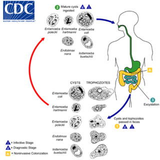
Entamoeba coli is a non-pathogenic species of Entamoeba that frequently exists as a commensal parasite in the human gastrointestinal tract. E. coli is important in medicine because it can be confused during microscopic examination of stained stool specimens with the pathogenic Entamoeba histolytica. This amoeba does not move much by the use of its pseudopod, and creates a "sur place (non-progressive) movement" inside the large intestine. Usually, the amoeba is immobile, and keeps its round shape. This amoeba, in its trophozoite stage, is only visible in fresh, unfixed stool specimens. Sometimes the Entamoeba coli have parasites as well. One is the fungus Sphaerita spp. This fungus lives in the cytoplasm of the E. coli. While this differentiation is typically done by visual examination of the parasitic cysts via light microscopy, new methods using molecular biology techniques have been developed. The scientific name of the amoeba, E. coli, is often mistaken for the bacterium, Escherichia coli. Unlike the bacterium, the amoeba is mostly harmless, and does not cause as many intestinal problems as some strains of the E. coli bacterium. To make the naming of these organisms less confusing, "alternate contractions" are used to name the species for the purpose making the naming easier; for example, using Esch. coli and Ent. coli for the bacterium and amoeba, instead of using E. coli for both.

Free-living amoebae are a group of protozoa that are important causes of infectious disease in humans and animals.

<span class="mw-page-title-main">Naegleriasis</span> Rare and usually fatal brain infection by a protist

Naegleriasis, also known as primary amoebic meningoencephalitis (PAM), is an almost invariably fatal infection of the brain by the free-living unicellular eukaryote Naegleria fowleri. Symptoms are meningitis-like and include headache, fever, nausea, vomiting, a stiff neck, confusion, hallucinations and seizures. Symptoms progress rapidly over around five days, and death usually results within one to two weeks of symptoms.

<i>Naegleria</i> Genus of protists

Naegleria is a free living amoebae protist genus consisting of 47 described species often found in warm aquatic environments as well as soil habitats worldwide. It has three life cycle forms: the amoeboid stage, the cyst stage, and the flagellated stage, and has been routinely studied for its ease in change from amoeboid to flagellated stages. The Naegleria genera became famous when Naegleria fowleri, a human pathogenic strain and the causative agent of primary amoebic meningoencephalitis (PAM), was discovered in 1965. Most species in the genus, however, are nonpathogenic, meaning they do not cause disease.

<span class="mw-page-title-main">Granulomatous amoebic encephalitis</span> Rare and usually fatal brain infection by certain amoebae

Granulomatous amoebic encephalitis (GAE) is a rare, often fatal, subacute-to-chronic central nervous system disease caused by certain species of free-living amoebae of the genera Acanthamoeba, Balamuthia and Sappinia. The term is most commonly used with Acanthamoeba. In more modern references, the term "balamuthia amoebic encephalitis" (BAE) is commonly used when Balamuthia mandrillaris is the cause.

<span class="mw-page-title-main">Miltefosine</span> Phospholipid drug

Miltefosine, sold under the trade name Impavido among others, is a medication mainly used to treat leishmaniasis and free-living amoeba infections such as Naegleria fowleri and Balamuthia mandrillaris. This includes the three forms of leishmaniasis: cutaneous, visceral and mucosal. It may be used with liposomal amphotericin B or paromomycin. It is taken by mouth.

<span class="mw-page-title-main">Nitroxoline</span> Antibiotic chemical compound

Nitroxoline is an antibiotic that has been in use in Europe for about fifty years, and has proven to be very effective at combating biofilm infections. Nitroxoline was shown to cause a decrease in the biofilm density of P. aeruginosa infections, which would allow access to the infection by the immune system in vivo. It was shown that nitroxoline functions by chelating Fe2+ and Zn2+ ions from the biofilm matrix; when Fe2+ and Zn2+ were reintroduced into the system, biofilm formation was reconstituted. The activity of biofilm degradation is comparable to EDTA, but has a history of human use in clinical settings and therefore has a precedent with which to allow its use against “slimy” biofilm infections.

<span class="mw-page-title-main">Amoebiasis</span> Human disease caused by amoeba protists

Amoebiasis, or amoebic dysentery, is an infection of the intestines caused by a parasitic amoeba Entamoeba histolytica. Amoebiasis can be present with no, mild, or severe symptoms. Symptoms may include lethargy, loss of weight, colonic ulcerations, abdominal pain, diarrhea, or bloody diarrhea. Complications can include inflammation and ulceration of the colon with tissue death or perforation, which may result in peritonitis. Anemia may develop due to prolonged gastric bleeding.

Sappinia diploidea is a free-living amoeba species.

<span class="mw-page-title-main">Amoebic brain abscess</span> Medical condition

Amoebic brain abscess is an affliction caused by the anaerobic parasitic protist Entamoeba histolytica. It is extremely rare; the first case being reported in 1849. Brain abscesses resulting from Entamoeba histolytica are difficult to diagnose and very few case reports suggest complete recovery even after the administration of appropriate treatment regimen.

Sappinia is a genus of heterotrophic, lobose amoebae within the family Thecamoebidae. A defining feature of Sappinia, which separates it from its sister genus Thecamoeba, is the presence of two closely apposed nuclei with a central, flattened connection. Sappinia species have two life cycle stages: a trophozoite and a cyst. Up until 2015, only two species had been discovered, Sappinia pedata and Sappinia diploidea. Sequencing of the small subunit rRNA of a particular isolate from a sycamore tree revealed a new species, Sappinia platani.Sappinia species were once thought to be coprozoic, as the first strains were isolated from animal dung. More research has shown that they are typical free-living amoebae, and can be found worldwide in soil, plant litter, and standing decaying plants, as well as freshwater ponds. In 2001, the first and only case of human pathogenesis in Sappinia was confirmed. The patient was a non-immunocompromised 38-year-old male who presented signs of amoebic encephalitis and who patient made a full recovery after treatment with several antimicrobials. The CDC initially classified the causative agent as S. diploidea based on morphological characteristics, but in 2009, Qvarnstrom et al. used molecular data to confirm that the true causative agent was S. pedata.

Cutaneous amoebiasis, refers to a form of amoebiasis that presents primarily in the skin. It can be caused by Acanthamoeba or Entamoeba histolytica. When associated with Acanthamoeba, it is also known as "cutaneous acanthamoebiasis". Balamuthia mandrillaris can also cause cutaneous amoebiasis, but can prove fatal if the amoeba enters the bloodstream It is characterized by ulcers. Diagnosis of amebiasis cutis calls for high degree of clinical suspicion. This needs to be backed with demonstration of trophozoites from lesions. Unless an early diagnosis can be made such patients can develop significant morbidity.

Balamuthia infection is a cutaneous condition resulting from Balamuthia that may result in various skin lesions.

Sappinia pedata is a free living amoeboid organism, first described by Pierre Augustin Dangeard in 1896. It belongs to the genus Sappinia within the Thecamoebida clade of Amoebozoa and is characterized by its unique monopodial locomotion and cell surface morphology. S. pedata has been found in various habitats worldwide, mostly on herbivore dung, decaying plant matter, and soil. The species has gained attention due to its potential medical relevance and has been the subject of most recent and emerging studies in Protistology and Eukaryotic Microbiology as a whole.

Sappinia amoebic encephalitis (SAE) is the name for amoebic encephalitis caused by species of Sappinia.

<i>Naegleria fowleri</i> Species of free-living excavate form of protist

Naegleria fowleri, also known as the brain-eating amoeba, is a species of the genus Naegleria. It belongs to the phylum Percolozoa and is technically classified as an amoeboflagellate excavate, rather than a true amoeba. This free-living microorganism primarily feeds on bacteria but can become pathogenic in humans, causing an extremely rare, sudden, severe, and usually fatal brain infection known as naegleriasis or primary amoebic meningoencephalitis (PAM).

Sarcocystis calchasi is an apicomplexan parasite. It has been identified to be the cause of Pigeon protozoal encephalitis (PPE) in the intermediate hosts, domestic pigeons. PPE is a central-nervous disease of domestic pigeons. Initially there have been reports of this parasite in Germany, with an outbreak in 2008 and in 2011 in the United States. Sarcocystis calchasi is transmitted by the definitive host Accipter hawks.

References

  1. Sarica, Feyzi Birol; Tufan, Kadir; Cekinmez, Melih; Erdoğan, Bülent; Altinörs, Mehmet Nur (2009). "A rare but fatal case of granulomatous amebic encephalitis with brain abscess: the first case reported from Turkey". Turkish Neurosurgery. 19 (3): 256–259. PMID   19621290.
  2. Cope, Jennifer R.; Landa, Janet; Nethercut, Hannah; Collier, Sarah A.; Glaser, Carol; Moser, Melanie; Puttagunta, Raghuveer; Yoder, Jonathan S.; Ali, Ibne K.; Roy, Sharon L. (2019-05-17). "The Epidemiology and Clinical Features of Balamuthia mandrillaris Disease in the United States, 1974 – 2016". Clinical Infectious Diseases. 68 (11): 1815–1822. doi:10.1093/cid/ciy813. ISSN   1058-4838. PMC   7453664 . PMID   30239654.
  3. Visvesvara, G S; Martinez, A J; Schuster, F L; Leitch, G J; Wallace, S V; Sawyer, T K; Anderson, M (1990-12-28). "Leptomyxid ameba, a new agent of amebic meningoencephalitis in humans and animals". Journal of Clinical Microbiology. 28 (12): 2750–2756. doi:10.1128/jcm.28.12.2750-2756.1990. ISSN   0095-1137. PMC   268267 . PMID   2280005.
  4. "Balamuthia mandrillaris ameba infection". Centers for Disease Control and Prevention. Retrieved 14 June 2014.
  5. Frederick L. Schuster; Thelma H. Dunnebacke; Gregory C. Booton; Shigeo Yagi; Candice K. Kohlmeier; Carol Glaser; Duc Vugia; Anna Bakardjiev; Parvin Azimi; Mary Maddux-Gonzalez; A. Julio Martinez; Govinda S. Visvesvara (July 2003). "Environmental Isolation of Balamuthia mandrillaris Associated with a Case of Amebic Encephalitis". J. Clin. Microbiol. 41 (7): 3175–3180. doi:10.1128/JCM.41.7.3175-3180.2003. PMC   165348 . PMID   12843060.
  6. Thelma H. Dunnebacke; Frederick L. Schuster; Shigeo Yagi; Gregory C. Booton (September 2004). "Balamuthia mandrillaris from soil samples". Microbiology. 150 (Pt 9): 2837–2842. doi: 10.1099/mic.0.27218-0 . PMID   15347743. Archived from the original (PDF) on 2018-07-27. Retrieved 2017-06-20.
  7. Kaneshiro, E. S.; Marciano-Cabral, F.; Moura, H. (2014). "Govinda S. Visvesvara: A Tribute". The Journal of Eukaryotic Microbiology. 62 (1): 1–2. doi:10.1111/jeu.12143. PMC   5674982 . PMID   25040661.
  8. Siddiqui, R.; Khan, N. A.; Jarroll, E. L. (2009). "The cyst wall carbohydrate composition of Balamuthia mandrillaris". Parasitology Research. 104 (6): 1439–1443. doi:10.1007/s00436-009-1346-8 (inactive 2024-05-03). PMID   19214576.{{cite journal}}: CS1 maint: DOI inactive as of May 2024 (link)
  9. Ruqaiyyah Siddiqui; Naveed Ahmed Khan (2015). "Balamuthia mandrillaris: Morphology, biology and virulence". Trop. Parasitol. 5 (1): 15–22. doi: 10.4103/2229-5070.149888 . PMC   4326988 . PMID   25709948.
  10. Guerrant RL, Walker DH, Weller PF (2011). Tropical Infectious Diseases:Principles, Pathogens and Practice (3rd ed.). Saunders. ISBN   978-0-7020-3935-5 . Retrieved 6 September 2016.
  11. Baig, A. M. (2015). "Pathogenesis of amoebic encephalitis: Are the amoebae being credited to an 'inside job' done by the host immune response?". Acta Tropica. 148: 72–76. doi:10.1016/j.actatropica.2015.04.022. PMID   25930186.
  12. Di Gregorio, C.; Rivasi, F.; Mongiardo, N.; De Rienzo, B.; Wallace, S.; Visvesvara, G. S. (December 1992). "Acanthamoeba meningoencephalitis in a patient with acquired immunodeficiency syndrome". Archives of Pathology & Laboratory Medicine. 116 (12): 1363–5. PMID   1456885.
  13. Mannan Baig, Abdul (Dec 2014). "Granulomatous amoebic encephalitis: ghost response of an immunocompromised host?". J Med Microbiol. 63 (12): 1763–6. doi: 10.1099/jmm.0.081315-0 . PMID   25239626. S2CID   28069984.
  14. 1 2 3 4 5 Bhosale, Namrata K.; Parija, Subhash Chandra (2021). "Balamuthia mandrillaris: An opportunistic, free-living ameba – An updated review". Tropical Parasitology. 11 (2): 78–88. doi: 10.4103/tp.tp_36_21 (inactive 31 January 2024). ISSN   2229-5070. PMC   8579774 . PMID   34765527.{{cite journal}}: CS1 maint: DOI inactive as of January 2024 (link)
  15. Martínez AJ, Visvesvara GS (March 2001). "Balamuthia mandrillaris infection". J. Med. Microbiol. 50 (3): 205–7. doi: 10.1099/0022-1317-50-3-205 . PMID   11232763.
  16. Greninger, Alexander L.; Messacar, Kevin; Dunnebacke, Thelma; Naccache, Samia N.; Federman, Scot; Bouquet, Jerome; Mirsky, David; Nomura, Yosuke; Yagi, Shigeo; Glaser, Carol; Vollmer, Michael; Press, Craig A.; Kleinschmidt-DeMasters, Bette K.; Dominguez, Samuel R.; Chiu, Charles Y. (2015). "Clinical metagenomic identification of Balamuthia mandrillaris encephalitis and assembly of the draft genome: the continuing case for reference genome sequencing". Genome Medicine. 7 (1): 113. doi: 10.1186/s13073-015-0235-2 . ISSN   1756-994X. PMC   4665321 . PMID   26620704.
  17. Ferrins L, Buskes MJ, Kapteyn MM, Engels HN, Enos SE, Lu C; et al. (2023). "Identification of novel anti-amoebic pharmacophores from kinase inhibitor chemotypes". Front Microbiol. 14: 1149145. doi: 10.3389/fmicb.2023.1149145 . PMC   10206040 . PMID   37234530.{{cite journal}}: CS1 maint: multiple names: authors list (link)
  18. Lares-Jiménez LF, Gámez-Gutiérrez RA, Lares-Villa F (2015). "Novel culture medium for the axenic growth of Balamuthia mandrillaris". Diagn Microbiol Infect Dis. 82 (4): 286–8. doi:10.1016/j.diagmicrobio.2015.04.007. PMID   25957459.{{cite journal}}: CS1 maint: multiple names: authors list (link)
  19. Deetz, T. R.; Sawyer, M. H.; Billman, G.; Schuster, F. L.; Visvesvara, G. S. (15 November 2003). "Successful Treatment of Balamuthia Amoebic Encephalitis: Presentation of 2 Cases". Clinical Infectious Diseases. 37 (10): 1304–1312. doi: 10.1086/379020 . PMID   14583863.
  20. Piper, Keenan H.; Foster, Haidn; Susanto, Daniel; Maree, Cynthia L.; Thornton, Sean D.; Cobbs, Charles S. (December 2018). "Fatal Balamuthia mandrillaris brain infection associated with improper nasal lavage". International Journal of Infectious Diseases. 77: 18–22. doi: 10.1016/j.ijid.2018.09.013 . PMID   30243910.
  21. Laurie, Matthew T.; White, Corin V.; Retallack, Hanna; Wu, Wesley; Moser, Matthew S.; Sakanari, Judy A.; Ang, Kenny; Wilson, Christopher; Arkin, Michelle R.; DeRisi, Joseph L.; Bassler, Bonnie (2018). "Functional Assessment of 2,177 U.S. and International Drugs Identifies the Quinoline Nitroxoline as a Potent Amoebicidal Agent against the Pathogen Balamuthia mandrillaris". mBio. 9 (5). doi:10.1128/mBio.02051-18. ISSN   2150-7511. PMC   6212833 . PMID   30377287.
  22. Kornei, Katherine (2023). "Repurposed drug battles 'brain-eating' amoeba". Science. doi:10.1126/science.adh0048 . Retrieved 10 February 2023.
  23. Spottiswoode, Natasha; Pet, Douglas; Kim, Annie; Gruenberg, Katherine; Shah, Maulik; Ramachandran, Amrutha; Laurie, Matthew T; Zia, Maham; Fouassier, Camille; Boutros, Christine L; Lu, Rufei; Zhang, Yueyuan; Servellita, Venice; Bollen, Andrew; Chiu, Charles Y; Wilson, Michael R; Valdivia, Liza; DeRisi, Joseph L (2023). "Successful Treatment of Balamuthia mandrillaris Granulomatous Amebic Encephalitis with Nitroxoline". Emerging Infectious Diseases. 29 (1): 197–201. doi:10.3201/eid2901.221531. PMC   9796214 . PMID   36573629.
  24. Centers for Disease Control Prevention (CDC) (17 September 2010). "Balamuthia mandrillaris transmitted through organ transplantation --- Mississippi, 2009". Morbidity and Mortality Weekly Report . 59 (36): 1165–70. PMID   20847719.
  25. Centers for Disease Control Prevention (CDC) (17 September 2010). "Notes from the field: transplant-transmitted Balamuthia mandrillaris --- Arizona, 2010". Morbidity and Mortality Weekly Report . 59 (36): 1182. PMID   20847722.