Cutaneous leishmaniasis

Last updated
Cutaneous leishmaniasis
Other namesOriental sore, Tropical sore, Chiclero ulcer, Chiclero's ulcer, Aleppo boil, Delhi Boil or Desert boil [1] [2] [3]
JerichoButtons.jpg
A man with cutaneous leishmaniasis in the Middle East, known then locally as "Jericho Buttons" for the frequency of cases near the ancient city of Jericho
Specialty Infectious diseases   OOjs UI icon edit-ltr-progressive.svg

Cutaneous leishmaniasis is the most common form of leishmaniasis affecting humans. [4] It is a skin infection caused by a single-celled parasite that is transmitted by the bite of a phlebotomine sand fly. There are about thirty species of Leishmania that may cause cutaneous leishmaniasis.

Contents

This disease is considered to be a zoonosis (an infectious disease that is naturally transmissible from animals to humans), with the exception of Leishmania tropica — which is often an anthroponotic disease (an infectious disease that is naturally transmissible from humans to vertebrate animals). [3]

Signs and symptoms

Skin lesions from cutaneous leishmaniasis may closely resemble those seen in leprosy Cut leishmaniasis.jpg
Skin lesions from cutaneous leishmaniasis may closely resemble those seen in leprosy

Post kala-azar dermal leishmaniasis

Post-kala-azar dermal leishmaniasis (PKDL) is a recurrence of kala-azar that may appear on the skin of affected individuals months and up to 20 years after being partially treated, untreated or even in those considered adequately treated. [5] [6] In Sudan, they can be demonstrated in up to 60% of treated cases. They manifest as hypopigmented skin lesions (such as macules, papules, nodules), or facial redness. Though any organism causing kala-azar can lead to PKDL, it is commonly associated with Leishmania donovani which gives different disease patterns in India and Sudan. In the Indian variant, nodules enlarge with time and form plaques but rarely ulcerate, but nodules from the African variety often ulcerate as they progress. Nerve involvement is common in African variety but rare in Indian subcontinent. [7] Histology demonstrates a mixture of chronic inflammatory cells; there can be macrophage or epitheloid granuloma. [8] Parasite concentration is not consistent among studies, perhaps reflecting low sensitivity of diagnostic methods used in earlier entries.[ citation needed ]

Current approach to diagnosis involves

  1. demonstration of parasite by microscopy, in vitro culture or animal inoculation;
  2. immunodiagnosis of parasite antigen;
  3. detection of parasite DNA in tissue.

Newer polymerase chain reaction (PCR) based tools have higher sensitivity and specificity. Emergence of PKDL has been reported in HIV affected individuals [9] and may become a problem in the future.

Sodium stibogluconate alone or in combination with rifampicin is used for the treatment of PKDL for a long course of up to 4 months. Compliance can be an issue for such a long course.[ citation needed ]

Mucocutaneous leishmaniasis

Mucocutaneous leishmaniasis is an especially disturbing form of cutaneous leishmaniasis, because it produces destructive and disfiguring lesions of the face. It is most often caused by Leishmania braziliensis, but cases caused by L. aethiopica have also been described. [10]

Mucocutaneous leishmaniasis is very difficult to treat. Treatment involves the use of pentavalent antimonial compounds, which are highly toxic (common side effects include thrombophlebitis, pancreatitis, cardiotoxicity and hepatotoxicity) and not very effective. For example, in one study, despite treatment with high doses of sodium stibogluconate for 28 days, only 30% of patients remained disease-free at 12 months follow-up. [11] Even in those patients who achieve an apparent cure, as many as 19% will relapse. [12] Several drug combinations with immunomodulators have been tested, for example, a combination of pentoxifylline (inhibitor of TNF-α) and a pentavalent antimonial at a high dose for 30 days in a small-scale (23 patients) randomised placebo-controlled study from Brazil achieved cure rates of 90% and reduced time to cure, [13] a result that should be interpreted cautiously in light of inherent limitations of small-scale studies. [14] In an earlier small-scale (12 patients) study, addition of imiquimod showed promising results [15] which need yet to be confirmed in larger trials.

Pathophysiology

Cutaneous leishmaniasis Cutaneous leishmaniasis3.jpg
Cutaneous leishmaniasis

Promastigotes of Leishmania are transmitted to human skin by the bite of a sandfly. Leishmania then invades human macrophages and replicates intracellularly. A raised, red lesion develops at the site of the bite (often weeks or sometimes years afterwards). The lesion then ulcerates and may become secondarily infected with bacteria. In many species (for example, L. major) the lesion often spontaneously heals with atrophic scarring. In some species (for example, L. braziliensis) the lesion may spontaneously heal with scarring, but then reappear elsewhere (especially as destructive mucocutaneous lesions). Lesions of other Leishmania species may spontaneously heal and then reappear as satellite lesions around the site of the original lesion, or along the route of lymphatic drainage.[ citation needed ]

Some species tend to cause cutaneous leishmaniasis (e.g., L. major and L.tropica), whereas some species tend to cause visceral leishmaniasis (e.g., L. infantum and L. donovani), though emerging research (due to high deployment rates of western countries to indigenous areas) is showing these species specific presentation lines are blurring.[ citation needed ]

Diagnosis

Cutaneous leishmaniasis in a man from French Guiana Leishmaniose cutanee - Guyane fr.JPG
Cutaneous leishmaniasis in a man from French Guiana

Diagnosis is based on the characteristic appearance of non-healing raised, scaling lesions that may ulcerate and become secondarily infected with organisms such as Staphylococcus aureus, in someone who has returned from an endemic area.[ citation needed ]

In resource limited settings, fine-needle aspiration of the lesion is confirmatory with identification of amastigote form of Leishmania. [16] The gold standard for diagnosis is a PCR test. [17]

Treatment

American cutaneous and mucocutaneous leishmaniasis

The best treatment for American cutaneous and mucocutaneous leishmaniasis (ACML) is not known. Pentavalent antimonial drugs (sodium stibogluconate (SSG) and meglumine antimonate (Glucantime, MA)) have been used since the 1940s, but they are expensive, toxic, and painful. [18] Treatments that work for one species of Leishmania may not work for another; therefore, it is recommended that the exact species be identified prior to initiating treatment. Unfortunately, leishmaniasis is an orphan disease in developed nations, and almost all the current treatment options are toxic with significant side effects. [18]

The best-studied treatments for ACML caused by two Leishmania species are listed below. However, one should note that most of the studies examining treatments of ACML were poorly designed. Therefore, no definitive treatment guidelines or recommendations are currently available, as large-scale and well-conducted studies are necessary to evaluate the long-term effects of current treatments. [18]

There is no strong evidence for the efficacy of surgery, oral itraconazole and fluconazole, oral antibiotics (rifampicin, metronidazole, cotrimoxazole), intravenous or topical amphotericin B, oral dapsone, photodynamic therapy, promoting healing therapies, laser, or cryotherapy treatments. [18]

Old World cutaneous leishmaniasis

Similar to ACML, the treatment recommendations for Old World cutaneous leishmaniasis (OWCL) are uncertain due to the variability of and inconsistencies within the research. [19]

Most studies done to assess treatments of OWCL included two species of parasites, Leishmania major and Leishmania tropica. The most well-studied treatments for OWCL are oral itraconazole and topical paromomycin. [19]

Patients treated with oral itraconazole for an average of 2.5 months had a higher cure rate compared to placebo, but they also had a higher rate of side effects, including gastrointestinal complaints, abnormal liver function, headaches, and dizziness. [19]

Patients treated with topical paromomycin showed no difference in cure rate compared to placebo, but patients treated with paromomycin had a higher rate of adverse skin reactions. [19]

The treatments for other Leishmania species responsible for OWCL, such as L. infantum, L. aethiopica, and L. donovani, have not been thoroughly studied. In addition, the effects of leishmaniasis treatment in children, women of childbearing age, patients with comorbidities, and immunocompromised patients have not been well established. [19]

Epidemiology

Cutaneous leishmaniasis in North Africa; Leishmania infantum=green, Leishmania major=blue, Leishmania tropica=red Parasite130072-fig1 Map of cutaneous leishmaniasis in North Africa.tif
Cutaneous leishmaniasis in North Africa; Leishmania infantum=green, Leishmania major=blue, Leishmania tropica=red

Cutaneous leishmaniasis is endemic in all tropical and subtropical areas of the world. [20] The distribution of this disease is very tightly linked to geography, and villages even 15 miles apart can have very different rates of cutaneous leishmaniasis.[ citation needed ]

Most species of Leishmania are capable of infecting humans and causing cutaneous leishmaniasis. In the New World, these organisms include L. amazonensis, L. braziliensis, L. guyanensis, L. lainsoni, L. lindenbergi, [21] L. mexicana, L. naiffi, L. panamensis, L. peruviana, L. shawi, and L. venezuelensis. Old World species that cause cutaneous leishmaniasis include L. aethiopica, L. infantum, L. major, and L. tropica. With the exception of L. tropica — which is commonly associated with human settlements and therefore considered to be an anthroponotic species — all of these organisms are zoonotic. [3] As demographic changes occur in developing nations, some species that have traditionally been considered to be zoonotic (e.g., L. panamensis) are becoming primarily human pathogens. [22]

Dogs and rodents serve as the primary animal reservoir hosts in the sylvatic cycle, but people with chronic PKDL can also serve as important reservoir hosts for cutaneous leishmaniasis. [23] The most common vectors for cutaneous leishmaniasis in the Old World are sandflies of the genus Phlebotomus , while Lutzomyia and those within the family Psychodidae (especially the genus Psychodopygus ) are the most common vectors in the New World. There are more than 600 species of phlebotomine sandflies, and only 30 of these are known vectors. [24] Cutaneous leishmaniasis has been seen in American and Canadian troops coming back from Afghanistan. [25]

Means of Prevention

The sand fly stings mainly at night, and it usually occurs about half a meter above the ground (so sleeping on high beds can prevent infection). To avoid stinging, apply mosquito repellent, and cover the body.[ citation needed ]

Studies conducted in recent years show that the plant Bougainvillea glabra may protect against the sand fly. The plant was found to be toxic to sand flies and that the life span of flies that ate from this plant was significantly shortened and sometimes led to their premature death before they could spread the disease. [26] [27]

Hebrew University study found that some plants attract sand flies. These plants often attract sand flies up to 14 times more than Bougainvillea glabra, but unlike Bougainvillea glabra, are not toxic to the sand flies. Based on this information, the dispersion of sand flies can be controlled by limiting the growth of these plants near populated areas. Alternatively, these plants may serve to capture and control sand flies by using their odor compounds or the plants themselves alongside simple glue traps, or by spraying them with deadly pesticides for sand flies which are safe for humans and mammals (e.g., boric acid or spinosad) thereby stopping the spread of the disease. Of the dozens of plants examined, the three plants that attracted especially sand flies are the Ochradenus baccatus, Prosopis farcta, and Tamarix nilotica. [28]

Outbreak in 2016

The Middle East, in 2016, seems to be experiencing an increase in the cutaneous leishmaniasis disease due to migrants fleeing the Islamic State of Iraq and the Levant. Reports of the increase in the disease have surfaced in Turkey, Lebanon, and elsewhere. [29] [30]

The huge increase in the spread of the disease is attributed to the refugee crises in the Middle East and North Africa over the past five years, particularly due to the displacement of millions of Syrian refugees. [31] The outbreak among Syrian refugees was documented by the World Health Organization (WHO) in 2012 and recognized as ongoing. [32]

Nepal

A recent study with large series of cases from Mid-western region of Nepal have demonstrated that cutaneous leishmaniasis is an under recognized medical condition posing health challenges mandating new guidelines for its elimination/ eradication. [16]

Other animals

A dog in Nicaragua with cutaneous leishmaniasis affecting the ear Dog with chiclero ulcer.JPG
A dog in Nicaragua with cutaneous leishmaniasis affecting the ear

Besides humans, cutaneous leishmaniasis often affects other animals, notably in dogs as canine leishmaniasis. [3]

Related Research Articles

<i>Leishmania</i> Genus of parasitic flagellate protist

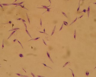
Leishmania is a parasitic protozoan, a single-celled organism of the genus Leishmania that is responsible for the disease leishmaniasis. They are spread by sandflies of the genus Phlebotomus in the Old World, and of the genus Lutzomyia in the New World. At least 93 sandfly species are proven or probable vectors worldwide. Their primary hosts are vertebrates; Leishmania commonly infects hyraxes, canids, rodents, and humans.

<span class="mw-page-title-main">Leishmaniasis</span> Disease caused by parasites of the Leishmania type

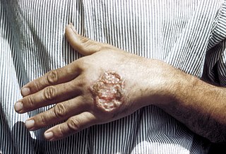
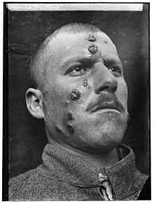
Leishmaniasis is a wide array of clinical manifestations caused by parasites of the Trypanosomatida genus Leishmania. It is generally spread through the bite of phlebotomine sandflies, Phlebotomus and Lutzomyia, and occurs most frequently in the tropics and sub-tropics of Africa, Asia, the Americas, and southern Europe. The disease can present in three main ways: cutaneous, mucocutaneous, or visceral. The cutaneous form presents with skin ulcers, while the mucocutaneous form presents with ulcers of the skin, mouth, and nose. The visceral form starts with skin ulcers and later presents with fever, low red blood cell count, and enlarged spleen and liver.

<span class="mw-page-title-main">Sandfly</span> Name of several types of blood-sucking fly

Sandfly or sand fly is a colloquial name for any species or genus of flying, biting, blood-sucking dipteran (fly) encountered in sandy areas. In the United States, sandfly may refer to certain horse flies that are also known as "greenheads", or to members of the family Ceratopogonidae. The bites usually result in a small, intensely itchy bump or welt, the strength of which intensifies over a period of 5-7 days before dissipating. Moderate relief is achieved with varying success through the application of over the counter products such as Benadryl (ingested) or an analgesic cream such as After Bite. Outside the United States, sandfly may refer to members of the subfamily Phlebotominae within the Psychodidae. Biting midges (Ceratopogonidae) are sometimes called sandflies or no-see-ums. New Zealand sandflies are in the genus of sand fly Austrosimulium, a type of black fly.

<i>Lutzomyia</i> Genus of flies

Lutzomyia is a genus of phlebotomine sand flies consisting of nearly 400 species, at least 33 of which have medical importance as vectors of human disease. Species of the genus Lutzomyia are found only in the New World, distributed in southern areas of the Nearctic and throughout the Neotropical realm. Lutzomyia is one of the two genera of the subfamily Phlebotominae to transmit the Leishmania parasite, with the other being Phlebotomus, found only in the Old World. Lutzomyia sand flies also serve as vectors for the bacterial Carrion's disease and a number of arboviruses.

<i>Phlebotomus</i> Genus of flies

Phlebotomus is a genus of "sand flies" in the Diptera family Psychodidae. In the past, they have sometimes been considered to belong in a separate family, Phlebotomidae, but this alternative classification has not gained wide acceptance.

<span class="mw-page-title-main">Visceral leishmaniasis</span> Human disease caused by protist parasites

Visceral leishmaniasis (VL), also known as kala-azar or "black fever", is the most severe form of leishmaniasis and, without proper diagnosis and treatment, is associated with high fatality. Leishmaniasis is a disease caused by protozoan parasites of the genus Leishmania.

<span class="mw-page-title-main">Miltefosine</span> Phospholipid drug

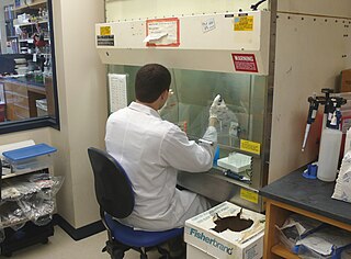
Miltefosine, sold under the trade name Impavido among others, is a medication mainly used to treat leishmaniasis and free-living amoeba infections such as Naegleria fowleri and Balamuthia mandrillaris. This includes the three forms of leishmaniasis: cutaneous, visceral and mucosal. It may be used with liposomal amphotericin B or paromomycin. It is taken by mouth.

A canine vector-borne disease (CVBD) is one of "a group of globally distributed and rapidly spreading illnesses that are caused by a range of pathogens transmitted by arthropods including ticks, fleas, mosquitoes and phlebotomine sandflies." CVBDs are important in the fields of veterinary medicine, animal welfare, and public health. Some CVBDs are of zoonotic concern.

<i>Leishmania infantum</i> Species of parasitic protist

Leishmania infantum is the causative agent of infantile visceral leishmaniasis in the Mediterranean region and in Latin America, where it has been called Leishmania chagasi. It is also an unusual cause of cutaneous leishmaniasis, which is normally caused by specific lineages. Wild canids and domestic dogs are the natural reservoir of this organism. The sandfly species Lutzomyia longipalpis serves as the primary vector for the transmission of the disease.

<i>Leishmania major</i> Species of parasitic protist

Leishmania major is a species of parasite found in the genus Leishmania, and is associated with the disease zoonotic cutaneous leishmaniasis. L. major is an intracellular pathogen which infects the macrophages and dendritic cells of the immune system. Though Leishmania species are found on every continent aside from Antarctica, Leishmania major is found only in the Eastern Hemisphere, specifically in Northern Africa, the Middle East, Northwestern China, and Northwestern India.

<span class="mw-page-title-main">Canine leishmaniasis</span> Disease affecting dogs

Canine leishmaniasis (LEESH-ma-NIGH-ah-sis) is a zoonotic disease caused by Leishmania parasites transmitted by the bite of an infected phlebotomine sandfly. Canine leishmaniasis was first identified in Europe in 1903, and in 1940, 40% of all dogs in Rome were determined to be positive for leishmaniasis. Traditionally thought of as a disease only found near the Mediterranean basin, 2008 research claims new findings are evidence that canine leishmaniasis is currently expanding in continental climate areas of northwestern Italy, far from the recognized disease-endemic areas along the Mediterranean coasts. Cases of leishmaniasis began appearing in North America in 2000, and, as of 2008, Leishmania-positive foxhounds have been reported in 22 U.S. states and two Canadian provinces.

<i>Leishmania donovani</i> Species of intracellular parasite

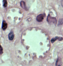
Leishmania donovani is a species of intracellular parasites belonging to the genus Leishmania, a group of haemoflagellate kinetoplastids that cause the disease leishmaniasis. It is a human blood parasite responsible for visceral leishmaniasis or kala-azar, the most severe form of leishmaniasis. It infects the mononuclear phagocyte system including spleen, liver and bone marrow. Infection is transmitted by species of sandfly belonging to the genus Phlebotomus in Old World and Lutzomyia in New World. The species complex it represents is prevalent throughout tropical and temperate regions including Africa, China, India, Nepal, southern Europe, Russia and South America. The species complex is responsible for thousands of deaths every year and has spread to 88 countries, with 350 million people at constant risk of infection and 0.5 million new cases in a year.

<i>Leishmania tropica</i> Species of protozoan parasite

Leishmania tropica is a flagellate parasite and the cause of anthroponotic cutaneous leishmaniasis in humans. This parasite is restricted to Afro-Eurasia and is a common cause of infection in Afghanistan, Iran, Syria, Yemen, Algeria, Morocco, and northern India.

Leishmania braziliensis is a Leishmania species.

<i>Leishmania mexicana</i> Species of parasitic protist

Leishmania mexicana is a species of obligate intracellular parasites of the protozoan genus Leishmania. In Mexico and Central America, this parasite is the primary cause of cutaneous leishmaniasis.

<span class="mw-page-title-main">Post-kala-azar dermal leishmaniasis</span>

Post-kala-azar dermal leishmaniasis (PKDL) is a complication of visceral leishmaniasis (VL); it is characterised by a macular, maculopapular, and nodular rash in a patient who has recovered from VL and who is otherwise well. The rash usually starts around the mouth from where it spreads to other parts of the body depending on severity.

Leishmania amazonensis is a parasite responsible for the disease leishmaniasis. This species has been known to spread by using sandflies as its vector and a vertebrate as its primary host, just like other species of Leishmania. They have also been known to spread throughout the Brazilian Amazon region due to their ecological niches needed for survival and reproduction. However, when the host is infected, the Leishmania can cause 3 different forms of Leishmaniasis. For this species, it has been known to cause cutaneous leishmaniasis and mucocutaneous leishmaniasis. Cutaneous leishmaniasis is commonly characterized with skin lesions, which can appear localized, or throughout the body. While mucocutaneous leishmaniasis is characterized with ulcers around the skin, mouth, and nose. This form of Leishmaniasis has also been known to can spread by metastasis and can be deadly.

<i>Lutzomyia longipalpis</i> Species of fly

Lutzomyia longipalpis is a species complex of sandfly belonging to the family Psychodidae. This species is primarily present in Central and South America, but has also appeared in Mexico. There have been reports of L. longipalpis as far south as Argentina, as they are found in a wide variety of ecological conditions. Both males and females feed on sugars from plants and aphids, but only adult females feed on the blood of other mammals. The species has recently begun appearing in urban areas throughout Brazil, and serves as a key vessel for the propagation of the parasite Leishmania infantum. The presence of these flies appears to be strongly correlated to the presence of domestic chickens in Latin America. The first major urban outbreak of the lethal Visceral leishmanias epidemic was detected in Teresina, Piauí State in the early 1980s following a massive planting of acacias.

Kala azar in India refers to the special circumstances of the disease kala azar as it exists in India. Kala azar is a major health problem in India with an estimated 146,700 new cases per year as of 2012. In the disease a parasite causes sickness after migrating to internal organs such as the liver, spleen and bone marrow. If left untreated the disease almost always results in the death. Signs and symptoms include fever, weight loss, fatigue, anemia, and substantial swelling of the liver and spleen.

<span class="mw-page-title-main">Leishmaniasis vaccine</span> Vaccine against leishmaniasis

A Leishmaniasis vaccine is a vaccine which would prevent leishmaniasis. As of 2017, no vaccine for humans was available. Currently some effective leishmaniasis vaccines for dogs exist.

References

  1. Calvopiña M, Martinez L, Hashiguchi Y (August 2013). "Cutaneous leishmaniasis "chiclero's ulcer" in subtropical Ecuador". The American Journal of Tropical Medicine and Hygiene. 89 (2): 195–6. doi:10.4269/ajtmh.12-0690. PMC   3741233 . PMID   23926136.
  2. Stowers JH (1920). "Case of Delhi Boil or Sore (Syn.: Oriental Sore; Aleppo Boil)". Proceedings of the Royal Society of Medicine. 13 (Dermatol Sect): 81–3. doi:10.1177/003591572001300351. PMC   2152205 . PMID   19980989.
  3. 1 2 3 4 The Institute for International Cooperation in Animal Biologics and the Center for Food Security and Public Health (October 2009). "Leishmaniasis (cutaneous and visceral)" (PDF). Ames, Iowa: College of Veterinary Medicine, Iowa State University. Retrieved 2015-01-04.
  4. James WD, Berger TG (2006). Andrews' Diseases of the Skin: clinical Dermatology. Saunders Elsevier. p. 423. ISBN   978-0-7216-2921-6.
  5. Banerjee N (December 1973). "Role of I.M.A. during natural calamities and national emergencies". Journal of the Indian Medical Association. 61 (11): 477–81. PMID   4600129.
  6. Rathi SK, Pandhi RK, Chopra P, Khanna N (2005). "Post-kala-azar dermal leishmaniasis: a histopathological study". Indian Journal of Dermatology, Venereology and Leprology. 71 (4): 250–3. doi: 10.4103/0378-6323.16616 . hdl: 1807/5537 . PMID   16394433.
  7. Salotra P, Singh R (March 2006). "Challenges in the diagnosis of post kala-azar dermal leishmaniasis". The Indian Journal of Medical Research. 123 (3): 295–310. PMID   16778312.
  8. Singh N, Ramesh V, Arora VK, Bhatia A, Kubba A, Ramam M (February 1998). "Nodular post-kala-azar dermal leishmaniasis: a distinct histopathological entity". Journal of Cutaneous Pathology. 25 (2): 95–9. doi:10.1111/j.1600-0560.1998.tb01696.x. PMID   9521498. S2CID   41796506.
  9. Stark D, Pett S, Marriott D, Harkness J (March 2006). "Post-kala-azar dermal leishmaniasis due to Leishmania infantum in a human immunodeficiency virus type 1-infected patient". Journal of Clinical Microbiology. 44 (3): 1178–80. doi:10.1128/JCM.44.3.1178-1180.2006. PMC   1393159 . PMID   16517925.
  10. van Griensven J, Gadisa E, Aseffa A, Hailu A, Beshah AM, Diro E (3 March 2016). "Treatment of Cutaneous Leishmaniasis Caused by Leishmania aethiopica: A Systematic Review". PLOS Neglected Tropical Diseases. 10 (3): e0004495. doi: 10.1371/journal.pntd.0004495 . PMC   4777553 . PMID   26938448.
  11. Franke ED, Wignall FS, Cruz ME, Rosales E, Tovar AA, Lucas CM, Llanos-Cuentas A, Berman JD (December 1990). "Efficacy and toxicity of sodium stibogluconate for mucosal leishmaniasis". Annals of Internal Medicine. 113 (12): 934–40. doi:10.7326/0003-4819-113-12-934. PMID   2173461.
  12. Netto EM, Marsden PD, Llanos-Cuentas EA, Costa JM, Cuba CC, Barreto AC, Badaró R, Johnson WD, Jones TC (1990). "Long-term follow-up of patients with Leishmania (Viannia) braziliensis infection and treated with Glucantime". Transactions of the Royal Society of Tropical Medicine and Hygiene. 84 (3): 367–70. doi:10.1016/0035-9203(90)90321-5. PMID   2260171.
  13. Machado PR, Lessa H, Lessa M, Guimarães LH, Bang H, Ho JL, Carvalho EM (March 2007). "Oral pentoxifylline combined with pentavalent antimony: a randomized trial for mucosal leishmaniasis". Clinical Infectious Diseases. 44 (6): 788–93. doi:10.1086/511643. PMID   17304449. S2CID   6875618.
  14. Ojha RP, Cervantes D, Fischbach LA (October 2007). "Oral pentoxifylline and pentavalent antimony for treatment of leishmaniasis: promising but inconclusive evidence of superiority, compared with antimony monotherapy". Clinical Infectious Diseases. 45 (8): 1104, author reply 1005–6. doi: 10.1086/521938 . PMID   17879933.
  15. Arevalo I, Ward B, Miller R, Meng TC, Najar E, Alvarez E, Matlashewski G, Llanos-Cuentas A (December 2001). "Successful treatment of drug-resistant cutaneous leishmaniasis in humans by use of imiquimod, an immunomodulator". Clinical Infectious Diseases. 33 (11): 1847–51. doi: 10.1086/324161 . PMID   11692295.
  16. 1 2 Ghimire PG, Shrestha R, Pandey S, Pokhrel K, Pande R (2018-03-29). "Cutaneous Leishmaniasis: A Neglected Vector Borne Tropical Disease in Midwestern Region of Nepal". Nepal Journal of Dermatology, Venereology & Leprology. 16 (1): 41–44. doi: 10.3126/njdvl.v16i1.19405 . ISSN   2091-167X.
  17. Reithinger R, Dujardin JC (January 2007). "Molecular diagnosis of leishmaniasis: current status and future applications". Journal of Clinical Microbiology. 45 (1): 21–5. doi:10.1128/JCM.02029-06. PMC   1828971 . PMID   17093038.
  18. 1 2 3 4 5 6 7 Pinart M, Rueda J, Romero GA, Pinzón-Flórez CE, Osorio-Arango K, Silveira Maia-Elkhoury AN, Reveiz L, Elias VM, Tweed JA (27 August 2020). "Interventions for American cutaneous and mucocutaneous leishmaniasis". The Cochrane Database of Systematic Reviews. 2020 (8): CD004834. doi:10.1002/14651858.CD004834.pub3. ISSN   1469-493X. PMC   8094931 . PMID   32853410.
  19. 1 2 3 4 5 Heras-Mosteiro J, Monge-Maillo B, Pinart M, Lopez Pereira P, Reveiz L, Garcia-Carrasco E, Campuzano Cuadrado P, Royuela A, Mendez Roman I, López-Vélez R (December 2017). "Interventions for Old World cutaneous leishmaniasis". The Cochrane Database of Systematic Reviews. 2017 (12): CD005067. doi:10.1002/14651858.CD005067.pub5. PMC   6485999 . PMID   29192424.
  20. 1 2 Aoun K, Bouratbine A (2014). "Cutaneous leishmaniasis in North Africa: a review". Parasite. 21: 14. doi:10.1051/parasite/2014014. PMC   3952656 . PMID   24626301.
  21. Cantanhêde LM, Mattos CB, de Souza Ronconi C, Filgueira CP, da Silva Júnior CF, Limeira C, de Jesus Silva HP, Ferreira GE, Porrozzi R, Ferreira Rd, Cupolillo E (2019). "First report of Leishmania (Viannia) lindenbergi causing tegumentary leishmaniasis in the Brazilian western Amazon region". Parasite. 26: 30. doi:10.1051/parasite/2019030. ISSN   1776-1042. PMC   6532396 . PMID   31120019. Open Access logo PLoS transparent.svg
  22. Vergel C, Palacios R, Cadena H, Posso CJ, Valderrama L, Perez M, Walker J, Travi BL, Saravia NG (August 2006). "Evidence for leishmania (viannia) parasites in the skin and blood of patients before and after treatment". The Journal of Infectious Diseases. 194 (4): 503–11. doi: 10.1086/505583 . PMID   16845635.
  23. Centers for Disease Control and Prevention (July 10, 2014). "Parasites - leishmaniasis". Resources for health professionals. Atlanta, Georgia: United States Department of Health and Human Services . Retrieved 2015-01-04.
  24. Connolly MA, World Health Organization (2005). Communicable disease control in emergencies: a field manual. World Health Organization. pp. 152–. ISBN   978-92-4-154616-4 . Retrieved 12 June 2016.
  25. "Canadian soldiers bring back Old World disease". Medical Post.[ permanent dead link ]
  26. Schlein Y, Jacobson RL, Müller GC (October 2001). "Sand fly feeding on noxious plants: a potential method for the control of leishmaniasis". The American Journal of Tropical Medicine and Hygiene. 65 (4): 300–3. doi: 10.4269/ajtmh.2001.65.300 . PMID   11693873.
  27. Kaldas RM, El Shafey AS, Shehata MG, Samy AM, Villinski JT (April 2014). "Experimental effect of feeding on Ricinus communis and Bougainvillea glabra on the development of the sand fly Phlebotomus papatasi (Diptera: Psychodidae) from Egypt". Journal of the Egyptian Society of Parasitology. 44 (1): 1–12. doi:10.12816/0006441. PMID   24961006. S2CID   44557398.
  28. Müller GC, Revay EE, Schlein Y (March 2011). "Relative attraction of the sand fly Phlebotomus papatasi to local flowering plants in the Dead Sea region". Journal of Vector Ecology. 36 (Suppl 1): S187–94. doi: 10.1111/j.1948-7134.2011.00130.x . PMID   21366774.
  29. Sims A (30 May 2016). "A disfiguring tropical disease is sweeping across the Middle East". The Independent. Retrieved 12 June 2016.
  30. Hiddleston S (2016). "An old disease rears its ugly head". Nature Middle East. doi:10.1038/nmiddleeast.2016.82.
  31. Du R, Hotez PJ, Al-Salem WS, Acosta-Serrano A (May 2016). "Old World Cutaneous Leishmaniasis and Refugee Crises in the Middle East and North Africa". PLOS Neglected Tropical Diseases. 10 (5): e0004545. doi: 10.1371/journal.pntd.0004545 . PMC   4882064 . PMID   27227772.
  32. Saroufim M, Charafeddine K, Issa G, Khalifeh H, Habib RH, Berry A, Ghosn N, Rady A, Khalifeh I (October 2014). "Ongoing epidemic of cutaneous leishmaniasis among Syrian refugees, Lebanon". Emerging Infectious Diseases. 20 (10): 1712–5. doi:10.3201/eid2010.140288. PMC   4193275 . PMID   25279543.