Trichomonas vaginalis

Last updated

Trichomonas vaginalis
Trichomonas vaginalis observed by scanning electron microscopy.jpg
Trichomonas vaginalis observed by scanning electron microscopy showing the axostyle (ax), the anterior flagella (af) and the undulating membrane (rf). [1]
Scientific classification OOjs UI icon edit-ltr.svg
Domain: Eukaryota
Phylum: Metamonada
Order: Trichomonadida
Family: Trichomonadidae
Genus: Trichomonas
Species:
T. vaginalis
Binomial name
Trichomonas vaginalis
(Donné 1836)

Trichomonas vaginalis is an anaerobic, flagellated protozoan parasite and the causative agent of a sexually transmitted disease called trichomoniasis. It is the most common pathogenic protozoan that infects humans in industrialized countries. [2] Infection rates in men and women are similar but women are usually symptomatic, while infections in men are usually asymptomatic. Transmission usually occurs via direct, skin-to-skin contact with an infected individual, most often through vaginal intercourse. The WHO has estimated that 160 million cases of infection are acquired annually worldwide. [3] The estimates for North America alone are between 5 and 8 million new infections each year, with an estimated rate of asymptomatic cases as high as 50%. [4] Usually treatment consists of metronidazole and tinidazole. [5]

Contents

Clinical

T. vaginalis protozoa. Colorized SEM Trichomonas vaginalis (20).png
T. vaginalis protozoa. Colorized SEM
T. vaginalis protozoa.SEM Trichomonas vaginalis (21).JPG
T. vaginalis protozoa.SEM
The structure of T. vaginalis Trichomonas vaginalis (02).png
The structure of T. vaginalis

History

Alfred Francois Donné (1801–1878) was the first to describe a procedure to diagnose trichomoniasis through "the microscopic observation of motile protozoa in vaginal or cervical secretions" in 1836. He published this in the article entitled, "Animalcules observés dans les matières purulentes et le produit des sécrétions des organes génitaux de l'homme et de la femme" in the journal, Comptes rendus de l'Académie des sciences. [6] With it, he created the binomial name of the parasite as Trichomonas vaginalis. [7]

Mechanism of infection

Trichomonas vaginalis from a vaginal swab. This is a heavy infection; there were probably thousands of trichomonads in the vagina.

Trichomonas vaginalis, a parasitic protozoan, is the etiologic agent of trichomoniasis, and is a sexually transmitted infection. [3] [8] More than 160 million people worldwide are annually infected by this protozoan. [3]

Symptoms

Pap smear, showing infection by Trichomonas vaginalis. Papanicolaou stain, 400x Pap test trichomonas.JPG
Pap smear, showing infection by Trichomonas vaginalis. Papanicolaou stain, 400×

Trichomoniasis, a sexually transmitted infection of the urogenital tract, is a common cause of vaginitis in women, while men with this infection can display symptoms of urethritis as well as symptoms of prostate infection. [9] 'Frothy', greenish vaginal discharge with a 'musty' malodorous smell is characteristic. [10]

Signs

Only 2% of women with the infection will have a "strawberry" cervix (colpitis macularis, an erythematous cervix with pinpoint areas of exudation) or vagina on examination. [11] [12] [13] This is due to capillary hemorrhage. [14]

Complications

Some of the complications of T. vaginalis in women include: preterm delivery, low birth weight, and increased mortality as well as predisposing to HIV infection, AIDS, and cervical cancer. [15] T. vaginalis has also been reported in the urinary tract, fallopian tubes, and pelvis and can cause pneumonia, bronchitis, and oral lesions. Condoms are effective at reducing, but not wholly preventing, transmission. [16]

Trichomonas vaginalis infection in males has been found to cause asymptomatic urethritis and prostatitis. [17] It has been proposed that it may increase the risk of prostate cancer; however, evidence is insufficient to support this association as of 2014. [17]

Diagnosis

A single trichomonas by phase contrast microscopy
Trichomonas vaginalis Gram stain (arrows) TV in Gram.jpg
Trichomonas vaginalis Gram stain (arrows)

Classically, with a cervical smear, infected women may have a transparent "halo" around their superficial cell nucleus but more typically the organism itself is seen with a slight cyanophilic tinge, faint eccentric nuclei, and fine acidophilic granules. [18] It is unreliably detected by studying a genital discharge or with a cervical smear because of their low sensitivity. T. vaginalis is also routinely diagnosed via a wet mount, in which "corkscrew" motility is observed. Currently, the most common method of diagnosis is via overnight culture, [19] [20] with a sensitivity range of 75–95%. [21] Newer methods, such as rapid antigen testing and transcription-mediated amplification, have even greater sensitivity, but are not in widespread use. [21] The presence of T. vaginalis can also be diagnosed by PCR, using primers specific for GENBANK/L23861. [22]

Treatment

Infection is treated and cured with metronidazole [23] or tinidazole. The CDC recommends a one time dose of 2 grams of either metronidazole or tinidazole as the first-line treatment; the alternative treatment recommended is 500 milligrams of metronidazole, twice daily, for seven days if there is failure of the single-dose regimen. [24] Medication should be prescribed to any sexual partner(s) as well because they may be asymptomatic carriers. [10] [25]

Morphology

Trichomonas vaginalis May-Grunwald-Giemsa staining. The spike-like axostyle can be seen on the left. Trichomonas vaginalis May-Grunwald-Giemsa staining.jpg
Trichomonas vaginalis May-Grünwald-Giemsa staining. The spike-like axostyle can be seen on the left.

Unlike other parasitic protozoa ( Giardia lamblia , Entamoeba histolytica, etc.), Trichomonas vaginalis exists in only one morphological stage, a trophozoite, and cannot encyst. The T. vaginalis trophozoite is oval as well as flagellated, or "pear" shaped as seen on a wet-mount. It is slightly larger than a white blood cell, measuring 9 × 7 μm. Five flagella arise near the cytostome; four of these immediately extend outside the cell together, while the fifth flagellum wraps backwards along the surface of the organism. [9] The functionality of the fifth flagellum is not known. In addition, a conspicuous barb-like axostyle projects opposite the four-flagella bundle. The axostyle may be used for attachment to surfaces and may also cause the tissue damage seen in trichomoniasis infections. [26] The nucleus is usually elongated, and the cytoplasm contains many hydrogenosomes. [9]

While T. vaginalis does not have a cyst form, organisms can survive for up to 24 hours in urine, semen, or even water samples. A nonmotile, round, pseudocystic form with internalized flagella has been observed under unfavorable conditions. [12] This form is generally regarded as a degenerate stage as opposed to a resistant form, [12] although viability of pseudocystic cells has been occasionally reported. [27] The ability to revert to trophozoite form, to reproduce and sustain infection has been described, [28] along with a microscopic cell staining technique to visually discern this elusive form. [29]

Trichomonas vaginalis from a human vagina x 400

Protein function

T. vaginalis lacks mitochondria and therefore necessary enzymes and cytochromes to conduct oxidative phosphorylation. It obtains nutrients by transport through the cell membrane and by phagocytosis. The organism is able to maintain energy requirements by the use of a small amount of enzymes to provide energy via glycolysis of glucose to glycerol and succinate in the cytoplasm, followed by further conversion of pyruvate and malate to hydrogen and acetate in an organelle called the hydrogenosome. [30]

Virulence factors

One of the hallmark features of T. vaginalis is the adherence factors that allow cervicovaginal epithelium colonization in women. The adherence that this organism illustrates is specific to vaginal epithelial cells (VECs) being pH, time and temperature dependent. A variety of virulence factors mediate this process some of which are the microtubules, microfilaments, bacterial adhesins (4), and cysteine proteinases. The adhesins are four trichomonad enzymes called AP65, AP51, AP33, and AP23 that mediate the interaction of the parasite to the receptor molecules on VECs. [31] Cysteine proteinases may be another virulence factor because not only do these 30 kDa proteins bind to host cell surfaces but also may degrade extracellular matrix proteins like hemoglobin, fibronectin or collagen IV. [32]

Genome sequencing and statistics

The T. vaginalis genome is approximately 160 megabases in size [33] – ten times larger than predicted from earlier gel-based chromosome sizing. [34] (The human genome is ~3.5 gigabases by comparison. [35] ) As much as two-thirds of the T. vaginalis sequence consists of repetitive and transposable elements, reflecting a massive, evolutionarily recent expansion of the genome. The total number of predicted protein-coding genes is ~98,000, which includes ~38,000 'repeat' genes (virus-like, transposon-like, retrotransposon-like, and unclassified repeats, all with high copy number and low polymorphism). Approximately 26,000 of the protein-coding genes have been classed as 'evidence-supported' (similar either to known proteins, or to ESTs), while the remainder have no known function. These extraordinary genome statistics are likely to change downward as the genome sequence, currently very fragmented due to the difficulty of ordering repetitive DNA, is assembled into chromosomes, and as more transcription data (ESTs, microarrays) accumulate. But it appears that the gene number of the single-celled parasite T. vaginalis is, at minimum, on par with that of its host H. sapiens.[ citation needed ]

In late 2007 TrichDB.org was launched as a free, public genomic data repository and retrieval service devoted to genome-scale trichomonad data. The site currently contains all of the T. vaginalis sequence project data, several EST libraries, and tools for data mining and display. TrichDB is part of the NIH/NIAID-funded EupathDB functional genomics database project. [36]

Genetic diversity

Recent studies into the genetic diversity of T. vaginalis has shown that there are two distinct lineages of the parasite found worldwide; both lineages are represented evenly in field isolates. The two lineages differ in whether or not T. vaginalis virus (TVV) infection is present. TVV infection in T. vaginalis is clinically relevant in that, when present, TVV has an effect on parasite resistance to metronidazole, a first line drug treatment for human trichomoniasis. [37]

Increased susceptibility to HIV

The damage caused by T. vaginalis to the vaginal epithelium increases a woman's susceptibility to an HIV infection. In addition to inflammation, the parasite also causes lysis of epithelial cells and RBCs in the area leading to more inflammation and disruption of the protective barrier usually provided by the epithelium. Having T. vaginalis also may increase the chances of the infected woman transmitting HIV to her sexual partner(s). [38] [39]

Evolution

The biology of T. vaginalis has implications for understanding the origin of sexual reproduction in eukaryotes. T. vaginalis is not known to undergo meiosis, a key stage of the eukaryotic sexual cycle. However, when Malik et al. [40] examined T. vaginalis for the presence of 29 genes known to function in meiosis, they found 27 such genes, including eight of nine genes that are specific to meiosis in model organisms. These findings suggest that the capability for meiosis, and hence sexual reproduction, was present in recent ancestors of T. vaginalis. 21 of the 27 meiosis genes were also found in another parasite Giardia lamblia (also called Giardia intestinalis), indicating that these meiotic genes were present in a common ancestor of T. vaginalis and G. intestinalis. Since these two species are descendants of lineages that are highly divergent among eukaryotes, and the meiotic genes were likely present in a common ancestor of all eukaryotes. [40]

See also

Related Research Articles

<span class="mw-page-title-main">Bacterial vaginosis</span> Excessive growth of bacteria in the vagina

Bacterial vaginosis (BV) is an infection of the vagina caused by excessive growth of bacteria. Common symptoms include increased vaginal discharge that often smells like fish. The discharge is usually white or gray in color. Burning with urination may occur. Itching is uncommon. Occasionally, there may be no symptoms. Having BV approximately doubles the risk of infection by a number of sexually transmitted infections, including HIV/AIDS. It also increases the risk of early delivery among pregnant women.

<span class="mw-page-title-main">Trichomoniasis</span> Medical condition

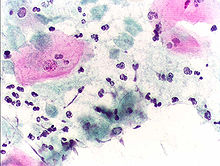
Trichomoniasis (trich) is an infectious disease caused by the parasite Trichomonas vaginalis. About 70% of affected people do not have symptoms when infected. When symptoms occur, they typically begin 5 to 28 days after exposure. Symptoms can include itching in the genital area, a bad smelling thin vaginal discharge, burning with urination, and pain with sex. Having trichomoniasis increases the risk of getting HIV/AIDS. It may also cause complications during pregnancy.

<i>Giardia</i> Genus of flagellate intestinal eukaryotes parasitic in various vertebrate

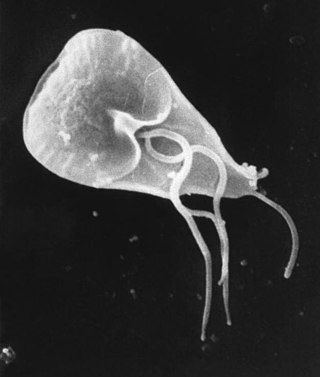
Giardia is a genus of anaerobic flagellated protozoan parasites of the phylum Metamonada that colonise and reproduce in the small intestines of several vertebrates, causing the disease giardiasis. Their life cycle alternates between a swimming trophozoite and an infective, resistant cyst. Giardia were first described by the Dutch microscopist Antonie van Leeuwenhoek in 1681. The genus is named after French zoologist Alfred Mathieu Giard.

<i>Giardia duodenalis</i> Parasitic microorganism that causes giardiasis

Giardia duodenalis, also known as Giardia intestinalis and Giardia lamblia, is a flagellated parasitic protozoan microorganism of the genus Giardia that colonizes the small intestine, causing a diarrheal condition known as giardiasis. The parasite attaches to the intestinal epithelium by an adhesive disc or sucker, and reproduces via binary fission. Giardiasis does not spread to other parts of the gastrointestinal tract, but remains confined to the lumen of the small intestine. The microorganism has an outer membrane that makes it possible to survive even when outside of its host, and which can render it tolerant to certain disinfectants. Giardia trophozoites are anaerobic, and absorb their nutrients from the intestinal lumen. If the organism is stained, its characteristic pattern resembles the familiar "smiley face" symbol.

<i>Gardnerella vaginalis</i> Species of bacterium

Gardnerella vaginalis is a species of Gram-variable-staining facultative anaerobic bacteria. The organisms are small non-spore-forming, nonmotile coccobacilli.

<span class="mw-page-title-main">Metronidazole</span> Antibiotic and antiprotozoal medication

Metronidazole, sold under the brand name Flagyl among others, is an antibiotic and antiprotozoal medication. It is used either alone or with other antibiotics to treat pelvic inflammatory disease, endocarditis, and bacterial vaginosis. It is effective for dracunculiasis, giardiasis, trichomoniasis, and amebiasis. It is an option for a first episode of mild-to-moderate Clostridioides difficile colitis if vancomycin or fidaxomicin is unavailable. Metronidazole is available orally, as a cream or gel, and by slow intravenous infusion.

Vaginitis, also known as vulvovaginitis, is inflammation of the vagina and vulva. Symptoms may include itching, burning, pain, discharge, and a bad smell. Certain types of vaginitis may result in complications during pregnancy.

<i>Acanthamoeba</i> Genus of protozoans

Acanthamoeba is a genus of amoebae that are commonly recovered from soil, fresh water, and other habitats. The genus Acanthamoeba has two stages in its life cycle, the metabolically active trophozoite stage and a dormant, stress-resistant cyst stage. In nature, Acanthamoeba species are generally free-living bacterivores. However, they are also opportunistic pathogens able to cause serious and sometimes fatal infections in humans and other animals.

<span class="mw-page-title-main">Giardiasis</span> Parasitic disease that results in diarrhea

Giardiasis is a parasitic disease caused by Giardia duodenalis. Infected individuals who experience symptoms may have diarrhoea, abdominal pain, and weight loss. Less common symptoms include vomiting and blood in the stool. Symptoms usually begin one to three weeks after exposure and, without treatment, may last two to six weeks or longer.

<span class="mw-page-title-main">Parabasalid</span> Group of flagellated protists

The parabasalids are a group of flagellated protists within the supergroup Excavata. Most of these eukaryotic organisms form a symbiotic relationship in animals. These include a variety of forms found in the intestines of termites and cockroaches, many of which have symbiotic bacteria that help them digest cellulose in woody plants. Other species within this supergroup are known parasites, and include human pathogens.

<i>Trichomonas</i> Genus of parasitic, flagellated protists

Trichomonas is a genus of anaerobic excavate parasites of vertebrates. It was first discovered by Alfred François Donné in 1836 when he found these parasites in the pus of a patient suffering from vaginitis, an inflammation of the vagina. Donné named the genus from its morphological characteristics. The prefix tricho- originates from the Ancient Greek word θρίξ (thrix) meaning hair, describing Trichomonas’s flagella. The suffix -monas, describes its similarity to unicellular organisms from the genus Monas.

<i>Tritrichomonas foetus</i> Species of protozoan parasite

Tritrichomonas foetus is a species of single-celled flagellated parasites that is known to be a pathogen of the bovine reproductive tract as well as the intestinal tract of cats. In cattle, the organism is transmitted to the female vagina and uterus from the foreskin of the bull where the parasite is known to reside. It causes infertility, and, at times, has caused spontaneous abortions in the first trimester. In the last ten years, there have been reports of Tritrichomonas foetus in the feces of young cats that have diarrhea and live in households with multiple cats. Tritrichomonas foetus looks similarly to Giardia and is often misdiagnosed for it when viewed under a microscope.

<span class="mw-page-title-main">Protozoan infection</span> Parasitic disease caused by a protozoan

Protozoan infections are parasitic diseases caused by organisms formerly classified in the kingdom Protozoa. These organisms are now classified in the supergroups Excavata, Amoebozoa, Harosa, and Archaeplastida. They are usually contracted by either an insect vector or by contact with an infected substance or surface.

<span class="mw-page-title-main">Sexually transmitted infection</span> Infection transmitted through human sexual behavior

A sexually transmitted infection (STI), also referred to as a sexually transmitted disease (STD) and the older term venereal disease (VD), is an infection that is spread by sexual activity, especially vaginal intercourse, anal sex, oral sex, or sometimes manual sex. STIs often do not initially cause symptoms, which results in a risk of passing the infection on to others. Symptoms and signs of STIs may include vaginal discharge, penile discharge, ulcers on or around the genitals, and pelvic pain. Some STIs can cause infertility.

Female genital disease is a disorder of the structure or function of the female reproductive system that has a known cause and a distinctive group of symptoms, signs, or anatomical changes. The female reproductive system consists of the ovaries, fallopian tubes, uterus, vagina, and vulva. Female genital diseases can be classified by affected location or by type of disease, such as malformation, inflammation, or infection.

<i>Trichomonas gallinae</i> Species of bird parasite

Trichomonas gallinae is a cosmopolitan parasite of birds including finches, pigeons, doves, turkeys, chickens, parrots, raptors. The condition in birds of prey is called frounce. It is believed to be an ancient pathogen causing frounce-like symptoms in theropod dinosaurs. The same condition in pigeons is commonly called canker.

In biology, a pathogen, in the oldest and broadest sense, is any organism or agent that can produce disease. A pathogen may also be referred to as an infectious agent, or simply a germ.

<i>Trichomonas tenax</i> Species of single-celled organism

Trichomonas tenax, or oral trichomonas, is a species of Trichomonas commonly found in the oral cavity of humans. Routine hygiene is generally not sufficient to eliminate the parasite, hence its Latin name, meaning "tenacious". The parasite is frequently encountered in periodontal infections, affecting more than 50% of the population in some areas, but it is usually considered insignificant. T. tenax is generally not found on the gums of healthy patients. It is known to play a pathogenic role in necrotizing ulcerative gingivitis and necrotizing ulcerative periodontitis, worsening preexisting periodontal disease. This parasite is also implicated in some chronic lung diseases; in such cases, removal of the parasite is sufficient to allow recovery.

Patricia Jean Johnson is a Professor of Microbiology at University of California, Los Angeles (UCLA). She works on the parasite Trichomonas vaginalis, which is responsible for the most prevalent sexually transmitted infections in the United States, Trichomoniasis. She was elected a member of the National Academy of Sciences (NAS) in 2019.

<span class="mw-page-title-main">Lactobacillus vaccine</span> Vaccine using an inactivated strain of Lactobacillus

Lactobacillus vaccines are used in the therapy and prophylaxis of non-specific bacterial vaginitis and trichomoniasis. The vaccines consist of specific inactivated strains of Lactobacilli, called "aberrant" strains in the relevant literature dating from the 1980s. These strains were isolated from the vaginal secretions of patients with acute colpitis. The lactobacilli in question are polymorphic, often shortened or coccoid in shape and do not produce an acidic, anti-pathogenic vaginal environment. A colonization with aberrant lactobacilli has been associated with an increased susceptibility to vaginal infections and a high rate of relapse following antimicrobial treatment. Intramuscular administration of inactivated aberrant lactobacilli provokes a humoral immune response. The production of specific antibodies both in serum and in the vaginal secretion has been demonstrated. As a result of the immune stimulation, the abnormal lactobacilli are inhibited, the population of normal, rod-shaped lactobacilli can grow and exert its defense functions against pathogenic microorganisms.

References

  1. Dias-Lopes G, Saboia-Vahia L, Margotti ET, Fernandes NS, Castro CL, Oliveira FO, Peixoto JF, Britto C, Silva FC, Cuervo P, Jesus JB (October 2017). "Morphologic study of the effect of iron on pseudocyst formation in Trichomonas vaginalis and its interaction with human epithelial cells". Memórias do Instituto Oswaldo Cruz. 112 (10): 664–673. doi:10.1590/0074-02760170032. PMC   5607515 . PMID   28953994.
  2. Soper, D (2004). "Trichomoniasis: under control or undercontrolled?". American Journal of Obstetrics and Gynecology. 190 (1): 281–90. doi:10.1016/j.ajog.2003.08.023. PMID   14749674.
  3. 1 2 3 Harp, Djana F.; Chowdhury, Indrajit (2011). "Trichomoniasis: Evaluation to execution". European Journal of Obstetrics & Gynecology and Reproductive Biology. 157 (1): 3–9. doi:10.1016/j.ejogrb.2011.02.024. PMC   4888369 . PMID   21440359.
  4. Hook, Edward W. (1999). "Trichomonas vaginalis—No Longer a Minor STD". Sexually Transmitted Diseases. 26 (7): 388–9. doi: 10.1097/00007435-199908000-00004 . PMID   10458631.
  5. W Evan Secor. "Trichomonas Vaginalis". MedScape.
  6. Donné, A. (19 September 1836). "Animalcules observés dans les matières purulentes et le produit des sécrétions des organes génitaux de l'homme et de la femme". Comptes Rendus Hebdomadaires des Séances de l'Académie des Sciences (in French). 3: 385–386.
  7. Diamantis, Aristidis; Magiorkinis, Emmanouil; Androutsos, George (2009). "Alfred Francois Donné (1801-78): a pioneer of microscopy, microbiology and haematology". Journal of Medical Biography. 17 (2): 81–87. doi:10.1258/jmb.2008.008040. ISSN   0967-7720. PMID   19401511. S2CID   9287263.
  8. Johnston VJ, Mabey DC (February 2008). "Global epidemiology and control of Trichomonas vaginalis". Current Opinion in Infectious Diseases. 21 (1): 56–64. doi:10.1097/QCO.0b013e3282f3d999. PMID   18192787. S2CID   39490032.
  9. 1 2 3 Ash, Lawrence; Orihel, Thomas (2007). Ash & Orihel's Atlas of Human Parasitology (5th ed.). American Society for Clinical Pathology Press. ISBN   9780891891673.
  10. 1 2 Nanda, N; Michel, RG; Kurdgelashvili, G; Wendel, KA (2006). "Trichomoniasis and its treatment". Expert Review of Anti-infective Therapy. 4 (1): 125–35. doi:10.1586/14787210.4.1.125. PMID   16441214. S2CID   43515177.
  11. Swygard H, Seña AC, Hobbs MM, Cohen MS (April 2004). "Trichomoniasis: clinical manifestations, diagnosis and management". Sex Transm Infect. 80 (2): 91–5. doi:10.1136/sti.2003.005124. PMC   1744792 . PMID   15054166.
  12. 1 2 3 Petrin D, Delgaty K, Bhatt R, Garber G (April 1998). "Clinical and microbiological aspects of Trichomonas vaginalis". Clin. Microbiol. Rev. 11 (2): 300–17. doi:10.1128/CMR.11.2.300. PMC   106834 . PMID   9564565.
  13. Garber GE (January 2005). "The laboratory diagnosis of Trichomonas vaginalis". Can J Infect Dis Med Microbiol. 16 (1): 35–8. doi: 10.1155/2005/373920 . PMC   2095007 . PMID   18159526.
  14. Parija, SubhashChandra; Preethi, V; Mandal, Jharna; Halder, Ajay (2011). "Trichomoniasis: An update". Tropical Parasitology. 1 (2). Medknow: 73–75. doi: 10.4103/2229-5070.86934 . ISSN   2229-5070. PMC   3593481 . PMID   23508486.
  15. Schwebke, J. R.; Burgess, D. (2004). "Trichomoniasis". Clinical Microbiology Reviews. 17 (4): 794–803, table of contents. doi:10.1128/CMR.17.4.794-803.2004. PMC   523559 . PMID   15489349.
  16. "Trichomoniasis". CDC Fact Sheet. Centers for Disease Control and Prevention. 2007-12-17. Retrieved 2010-06-11.
  17. 1 2 Caini, Saverio; Gandini, Sara; Dudas, Maria; Bremer, Viviane; Severi, Ettore; Gherasim, Alin (2014). "Sexually transmitted infections and prostate cancer risk: A systematic review and meta-analysis". Cancer Epidemiology. 38 (4): 329–338. doi:10.1016/j.canep.2014.06.002. ISSN   1877-7821. PMID   24986642.
  18. Powers, Celeste N. (1998). "Diagnosis of Infectious Diseases: a Cytopathologist's Perspective". Clinical Microbiology Reviews. 11 (2): 341–65. doi:10.1128/CMR.11.2.341. PMC   106836 . PMID   9564567.
  19. Ohlemeyer, C; Hornberger, L; Lynch, D; Swierkosz, E (March 1998). "Diagnosis of Trichomonas vaginalis in adolescent females: InPouch TV culture versus wet-mount microscopy". Journal of Adolescent Health. 22 (3): 205–8. doi:10.1016/S1054-139X(97)00214-0. PMID   9502007.
  20. Sood, Seema; Mohanty, Srujana; Kapil, Arti; Tolosa, Jorge; Mittal, Suneeta (2007). "InPouch TV culture for detection of Trichomonas vaginalis" (PDF). The Indian Journal of Medical Research. 125 (4): 567–71. PMID   17598943.
  21. 1 2 Huppert, Jill S.; Mortensen, Joel E.; Reed, Jennifer L.; Kahn, Jessica A.; Rich, Kimberly D.; Miller, William C.; Hobbs, Marcia M. (2007). "Rapid Antigen Testing Compares Favorably with Transcription-Mediated Amplification Assay for the Detection of Trichomonas vaginalis in Young Women". Clinical Infectious Diseases. 45 (2): 194–8. doi: 10.1086/518851 . PMID   17578778.
  22. Schirm, Jurjen; Bos, Petra A.J.; Roozeboom-Roelfsema, Irene K.; Luijt, Dirk S.; Möller, Lieke V. (2007). "Trichomonas vaginalis detection using real-time TaqMan PCR". Journal of Microbiological Methods. 68 (2): 243–7. doi:10.1016/j.mimet.2006.08.002. PMID   17005275.
    "Trichomonas vaginalis repeated DNA target for PCR identification". GenBank Nucleotide Database. 2002-04-16. L23861.1.
    Kengne P, Veas F, Vidal N, Rey JL, Cuny G (September 1994). "Trichomonas vaginalis: repeated DNA target for highly sensitive and specific polymerase chain reaction diagnosis". Cell. Mol. Biol. (Noisy-le-grand). 40 (6): 819–31. PMID   7812190.
  23. "Metronidazole". Drugs.com. Retrieved 23 February 2018.
  24. Workowski, K.A.; Berman, S.; Centers for Disease Control and Prevention (CDC) (17 December 2010). "Sexually transmitted diseases treatment guidelines, 2010" (PDF). MMWR Recomm Rep. 59 (RR-12): 1–110. PMID   21160459. Erratum in: MMWR Recomm Rep. 2011 Jan 14;60(1):18.
  25. Cudmore, S. L.; Delgaty, K. L.; Hayward-Mcclelland, S. F.; Petrin, D. P.; Garber, G. E. (2004). "Treatment of Infections Caused by Metronidazole-Resistant Trichomonas vaginalis". Clinical Microbiology Reviews. 17 (4): 783–93, table of contents. doi:10.1128/CMR.17.4.783-793.2004. PMC   523556 . PMID   15489348.
  26. Ryan, Kenneth James; Ray, C. George; Sherris, John C., eds. (2004). Sherris Medical Microbiology (4th ed.). McGraw Hill. ISBN   978-0-8385-8529-0.[ page needed ]
  27. Mielczarek E, Blaszkowska J (November 2015). "Trichomonas vaginalis: pathogenicity and potential role in human reproductive failure". Infection. 44 (4): 447–458. doi:10.1007/s15010-015-0860-0. PMID   26546373. S2CID   33823038.
  28. Hussein EM, Atwa MM (December 2008). "Infectivity of Trichomonas vaginalis pseudocysts inoculated intra-vaginally in mice". Journal of the Egyptian Society of Parasitology. 38 (3): 749–762. PMID   19209760.
  29. Verges J (June 1979). "Les prostatites à trichomonas formes rondes (T.F.R.)" [Prostatitis due to the circular form of trichomonas]. Journal d'Urologie et de Nephrologie (in French). 85 (6): 357–361. ISSN   0021-8200. PMID   490766.
  30. Upcroft, P.; Upcroft, J. A. (2001). "Drug Targets and Mechanisms of Resistance in the Anaerobic Protozoa". Clinical Microbiology Reviews. 14 (1): 150–64. doi:10.1128/CMR.14.1.150-164.2001. PMC   88967 . PMID   11148007.
  31. Arroyo, R.; Engbring, J.; Alderete, J. F. (1992). "Molecular basis of host epithelial cell recognition by Trichomonas vaginalis". Molecular Microbiology. 6 (7): 853–862. doi:10.1111/j.1365-2958.1992.tb01536.x. PMID   1602965. S2CID   44364966.
  32. Mendoza-Lopez, M. R.; Becerril-Garcia, C.; Fattel-Facenda, L. V.; Avila-Gonzalez, L.; Ruiz-Tachiquin, M. E.; Ortega-Lopez, J.; Arroyo, R. (2000). "CP30, a Cysteine Proteinase Involved in Trichomonas vaginalis Cytoadherence". Infection and Immunity. 68 (9): 4907–12. doi:10.1128/IAI.68.9.4907-4912.2000. PMC   101697 . PMID   10948104.
  33. Carlton, J. M.; Hirt, R. P.; Silva, J. C.; Delcher, A. L.; Schatz, M.; Zhao, Q.; Wortman, J. R.; Bidwell, S. L.; et al. (2007). "Draft Genome Sequence of the Sexually Transmitted Pathogen Trichomonas vaginalis". Science. 315 (5809): 207–12. Bibcode:2007Sci...315..207C. doi:10.1126/science.1132894. PMC   2080659 . PMID   17218520.
  34. Lehker, M. W.; Alderete, J. F. (1999). "Resolution of Six Chromosomes of Trichomonas vaginalis and Conservation of Size and Number among Isolates". The Journal of Parasitology. 85 (5): 976–9. doi:10.2307/3285842. JSTOR   3285842. PMID   10577741.
  35. Zimmer, C. (2007). "EVOLUTION: Jurassic Genome". Science. 315 (5817): 1358–9. doi:10.1126/science.315.5817.1358. PMID   17347424. S2CID   34154189.
  36. Aurrecoechea, Cristina; Brestelli, John; Brunk, Brian P.; Carlton, Jane M.; Dommer, Jennifer; Fischer, Steve; Gajria, Bindu; Gao, Xin; et al. (2009). "GiardiaDB and TrichDB: integrated genomic resources for the eukaryotic protist pathogens Giardia lamblia and Trichomonas vaginalis". Nucleic Acids Research. 37 (Database issue): D526–30. doi:10.1093/nar/gkn631. PMC   2686445 . PMID   18824479.
  37. Conrad, Melissa D.; Gorman, Andrew W.; Schillinger, Julia A.; Fiori, Pier Luigi; Arroyo, Rossana; Malla, Nancy; Dubey, Mohan Lal; Gonzalez, Jorge; Blank, Susan (2012). "Extensive Genetic Diversity, Unique Population Structure and Evidence of Genetic Exchange in the Sexually Transmitted Parasite Trichomonas vaginalis". PLOS Neglected Tropical Diseases. 6 (3): e1573. doi: 10.1371/journal.pntd.0001573 . PMC   3313929 . PMID   22479659.
  38. Mulla, Summaiyaa; Kosambiya, JK; Desai, Vikask; Shethwala, Nimishad (2009). "Sexually transmitted infections and reproductive tract infections in female sex workers". Indian Journal of Pathology and Microbiology. 52 (2): 198–9. doi: 10.4103/0377-4929.48916 . PMID   19332911.
  39. Mavedzenge, Sue Napierala; Van der Pol, Barbara; Cheng, Helen; Montgomery, Elizabeth T.; Blanchard, Kelly; de Bruyn, Guy; Ramjee, Gita; Van der Straten, Ariane (2010). "Epidemiological Synergy of Trichomonas vaginalis and HIV in Zimbabwean and South African Women". Sexually Transmitted Diseases. 37 (7): 460–6. doi: 10.1097/OLQ.0b013e3181cfcc4b . PMID   20562586. S2CID   19708165.
  40. 1 2 Malik SB, Pightling AW, Stefaniak LM, Schurko AM, Logsdon JM (2008). "An expanded inventory of conserved meiotic genes provides evidence for sex in Trichomonas vaginalis". PLOS ONE. 3 (8): e2879. Bibcode:2008PLoSO...3.2879M. doi: 10.1371/journal.pone.0002879 . PMC   2488364 . PMID   18663385.

Further reading