Trichomonas

Last updated

Contents

Trichomonas
Trichomonas Giemsa DPDx.JPG
Two trophozoites of Trichomonas vaginalis stained with Giemsa
Scientific classification OOjs UI icon edit-ltr.svg
Domain: Eukaryota
Phylum: Metamonada
Subphylum: Trichozoa
(unranked): Parabasalia
Order: Trichomonadida
Family: Trichomonadidae
Genus: Trichomonas
Species [1]

Trichomonas is a genus of anaerobic excavate parasites of vertebrates. It was first discovered by Alfred François Donné in 1836 when he found these parasites in the pus of a patient suffering from vaginitis, an inflammation of the vagina. Donné named the genus from its morphological characteristics. The prefix tricho- originates from the Ancient Greek word θρίξ (thrix) meaning hair, describing Trichomonas’s flagella. The suffix -monas (μονάς – single unit), describes its similarity to unicellular organisms from the genus Monas . [2]

Habitat and ecology

Trichomonas is typically found in anaerobic environments. It is a known parasite of many different animals including humans, birds, dogs, and cats. [3] [4] [5] [6] In humans, it can be found in the urogenital tract and in the oral cavity. It is estimated that 276 million new cases of urogenital infections occur each year. [3] [5] [6] Depending on the Trichomonas species, it can either be transmitted through direct sexual contact or through contaminated water sources. [3] [5] [6] In birds, it can be found in the upper digestive tract and is transmitted when adult birds regurgitate food to feed their young, when a bird of prey feeds on an infected bird, and through contaminated food or water. [7]

Morphology

Trichomonas is around 10 µm in length and is normally pear-shaped. It has four flagella at its anterior end, distinguishing itself from closely related organisms that have different numbers of anterior flagella. At the base of these flagella are the parabasal bodies, kinetosomes accompanied by Golgi stacks. The pelta is a sheet of microtubules that curve around the flagellar bases. Posterior to the pelta is the axostyle, is a bundle of microtubules that extends from the anterior end of the organism all the way to the posterior end. The nucleus of Trichomonas is situated close to where the pelta and axostyle meet. [7] [6] [8] [9]

Another distinguishing feature of Trichomonas is the presence of an undulating membrane. The undulating membrane is a fin-like extension of the plasma membrane located on the side of the organism. A flagellum that extends to the posterior end of the organism is attached to the outer edge of the undulating membrane. At the base of the undulating membrane is a striated fiber called the costa which is thought to exist for structural support. [7] [10] [6] [8] [9]

Trichomonas has a very interesting organelle: the hydrogenosome. [6] Hydrogenosomes are double-membraned organelles used by trichomonads, instead of mitochondria, to produce ATP. They do not require oxygen and instead use pyruvate:ferredoxin oxido-reductase and hydrogenase to produce ATP from pyruvate, generating hydrogen gas as a by-product. [11]

Genetics

Trichomonas vaginalis , being the species that causes the most complications in humans, is the only fully sequenced Trichomonas species. Through whole-genome shotgun sequencing, the Trichomonas vaginalis genome is estimated to be around 160 Mb long, divided into six chromosomes. However, at least 65% of its genome was found to be redundant. The redundant genetic material is hypothesized to have emerged during Trichomonas's transition from aerobic to anaerobic environments. [12]

In addition to discovering the large proportion of repetitive DNA in Trichomonas vaginalis genome, the sequenced genes were also characterized. Approximately 60,000 protein-coding genes were found. Transfer RNAs for all 20 amino acids and approximately 250 ribosomal RNA were all found on the same chromosome. [12]

Life cycle

Trichomonas has a trophozoite form, its pear-shaped form, which is most commonly observed, and an amoeboid form, which appears during host colonization. [12] It lacks a cyst form, but many studies have noted a unique form in which Trichomonas appears ovoidal rather than its typical pear-shaped form. In this ovoidal form, all its flagella are retracted in endocytic vacuoles, giving the impression of a cystic form. However, due to the lack of a cystic wall surrounding the organism, many studies describe this form as a pseudocystic form. [7] [6]

In its trophozoite form, Trichomonas undergoes cell division through an interesting process called cryptopleuromitosis. There are three common forms of mitosis: open, closed, and semi-open. In open mitosis, the nuclear envelope disappears so that mitotic spindles can interact with the chromosomes. In closed mitosis, the nuclear envelope does not disappear but mitotic spindles appear within the nucleus to separate the chromosomes. In semi-open mitosis, the nuclear envelope remains intact but the mitotic spindles pierce through the nuclear envelope to divide the chromosomes. Cryptopleuromitosis is different from all the other more commonly known methods of cell division. In cryptopleuromitosis, the chromosomes divide without the breakdown of the nuclear envelope and without the entry of mitotic spindles into the nucleus. [13]

Diseases

Trichomonas causes disease in humans and in birds. In humans, the causative species is Trichomonas vaginalis and Trichomonas tenax . [3] [5] [6] In birds, the causative species are Trichomonas gallinae , Trichomonas gypactinii, and Trichomonas stableri. [14] [15] [7]

In humans

Trichomonas vaginalis is a sexually transmitted disease and causes trichomoniasis. It resides on squamous epithelium of the urogenital tract. Many carriers of Trichomonas vaginalis, especially men, are asymptomatic. Complications for symptomatic women include vaginitis, endometritis, infertility, and cervical cancer. Complications for symptomatic men include urethritis, prostatitis, epididymitis, and infertility. It is also associated with increased risk of transmission and acquisition of HIV. [5] [6]

Trichomonas tenax is transmitted through exchange of saliva and contaminated water sources. It is an opportunistic pathogen and may cause pulmonary trichomoniasis. [3]

In birds

Trichomonas in birds inhabit the upper digestive tract and also cause trichomoniasis. It creates lesions in the trachea and esophagus, occupying space and eventually causing emaciation and asphyxiation. [14] [15] [7]

Species

Related Research Articles

<span class="mw-page-title-main">Mitosis</span> Process in which chromosomes are replicated and separated into two new identical nuclei

Mitosis is a part of the cell cycle in which replicated chromosomes are separated into two new nuclei. Cell division by mitosis is an equational division which gives rise to genetically identical cells in which the total number of chromosomes is maintained. Mitosis is preceded by the S phase of interphase and is followed by telophase and cytokinesis, which divide the cytoplasm, organelles, and cell membrane of one cell into two new cells containing roughly equal shares of these cellular components. The different stages of mitosis altogether define the mitotic phase of a cell cycle—the division of the mother cell into two daughter cells genetically identical to each other.

<span class="mw-page-title-main">Trichomoniasis</span> Medical condition

Trichomoniasis (trich) is an infectious disease caused by the parasite Trichomonas vaginalis. About 70% of affected people do not have symptoms when infected. When symptoms occur, they typically begin 5 to 28 days after exposure. Symptoms can include itching in the genital area, a bad smelling thin vaginal discharge, burning with urination, and pain with sex. Having trichomoniasis increases the risk of getting HIV/AIDS. It may also cause complications during pregnancy.

<i>Trichomonas vaginalis</i> Species of parasite that causes sexually transmitted infections

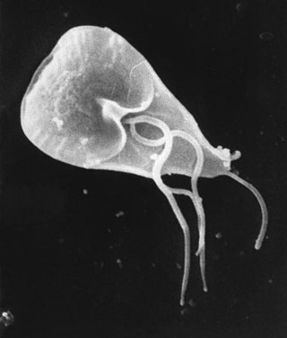
Trichomonas vaginalis is an anaerobic, flagellated protozoan parasite and the causative agent of a sexually transmitted disease called trichomoniasis. It is the most common pathogenic protozoan that infects humans in industrialized countries. Infection rates in men and women are similar but women are usually symptomatic, while infections in men are usually asymptomatic. Transmission usually occurs via direct, skin-to-skin contact with an infected individual, most often through vaginal intercourse. The WHO has estimated that 160 million cases of infection are acquired annually worldwide. The estimates for North America alone are between 5 and 8 million new infections each year, with an estimated rate of asymptomatic cases as high as 50%. Usually treatment consists of metronidazole and tinidazole.

<span class="mw-page-title-main">Parabasalid</span> Group of flagellated protists

The parabasalids are a group of flagellated protists within the supergroup Excavata. Most of these eukaryotic organisms form a symbiotic relationship in animals. These include a variety of forms found in the intestines of termites and cockroaches, many of which have symbiotic bacteria that help them digest cellulose in woody plants. Other species within this supergroup are known parasites, and include human pathogens.

<span class="mw-page-title-main">Hydrogenosome</span> Mitochondrion-derived organelle

A hydrogenosome is a membrane-enclosed organelle found in some anaerobic ciliates, flagellates, and fungi. Hydrogenosomes are highly variable organelles that have presumably evolved from protomitochondria to produce molecular hydrogen and ATP in anaerobic conditions.

<span class="mw-page-title-main">Trichomonadida</span> Order of flagellated protists

Trichomonadida is an order of anaerobic protists, included with the parabasalids. Members of this order are referred to as trichomonads.

An axostyle is a sheet of microtubules found in certain protists. It arises from the bases of the flagella, sometimes projecting beyond the end of the cell, and is often flexible or contractile, and so may be involved in movement and provides support for the cell. Axostyles originate in association with a flagellar microtubular root and occur in two groups, the oxymonads and parabasalids; they have different structures and are not homologous. Within Trichomonads the axostyle has been theorised to participate in locomotion and cell adhesion, but also karyokinesis during cell division.

<i>Tritrichomonas foetus</i> Species of protozoan parasite

Tritrichomonas foetus is a species of single-celled flagellated parasites that is known to be a pathogen of the bovine reproductive tract as well as the intestinal tract of cats. In cattle, the organism is transmitted to the female vagina and uterus from the foreskin of the bull where the parasite is known to reside. It causes infertility, and, at times, has caused spontaneous abortions in the first trimester. In the last ten years, there have been reports of Tritrichomonas foetus in the feces of young cats that have diarrhea and live in households with multiple cats. Tritrichomonas foetus looks similarly to Giardia and is often misdiagnosed for it when viewed under a microscope.

<span class="mw-page-title-main">Protozoan infection</span> Parasitic disease caused by a protozoan

Protozoan infections are parasitic diseases caused by organisms formerly classified in the kingdom Protozoa. These organisms are now classified in the supergroups Excavata, Amoebozoa, Harosa, and Archaeplastida. They are usually contracted by either an insect vector or by contact with an infected substance or surface.

<i>Trichomonas gallinae</i> Species of bird parasite

Trichomonas gallinae is a cosmopolitan parasite of birds including finches, pigeons, doves, turkeys, chickens, parrots, raptors. The condition in birds of prey is called frounce. It is believed to be an ancient pathogen causing frounce-like symptoms in theropod dinosaurs. The same condition in pigeons is commonly called canker.

Jane M. Carlton is a biologist at New York University whose research centers on the genomics of two groups of single-celled parasites: those which cause malaria, and trichomonads, which include the common sexually transmitted parasite Trichomonas vaginalis.

<i>Trichomonas tenax</i> Species of single-celled organism

Trichomonas tenax, or oral trichomonas, is a species of Trichomonas commonly found in the oral cavity of humans. Routine hygiene is generally not sufficient to eliminate the parasite, hence its Latin name, meaning "tenacious". The parasite is frequently encountered in periodontal infections, affecting more than 50% of the population in some areas, but it is usually considered insignificant. T. tenax is generally not found on the gums of healthy patients. It is known to play a pathogenic role in necrotizing ulcerative gingivitis and necrotizing ulcerative periodontitis, worsening preexisting periodontal disease. This parasite is also implicated in some chronic lung diseases; in such cases, removal of the parasite is sufficient to allow recovery.

The Mastigont system is a series of structures found in several Protists such as thrichomonads and amoebae. It is formed by the basal bodies and several other structures composed of fibrils. Their function is not fully understood. The system is studied and visualised mainly through techniques such as plasma membrane extraction, high-voltage electron microscopy, field emission scanning electron microscopy, the cell-sandwich technique, freeze-etching, and immunocytochemistry.

Tritrichomonas blagburni is a genus of parasite that infects the digestive system of cats.

Monocercomonoides is a genus of flagellate Excavata belonging to the order Oxymonadida. It was established by Bernard V. Travis and was first described as those with "polymastiginid flagellates having three anterior flagella and a trailing one originating at a single basal granule located in front of the anteriorly positioned nucleus, and a more or less well-defined axostyle". It is the first eukaryotic genus to be found to completely lack mitochondria, and all hallmark proteins responsible for mitochondrial function. The genus also lacks any other mitochondria related organelles (MROs) such as hydrogenosomes or mitosomes. Data suggests that the absence of mitochondria is not an ancestral feature, but rather due to secondary loss. Monocercomonoides sp. was found to obtain energy through an enzymatic action of nutrients absorbed from the environment. The genus has replaced the iron-sulfur cluster assembly pathway with a cytosolic sulfur mobilization system, likely acquired by horizontal gene transfer from a eubacterium of a common ancestor of oxymonads. These organisms are significant because they undermine assumptions that eukaryotes must have mitochondria to properly function. The genome of Monocercomonoides exilis has approximately 82 million base pairs, with 18 152 predicted protein-coding genes.

Patricia Jean Johnson is a Professor of Microbiology at University of California, Los Angeles (UCLA). She works on the parasite Trichomonas vaginalis, which is responsible for the most prevalent sexually transmitted infections in the United States, Trichomoniasis. She was elected a member of the National Academy of Sciences (NAS) in 2019.

<span class="mw-page-title-main">Lactobacillus vaccine</span> Vaccine using an inactivated strain of Lactobacillus

Lactobacillus vaccines are used in the therapy and prophylaxis of non-specific bacterial vaginitis and trichomoniasis. The vaccines consist of specific inactivated strains of Lactobacilli, called "aberrant" strains in the relevant literature dating from the 1980s. These strains were isolated from the vaginal secretions of patients with acute colpitis. The lactobacilli in question are polymorphic, often shortened or coccoid in shape and do not produce an acidic, anti-pathogenic vaginal environment. A colonization with aberrant lactobacilli has been associated with an increased susceptibility to vaginal infections and a high rate of relapse following antimicrobial treatment. Intramuscular administration of inactivated aberrant lactobacilli provokes a humoral immune response. The production of specific antibodies both in serum and in the vaginal secretion has been demonstrated. As a result of the immune stimulation, the abnormal lactobacilli are inhibited, the population of normal, rod-shaped lactobacilli can grow and exert its defense functions against pathogenic microorganisms.

J. Richard McIntosh is a Distinguished Professor Emeritus in Molecular, Cellular, and Developmental Biology at the University of Colorado Boulder. McIntosh first graduated from Harvard with a BA in Physics in 1961, and again with a Ph.D. in Biophysics in 1968. He began his teaching career at Harvard but has spent most of his career at the University of Colorado Boulder. At the University of Colorado Boulder, McIntosh taught biology courses at both the undergraduate and graduate levels. Additionally, he created an undergraduate course in the biology of cancer towards the last several years of his teaching career. McIntosh's research career looks at a variety of things, including different parts of mitosis, microtubules, and motor proteins.

Holomastigotoides is a genus of parabasalids found in the hindgut of lower termites. It is characterized by its dense, organized arrangement of flagella on the cell surface and the presence of a mitotic spindle outside its nucleus during the majority of its cell cycle. As a symbiont of termites, Holomastigotoides is able to ingest wood and aid its host in digestion. In return, Holomastigotoides is supplied with a stable habitat and steady supply of food. Holomastigotoides has notably been studied to observe the mechanisms of chromosomal pairing and segregation in haploid and diploid cells.

Monocercomonas is a Parabasalian genus belonging to the order Trichomonadida. It presents four flagella, three forward-facing and one trailing, without the presence of a costa or any kind of undulating membrane. Monocercomonas is found in animal guts. and is susceptible to cause Monocercomoniasis in reptiles

References

  1. "Trichomonas". NCBI taxonomy. Bethesda, MD: National Center for Biotechnology Information. Retrieved 4 February 2019.
  2. Donné, A. (19 September 1836). "Animalcules observés dans les matières purulentes et le produit des sécrétions des organes génitaux de l'homme et de la femme". Comptes Rendus Hebdomadaires des Séances de l'Académie des Sciences (in French). 3: 385–386.
  3. 1 2 3 4 5 6 Hersh, S. M. (1 August 1985). "Pulmonary trichomoniasis and Trichomonas tenax". Journal of Medical Microbiology. 20 (1): 1–10. doi: 10.1099/00222615-20-1-1 . PMID   3894667.
  4. 1 2 Kellerová, Pavlína; Tachezy, Jan (April 2017). "Zoonotic Trichomonas tenax and a new trichomonad species, Trichomonas brixi n. sp., from the oral cavities of dogs and cats". International Journal for Parasitology. 47 (5): 247–255. doi:10.1016/j.ijpara.2016.12.006. PMID   28238869.
  5. 1 2 3 4 5 6 Menezes, Camila Braz; Frasson, Amanda Piccoli; Tasca, Tiana (5 September 2016). "Trichomoniasis – are we giving the deserved attention to the most common non-viral sexually transmitted disease worldwide?". Microbial Cell. 3 (9): 404–418. doi:10.15698/mic2016.09.526. PMC   5354568 . PMID   28357378.
  6. 1 2 3 4 5 6 7 8 9 10 Schwebke, Jane R.; Burgess, Donald (15 October 2004). "Trichomoniasis". Clinical Microbiology Reviews. 17 (4): 794–803. doi:10.1128/CMR.17.4.794-803.2004. PMC   523559 . PMID   15489349.
  7. 1 2 3 4 5 6 7 Amin, Aziza; Bilic, Ivana; Liebhart, Dieter; Hess, Michael (May 2014). "Trichomonads in birds — a review". Parasitology. 141 (6): 733–47. doi:10.1017/S0031182013002096. PMID   24476968. S2CID   206247917.
  8. 1 2 Tasca, Tiana; De Carli, Geraldo A. (December 2003). "Scanning electron microscopy study of Trichomonas gallinae". Veterinary Parasitology. 118 (1–2): 37–42. doi:10.1016/j.vetpar.2003.09.009. PMID   14651873.
  9. 1 2 Wartoń, A; Honigberg, BM (February 1979). "Structure of trichomonads as revealed by scanning electron microscopy". The Journal of Protozoology. 26 (1): 56–62. doi:10.1111/j.1550-7408.1979.tb02732.x. PMID   314517.
  10. Kulda, Jaroslav; Nohýnková, Eva; Ludvík, Jiří (October 1987). "Basic structure and function of the trichomonad cell". Acta Universitatis Carolinae - Biologica. 30: 181–198. ISSN   0001-7124. OCLC   967953815.
  11. Müller, Miklós; Mentel, Marek; van Hellemond, Jaap J.; Henze, Katrin; Woehle, Christian; Gould, Sven B.; Yu, Re-Young; van der Giezen, Mark; Tielens, Aloysius G. M.; Martin, William F. (11 June 2012). "Biochemistry and evolution of anaerobic energy metabolism in eukaryotes". Microbiology and Molecular Biology Reviews. 76 (2): 444–495. doi:10.1128/MMBR.05024-11. PMC   3372258 . PMID   22688819.
  12. 1 2 3 Carlton, JM; Hirt, RP; Silva, JC; Delcher, AL; Schatz, M; Zhao, Q; Wortman, JR; Bidwell, SL; Alsmark, UC; Besteiro, S; Sicheritz-Ponten, T; Noel, CJ; Dacks, JB; Foster, PG; Simillion, C; Van de Peer, Y; Miranda-Saavedra, D; Barton, GJ; Westrop, GD; Müller, S; Dessi, D; Fiori, PL; Ren, Q; Paulsen, I; Zhang, H; Bastida-Corcuera, FD; Simoes-Barbosa, A; Brown, MT; Hayes, RD; Mukherjee, M; Okumura, CY; Schneider, R; Smith, AJ; Vanacova, S; Villalvazo, M; Haas, BJ; Pertea, M; Feldblyum, TV; Utterback, TR; Shu, CL; Osoegawa, K; de Jong, PJ; Hrdy, I; Horvathova, L; Zubacova, Z; Dolezal, P; Malik, SB; Logsdon JM, Jr; Henze, K; Gupta, A; Wang, CC; Dunne, RL; Upcroft, JA; Upcroft, P; White, O; Salzberg, SL; Tang, P; Chiu, CH; Lee, YS; Embley, TM; Coombs, GH; Mottram, JC; Tachezy, J; Fraser-Liggett, CM; Johnson, PJ (12 January 2007). "Draft genome sequence of the sexually transmitted pathogen Trichomonas vaginalis". Science. 315 (5809): 207–12. Bibcode:2007Sci...315..207C. doi:10.1126/science.1132894. PMC   2080659 . PMID   17218520.
  13. Ribeiro, Karla C.; Monteiro-Leal, Luiz Henrique; Benchimol, Marlene (2000). "Contributions of the axostyle and flagella to closed mitosis in the protists Tritrichomonas foetus and Trichomonas vaginalis". The Journal of Eukaryotic Microbiology. 47 (5): 481–92. doi:10.1111/j.1550-7408.2000.tb00077.x. PMID   11001145. S2CID   5852015.
  14. 1 2 3 Girard, Yvette A.; Rogers, Krysta H.; Gerhold, Richard; Land, Kirkwood M.; Lenaghan, Scott C.; Woods, Leslie W.; Haberkern, Nathan; Hopper, Melissa; Cann, Jeff D.; Johnson, Christine K. (April 2014). "Trichomonas stableri n. sp., an agent of trichomonosis in Pacific Coast band-tailed pigeons (Patagioenas fasciata monilis)". International Journal for Parasitology: Parasites and Wildlife. 3 (1): 32–40. doi:10.1016/j.ijppaw.2013.12.002. PMC   4047957 . PMID   24918075.
  15. 1 2 3 Martínez-Díaz, Rafael Alberto; Ponce-Gordo, Francisco; Rodríguez-Arce, Irene; del Martínez-Herrero, María Carmen; González, Fernando González; Molina-López, Rafael Ángel; Gómez-Muñoz, María Teresa (2 October 2014). "Trichomonas gypaetinii n. sp., a new trichomonad from the upper gastrointestinal tract of scavenging birds of prey". Parasitology Research. 114 (1): 101–112. doi:10.1007/s00436-014-4165-5. PMID   25273632. S2CID   18438912.