Retortamonas

Last updated

Contents

Retortamonas
Metamonada retortamonas hexamita giardia.svg
Retortamonas (Retortamonadida), on left; Hexamita , on center, and Giardia , on right (both Diplomonadida)
Scientific classification
Domain:
(unranked):
Phylum:
Order:
Family:
Genus:
Retortamonas

Grassi, 1879
Species
  • Insect clade
    • R. agilis(Mackinon 1911)
    • R. alexeiffi(Mackinon 1912)
    • R. belostomae(Brug 1922)
    • R. blattae(Bishop 1931)
    • R. caudacusGeiman 1932
    • R. gryllotalpaeGrassi 1879
    • R. hodotermitisBrugerolle 2006
    • R. phyllophagae(Travis & Becker 1931) Wenrich 1932
    • R. termitusKirby 1932
    • toddiRao 1968
  • Vertebrate clade
    • R. boaeKulda 1954
    • R. bradypi(Hegner & Schumaker 1928)
    • R. caviae(Hegner & Schumaker 1928)
    • R. cheloniiJanakidevi 1962
    • R. cuniculi(Collier & Boeck 1926) Wenrich 1932
    • R. dobelli(Bishop 1931)
    • R. intestinalis (Wenyon & O'Connor 1917) Wenrich 1932
    • R. kirbiiGabel 1954
    • R. mitrulaeKirby & Honigberg 1950
    • R. ovis(Hegner & Schumaker 1928)
    • R. ruminatumKnowtes & Das Gupta 1931
    • R. saurarumGrassé 1926
    • R. testudaeAnsari 1955
    • R. viperaeKrishnamurthy & Madre 1976
  • Uncertain
    • R. aurangabadensisSultana 1976
    • R. grassi
    • R. indicaKrishnamurthy & Sultana 1977
    • R. masoodiMali 1993
    • R. nagabhushaniSultana 1976
    • R. orthopterorum(Parisi 1910) Wenyon 1926
    • R. pericoptiLaird 1956
    • R. rotundaBishop 1932
    • R. sinensis(Faust & Wassell 1921)
    • R. wenrichiStabler 1944
    • R. wenyoni(da Fonseca 1917)
Synonyms

Retortamonas is a genus of flagellated excavates. [1] [2] It is one of only two genera belonging to the family Retortamonadidae along with the genus Chilomastix . [3] The genus parasitizes a large range of hosts including humans. [4] Species within this genus are considered harmless commensals which reside in the intestine of their host. [5] [4] The wide host diversity is a useful factor given that species are distinguished based on their host rather than morphology. [3] This is because all species share similar morphology, which would present challenges when trying to make classifications based on structural anatomy. [3] Although Retortamonas currently includes over 25 known species, it is possible that some defined species are synonymous, given that such overlapping species have been discovered in the past. [3] Further efforts into learning about this genus must be done such as cross-transmission testing as well as biochemical and genetic studies. [3] One of the most well-known species within this genus is Retortamonas intestinalis , a human parasite that lives in the large intestine of humans. [5]

Etymology

Retort- is derived from the Latin re-torqueo – “to twist back” while the Greek term monas- “one unit, single” refers to its singularity. [6] During locomotion the anterior region of the body twists in a clock-wise direction about its axis, propelling the cell to swim forward, and therefore, the body structure often appears twisted. [1]

History of knowledge

First discovery of the genus

The human parasite Retortamonas intestinalis was first discovered in 1917 Egypt by Wenyon and O’Connor. [5] This case was reported by a soldier who was diagnosed to be infected with Waskia intestinalis, a species which was later renamed Embadomonas intestinalis by Chalmers and Pekkola in 1918. [7] Afterwards, both Waskia and Embadomonas species were both revised to Retortamonas by Wenrich in 1932. [4]

Modern research

In more recent findings, a study on the presence of parasites in the intestinal contents of ostriches conducted by a team in Spain 2001 discovered that this genus infected these birds. [3] This was the first time in history that Retortamonas was found to be a parasite of the taxon Aves. [3] There is a chance that the Retortamonas species discovered by this team is one that has already been identified in other hosts, and that other species of birds are also infected.

In previous studies, it was determined that Retortamonas species infect a wide range of hosts including mammals, amphibians, reptiles and insects. [3] Since species are classified on host-type, rather than morphology, this gave rise to many species within this genus. [3] This knowledge of the genus parasitizing a wide range of hosts was used by a team in Asia in 2018 as a tool to assess the phylogeny of Retortamonas species and determine if overlap of synonymous species exists. Through comparison of Retortamonas haplotypes of ribosomal RNA subunits and network analysis between mammal-, amphibian-, and insect-infecting species, a discovery about the species was made by the research team. [4] Results showed that while haplotypes from vertebrate-infecting species formed a three-cluster formation, the insect species were separated, in an isolated cluster with Chilomastix. [4] This indicated insect-infecting species are in fact closer-related to Chilomastix than they are to other Retortamonas. [4] In addition, it was found that Retortamonas infecting humans (R. intestinalis) and other mammals are most likely the same species, and transfer between mammalian hosts likely occurs through zoonotic transmission. [4] Strong evidence showcased this through all mammalian-infecting species grouping together in one monophyphyletic cluster. [4] This discovery is a major piece of evidence towards promoting the reassessment of the current Retortamonas phylogeny. In addition, it addresses the issue that synonymous species are most likely present. It is possible that the three clusters recognized by the team is a telling piece of evidence that only three species of Retortamonas exists.

Habitat and ecology

Geographical distribution

Retortamonas has a worldwide geographical distribution though presence of infection are commonly found in warmer environments, particularly in Retortamonas intestinalis, suggesting that this is the favourable climate for the genus. [5] The rate of infection of Retortamonas was found to be higher during the summer months in one study conducted in Spain, supporting this idea. [3] In another parasitological study spanning across multiple areas, Retortamonas did not show any significant variation in prevalence when tested in rural and urban areas, thus indicating it has no preference between the two environments. [8] However, the study did show overcrowded areas and regions of poorer sanitation act as environmental factors that play a key role in the prevalence of the parasite [8] A lack of sanitation practices often results in a higher probability of exposure to contaminated feces or other objects likely to carry infection, and paired with high-density populations, this can allow for more rapid transmission between individuals.

Microenvironment

Retortamonas is adapted to low-oxygen environments, and thus live in such habitats like the intestinal tract of organisms where oxygen levels are extremely low. [2] Retortamonas trophozoites live within the intestinal tract of its host while cysts are more resilient and can survive in the external environment in habitats such as water, awaiting to be ingested by the next host. [2]

Parasitic hosts

Retortamonas trophozoites have been found to feed on the intestinal bacteria of a wide variety of vertebrates including mammalian, avian, and amphibian hosts, as well as invertebrates, such as insects. [4] Recent evidence however, suggests that species infecting insects are in fact Chilomastix. [4]

Life cycle

Retortamonas has a direct life cycle as it infects a single host without requiring an intermediate or vector to complete its life cycle. [9] Cysts live in the external environment and possess a thickened cyst wall which functions to protect the cell in the harsher habitats during its time away from a host. [2] Cysts are ingested via the fecal-oral route, commonly from contaminated food, water or other materials likely to carry infection. [5] Once in the intestine of its host, cysts undergo excystation into the trophozoite stage that feeds on intestinal bacteria. [3] Each cyst gives rise to only one trophozoite. [9] Retortamonas trophozoites then multiply asexually through lateral binary fission. [2] [9] Eventually the trophozoites encyst, and the cysts are discharged via feces. [2] [5] The cysts are usually found living in food and water while they wait to be taken up by the next host and continue the spread of transmission via the fecal-oral route. [2]

Feeding mechanisms

Retortamonas feed on intestinal bacteria in the gut of the host. [3] The trophozoites feed by endocytosis with the help of the posterior, vaned flagellum. [1] Movement in a waving-like motion by the flagellum brings food particles inwards, towards the cell. [1] Retortamonas is then able to engulf them into the cell and form an internal food vacuole. [3] The cyst stage's main function is to survive in the external environment away from the host and spread transmission rather than feeding, coinciding with the fact it has no availability to a food source as it is located away from the intestine of its host. [2]

Pathogenesis

Retortamonas is non-pathogenic. [5] Since this genus has not been discovered to cause any disease, it has been classified as a harmless commensal. [4]

Diagnosis

Infection is commonly detectable by the presence of cysts or trophozoites in fecal samples. The probability of finding either or both the cyst and trophozoite stage in feces is very low, and often up to thousands of samples are required before detection is achieved. [5] Since this genus does not cause any disease-like symptoms, diagnosis usually only occurs when it is being specifically looked for, such as in studies targeting presence of Retortamonas in patients. In other cases, it is found in patients who are infected with additional pathogenic parasites and being treated for those symptoms. This is common because transmission via the fecal-oral route is the mode used by many parasites, and so individuals are often infected with multiple species at a time.

Treatment

There is no known treatment for infection of this parasite, most likely due to the fact that it is non-pathogenic and requires no intensive medical attention.

Morphology and anatomy

Trophozoites

The trophozoite shape varies among Retortamonas species. Pyriform, fusiform, and rounded structures have all been found. [3] Many are described to have a blunt anterior region and a pointed posterior, ending in a needle-like tail. [1] A notable difference in cell length among species has also been recognized. [7] All species have an enlarged feeding groove acting as the mouth of the organism and a small nucleus located in the anterior region of the cell body. [1] The feeding groove, located near the nucleus, is divided into two sections; an anterior and a posterior pocket positioned in the upper region and deeper within the cell respectively. [10] Retortmonas species lack of an axostyle, kinetoplast, and parabasal body. [5] In addition, species of this genus also lack mitochondria, Golgi apparatus, and an undulating membrane, a shared characteristic with the other genus of the family Retortamonadidae. [11] All species of Retortamonas possess two flagella; one free, anteriorly-directed flagellum, and one posteriorly-directed, vaned, trailing flagellum located in the feeding groove. [10] While in some species the posterior flagellum is confined within the feeding groove, in others it extends far out beyond this oral pouch. [7] The functionality of both the anterior and posterior flagella is consistent across all Retortamonas species. [7] The anterior flagellum aids in locomotion by moving in a whip-like motion, allowing the cell body to propel forward. [1] The trailing, recurrent flagellum extends out from the feeding groove and is most likely involved only in feeding mechanisms such as ingestion. [3] A single, spherical nucleus is present in the anterior region of the cell body. [1]

Cysts

Retortamonas cysts are pear-shaped, and smaller than the trophozoite. [1] In addition, the cysts are also rounder than trophozoites, having been described as oval-shaped with a rounded posterior while the anterior end tapers only slightly, forming a cap of the cell with a thickened cyst wall. [12] The cysts also contain a distinct anterior nucleus, but the flagella thicken and shorten during encystation, becoming indistinguishable within the cytoplasm. [1]

Related Research Articles

<i>Entamoeba</i> Genus of internal parasites

Entamoeba is a genus of Amoebozoa found as internal parasites or commensals of animals. In 1875, Fedor Lösch described the first proven case of amoebic dysentery in St. Petersburg, Russia. He referred to the amoeba he observed microscopically as Amoeba coli; however, it is not clear whether he was using this as a descriptive term or intended it as a formal taxonomic name. The genus Entamoeba was defined by Casagrandi and Barbagallo for the species Entamoeba coli, which is known to be a commensal organism. Lösch's organism was renamed Entamoeba histolytica by Fritz Schaudinn in 1903; he later died, in 1906, from a self-inflicted infection when studying this amoeba. For a time during the first half of the 20th century the entire genus Entamoeba was transferred to Endamoeba, a genus of amoebas infecting invertebrates about which little is known. This move was reversed by the International Commission on Zoological Nomenclature in the late 1950s, and Entamoeba has stayed 'stable' ever since.

<i>Giardia duodenalis</i> Parasitic microorganism that causes giardiasis

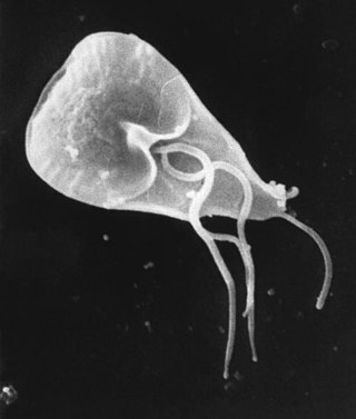
Giardia duodenalis, also known as Giardia intestinalis and Giardia lamblia, is a flagellated parasitic protozoan microorganism of the genus Giardia that colonizes the small intestine, causing a diarrheal condition known as giardiasis. The parasite attaches to the intestinal epithelium by an adhesive disc or sucker, and reproduces via binary fission. Giardiasis does not spread to other parts of the gastrointestinal tract, but remains confined to the lumen of the small intestine. The microorganism has an outer membrane that makes it possible to survive even when outside of its host, and which can render it tolerant to certain disinfectants. Giardia trophozoites are anaerobic, and absorb their nutrients from the intestinal lumen. If the organism is stained, its characteristic pattern resembles the familiar "smiley face" symbol.

<i>Entamoeba histolytica</i> Anaerobic parasitic protist

Entamoeba histolytica is an anaerobic parasitic amoebozoan, part of the genus Entamoeba. Predominantly infecting humans and other primates causing amoebiasis, E. histolytica is estimated to infect about 35-50 million people worldwide. E. histolytica infection is estimated to kill more than 55,000 people each year. Previously, it was thought that 10% of the world population was infected, but these figures predate the recognition that at least 90% of these infections were due to a second species, E. dispar. Mammals such as dogs and cats can become infected transiently, but are not thought to contribute significantly to transmission.

<span class="mw-page-title-main">Giardiasis</span> Parasitic disease that results in diarrhea

Giardiasis is a parasitic disease caused by Giardia duodenalis. Infected individuals who experience symptoms may have diarrhoea, abdominal pain, and weight loss. Less common symptoms include vomiting and blood in the stool. Symptoms usually begin one to three weeks after exposure and, without treatment, may last two to six weeks or longer.

<i>Entamoeba coli</i> Species of parasitic amoeba

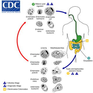
Entamoeba coli is a non-pathogenic species of Entamoeba that frequently exists as a commensal parasite in the human gastrointestinal tract. E. coli is important in medicine because it can be confused during microscopic examination of stained stool specimens with the pathogenic Entamoeba histolytica. This amoeba does not move much by the use of its pseudopod, and creates a "sur place (non-progressive) movement" inside the large intestine. Usually, the amoeba is immobile, and keeps its round shape. This amoeba, in its trophozoite stage, is only visible in fresh, unfixed stool specimens. Sometimes the Entamoeba coli have parasites as well. One is the fungus Sphaerita spp. This fungus lives in the cytoplasm of the E. coli. While this differentiation is typically done by visual examination of the parasitic cysts via light microscopy, new methods using molecular biology techniques have been developed. The scientific name of the amoeba, E. coli, is often mistaken for the bacterium, Escherichia coli. Unlike the bacterium, the amoeba is mostly harmless, and does not cause as many intestinal problems as some strains of the E. coli bacterium. To make the naming of these organisms less confusing, "alternate contractions" are used to name the species for the purpose making the naming easier; for example, using Esch. coli and Ent. coli for the bacterium and amoeba, instead of using E. coli for both.

Hymenolepiasis is infestation by one of two species of tapeworm: Hymenolepis nana or H. diminuta. Alternative names are dwarf tapeworm infection and rat tapeworm infection. The disease is a type of helminthiasis which is classified as a neglected tropical disease.

<span class="mw-page-title-main">Protozoan infection</span> Parasitic disease caused by a protozoan

Protozoan infections are parasitic diseases caused by organisms formerly classified in the kingdom Protozoa. These organisms are now classified in the supergroups Excavata, Amoebozoa, Harosa, and Archaeplastida. They are usually contracted by either an insect vector or by contact with an infected substance or surface.

<span class="mw-page-title-main">Amoebiasis</span> Human disease caused by amoeba protists

Amoebiasis, or amoebic dysentery, is an infection of the intestines caused by a parasitic amoeba Entamoeba histolytica. Amoebiasis can be present with no, mild, or severe symptoms. Symptoms may include lethargy, loss of weight, colonic ulcerations, abdominal pain, diarrhea, or bloody diarrhea. Complications can include inflammation and ulceration of the colon with tissue death or perforation, which may result in peritonitis. Anemia may develop due to prolonged gastric bleeding.

<i>Colpodella</i> Genus of single-celled organisms

Colpodella is a genus of alveolates comprising 5 species, and two further possible species: They share all the synapomorphies of apicomplexans, but are free-living, rather than parasitic. Many members of this genus were previously assigned to a different genus - Spiromonas.

<i>Proteromonas</i> Genus of single-celled organisms

Proteromonas is a genus of single-celled biflagellated microbial eukaryotes belonging to the Superphylum Stramenopiles which are characterized by the presence of tripartite, hair-like structures on the anteriorly-directed larger of the two flagella. Proteromonas on the other hand are notable by having tripartite hairs called somatonemes not on the flagella but on the posterior of the cell. Proteromonas are closely related to Karotomorpha and Blastocystis, which belong to the Opalines group.

<i>Katabia</i> Genus of heterotrophic protists

Katabia is a genus of soil-dwelling heterotrophic flagellate cercozoans containing the single species Katabia gromovi, and the only member of family Katabiidae.

<i>Trichomonas gallinae</i> Species of bird parasite

Trichomonas gallinae is a cosmopolitan parasite of birds including finches, pigeons, doves, turkeys, chickens, parrots, raptors. The condition in birds of prey is called frounce. It is believed to be an ancient pathogen causing frounce-like symptoms in theropod dinosaurs. The same condition in pigeons is commonly called canker.

<i>Dientamoeba fragilis</i> Parasite of humans, pigs and gorillas

Dientamoeba fragilis is a species of single-celled excavates found in the gastrointestinal tract of some humans, pigs and gorillas. It causes gastrointestinal upset in some people, but not in others. It is an important cause of travellers diarrhoea, chronic diarrhoea, fatigue and, in children, failure to thrive. Despite this, its role as a "commensal, pathobiont, or pathogen" is still debated. D. fragilis is one of the smaller parasites that are able to live in the human intestine. Dientamoeba fragilis cells are able to survive and move in fresh feces but are sensitive to aerobic environments. They dissociate when in contact or placed in saline, tap water or distilled water.

<i>Cystoisospora belli</i> Species of single-celled organism

Cystoisospora belli, previously known as Isospora belli, is a parasite that causes an intestinal disease known as cystoisosporiasis. This protozoan parasite is opportunistic in immune suppressed human hosts. It primarily exists in the epithelial cells of the small intestine, and develops in the cell cytoplasm. The distribution of this coccidian parasite is cosmopolitan, but is mainly found in tropical and subtropical areas of the world such as the Caribbean, Central and S. America, India, Africa, and S.E. Asia. In the U.S., it is usually associated with HIV infection and institutional living.

Phytomonas is a genus of trypanosomatids that infect plant species. Initially described using existing genera in the family Trypanosomatidae, such as Trypanosoma or Leishmania, the nomenclature of Phytomonas was proposed in 1909 in light of their distinct hosts and morphology. When the term was originally coined, no strict criterion was followed, and the term was adopted by the scientific community to describe flagellate protozoa in plants as a matter of convenience. Members of the taxon are globally distributed and have been discovered in members of over 24 plant families. Of these 24, the two main families that are infected by Phytomonas are Euphorbiaceae and Asclepiadiacae. These protists have been found in hosts between 50° latitude North and South, and thus they can be found on all continents save for Antarctica.

<i>Chilomastix</i>

Chilomastix is a genus of pyriform excavates within the family Retortamonadidae All species within this genus are flagellated, structured with three flagella pointing anteriorly and a fourth contained within the feeding groove. Chilomastix also lacks Golgi apparatus and mitochondria but does possess a single nucleus. The genus parasitizes a wide range of vertebrate hosts, but is known to be typically non-pathogenic, and is therefore classified as harmless. The life cycle of Chilomastix lacks an intermediate host or vector. Chilomastix has a resistant cyst stage responsible for transmission and a trophozoite stage, which is recognized as the feeding stage. Chilomastix mesnili is one of the more studied species in this genus due to the fact it is a human parasite. Therefore, much of the information on this genus is based on what is known about this one species.

Cochlosoma is a genus of flagellated protozoa in the order Trichomonadida created by A. Kotlán (1923). Some of their typical features include a prominent adhesive disc, axostyle, costa, and six flagella – one of which is attached to an undulating membrane that runs laterally along the body.

Colponema is a genus of single-celled flagellates that feed on eukaryotes in aquatic environments and soil. The genus contains 6 known species and has not been thoroughly studied. Colponema has two flagella which originate just below the anterior end of the cell. One extends forwards and the other runs through a deep groove in the surface and extends backwards. Colponema is a predator that feeds on smaller flagellates using its ventral groove. Like many other alveolates, they possess trichocysts, tubular mitochondrial cristae, and alveoli. It has been recently proposed that Colponema may be the sister group to all other alveolates. The genus could help us understand the origin of alveolates and shed light on features that are ancestral to all eukaryotes.

Euduboscquella (juˌduːboʊˈskwɛlə) is a genus of early branching dinoflagellates found in coastal waters around the globe. The members of this genus are all intracellular parasites that primarily infect Tintinnids. Euduboscquella are commonly found in marine environments, either infecting a host or in a resting stage in search of a new host, but there are a few freshwater and terrestrial species. Euduboscquella possess a multi-grooved shield separating their cytoplasm from the host’s cytoplasm, which is used by researchers to taxonomically identify them. The genus Euduboscquella contains nine species.

Monocercomonas is a Parabasalian genus belonging to the order Trichomonadida. It presents four flagella, three forward-facing and one trailing, without the presence of a costa or any kind of undulating membrane. Monocercomonas is found in animal guts. and is susceptible to cause Monocercomoniasis in reptiles

References

  1. 1 2 3 4 5 6 7 8 9 10 Geiman, Q. M. 1932: Retortamonas caudacus (n. sp.), an Intestinal Flagellate from a Beetle Larva, Gyrinidae sp. Transactions of the American Microscopical Society 51(4), 219-224. Doi: DOI: 10.2307/3222268
  2. 1 2 3 4 5 6 7 8 Kulda J., Nohýnková E., Čepička I. 2017: Retortamonadida (with Notes on Carpediemonas-Like Organisms and Caviomonadidae). In: Archibald J., Simpson A., Slamovits C. (eds) Handbook of the Protists. Springer, Cham: 1247-1278. doi: https://doi.org/10.1007/978-3-319-28149-0_3
  3. 1 2 3 4 5 6 7 8 9 10 11 12 13 14 15 Martinez-Diaz, R. A., Castro, A. T., Herrera, S. and Ponce, F. 2001: First Report of the Genus Retortamonas (Sarcomastigophora: Retortamonadidae) in Birds. Memórias do Instituto Oswalda Cruz, 96(7), 961-963. doi: https://doi.org/10.1590/S0074-02762001000700013
  4. 1 2 3 4 5 6 7 8 9 10 11 Hendarto, J., Mizuno, T., Hidayati, A. P.N., Rozi, I. E., Asih, P. B.S., Syafruddin, D., Yashikawa, H., Matsubayashi, M. and Tokoro, M. 2018: Three monophyletic clusters in Retortamonas species isolated from vertebrates. Parasitology International 69, 93-98. doi: https://doi.org/10.1016/j.parint.2018.12.004
  5. 1 2 3 4 5 6 7 8 9 Kawamura, O., Kon, Y., Naganuma, A., Iwami, T., Maruyama H., Yamada, T., Sonobe, K., Horikoshi, T., Kusano, M. and Mori, M. 2001: Retortamonas intestinalis in the pancreatic juice of a patient with small nodular lesions of the main pancreatic duct. Gastrointestinal Endoscopy 53(4): 508-510. doi: https://doi.org/10.1067/mge.2001.112746
  6. 2012: The Free Dictionary: Retortamonas. Farlex. Retrieved from https://medical-dictionary.thefreedictionary.com/Retortamonas
  7. 1 2 3 4 Wenrich, D. H. (October 1932). "The Relation of the Protozoan Flagellate, Retortamonas gryllotalpae (Grassi, 1879) Stiles, 1902 to the Species of the Genus Embadomonas Mackinnon, 1911". Transactions of the American Microscopical Society. 51 (4): 225–238. doi:10.2307/3222269. JSTOR   3222269.
  8. 1 2 Paradisi, F.; Nuñez, L.; Bartoloni, A.; Cancrini, G. (1988). "Intestinal parasites in the Camiri, Gutierrez and Boyuibe areas, Santa Cruz Department, Bolivia". Parassitologia. 30 (2–3): 263–269. ISSN   0048-2951. PMID   3271990.
  9. 1 2 3 Melvin, Dorothy M.; Brooke, Marion Murphy; Healy, George R. (1965). Common blood and tissue parasites of man : life cycle charts /. Atlanta, Ga.: U.S. Department of Health, Education, and Welfare, Public Health Service, Communicable Disease Center. doi:10.5962/bhl.title.115055. hdl:2027/pur1.32754075989495.
  10. 1 2 Brugerolle, Guy (2005-12-21). "The symbiotic fauna of the African termite Hodotermes mossambicus identification of four flagellate species of the genera Spironympha, Trichomonoides and Retortamonas". Parasitology Research. 98 (3): 257–263. doi:10.1007/s00436-005-0002-1. ISSN   0932-0113. PMID   16369849. S2CID   172579.
  11. Singleton, Paul; Sainsbury, Diana (September 2007). "Dictionary of Microbiology and Molecular Biology. Third Edition, Revised. By Paul Singleton and, Diana Sainsbury. Chichester (United Kingdom) and Hoboken (New Jersey): John Wiley & Sons. $110.00 (paper). xi + 895 p; ill.; no index". The Quarterly Review of Biology. 82 (3): 275. doi:10.1086/523143. ISBN   978-0-470-03545-0. ISSN   0033-5770.
  12. Stabler, Robert M. (June 1944). "A New Species of Retortamonas (Protozoa) from the Common Mole Cricket, Gryllotalpa hexadactyla". The Journal of Parasitology. 30 (3): 173–176. doi:10.2307/3272792. JSTOR   3272792.