Giardia

Last updated

Contents

Giardia
Giardia lamblia SEM 8698 lores.jpg
Giardia trophozoite, SEM
Scientific classification OOjs UI icon edit-ltr.svg
Domain: Eukaryota
Phylum: Metamonada
Order: Diplomonadida
Family: Hexamitidae
Subfamily: Giardiinae
Genus: Giardia
Künstler, 1882 [1]
Species
Synonyms
  • LambliaR. Blanchard, 1888 [2]

Giardia ( /ˈɑːrdiə/ or /ˈɑːrdiə/ ) is a genus of anaerobic flagellated protozoan parasites of the phylum Metamonada that colonise and reproduce in the small intestines of several vertebrates, causing the disease giardiasis. Their life cycle alternates between a swimming trophozoite and an infective, resistant cyst. Giardia were first described by the Dutch microscopist Antonie van Leeuwenhoek in 1681. [3] The genus is named after French zoologist Alfred Mathieu Giard. [4]

Characteristics

Like other diplomonads, Giardia have two nuclei, each with four associated flagella, and were thought to lack both mitochondria and Golgi apparatuses. However, they are now known to possess a complex endomembrane system as well as mitochondrial remnants, called mitosomes, through mitochondrial reduction. [5] [6] [7] [8] The mitosomes are not used in ATP synthesis the way mitochondria are, but are involved in the maturation of iron-sulfur proteins. [9] The synapomorphies of genus Giardia include cells with duplicate organelles, absence of cytostomes, and ventral adhesive disc. [10]

Systematics

About 40 species have been described, but most of them are probably synonyms. [11] Currently, five to six morphologically distinct species are recognised. [12] Giardia duodenalis (=G. intestinalis, =G. lamblia) infect humans and other mammals, G. microti infects voles, G. muris is found in other mammals, G. ardeae and G. psittaci in birds, and G. agilis in amphibians. [4] Other described (but not certainly valid), species include: [13]

Real-time polymerase chain reaction (PCR) tests have been developed to detect specific species of Giardia. Gene probe can also used to differentiate between species of Giardia. A more common and less time-consuming means of identifying different species of Giardia includes microscopy and immunofluorescence techniques. [14]

Genetic and biochemical studies have revealed the heterogeneity of Giardia duodenalis , which contains probably at least eight lineages or cryptic species. [15]

Phylogeny

The phylogeny of Giardia is unclear, but two main theories exist. Firstly, Giardia may be extremely primitive eukaryotes that branched off early from other members of their group. This theory is supported by several features: their lack of complete mitochondria (see Characteristics) and other organelles, their primitive metabolic pathways, and their position on a phylogenetic tree. [16] However, many of these differences have been refuted in recent years, and many researchers are supporting a second theory: that Giardia are highly evolved parasites, which have lost ancestral characteristics. [17]

Genome

A Giardia isolate (WB) was the first diplomonad to have its genome sequenced. Its 11.7 million basepair genome is compact in structure and content with simplified basic cellular machineries and metabolism. Currently the genomes of several other Giardia isolates and diplomonads (the fish pathogens Spironucleus vortens and S. salmonicida) are being sequenced. [18]

A second isolate (the B assemblage) from humans has been sequenced along with a species from a pig (the E assemblage). [19] There are ~5000 genes in the genome. The E assemblage is more closely related to the A assemblage than is the B. A number of chromosomal rearrangements are present.

Infection

An SEM micrograph of the small intestine of a gerbil infested with Giardia reveals a mucosa surface almost entirely obscured by attached trophozoites Giardia-spp.--infected--gerbil-intestine.jpg
An SEM micrograph of the small intestine of a gerbil infested with Giardia reveals a mucosa surface almost entirely obscured by attached trophozoites

Giardia lives in the intestines of infected humans or other animals, individuals of which become infected by ingesting or coming into contact with contaminated foods, soil, or water tainted by the feces of an infected carrier. [20]

The symptoms of Giardia, which may begin to appear 2 days after infection, can include mild to violent diarrhoea, excess gas, stomach or abdominal cramps, upset stomach, and nausea. Resulting dehydration and nutritional loss may need immediate treatment. A typical infection can be slight, resolve without treatment, and last between 2 and 6 weeks, although it can sometimes last longer and/or be more severe. Coexistence with the parasite is possible (symptoms fade), but an infected individual can remain a carrier and transmit it to others. Medication containing tinidazole or metronidazole decreases symptoms and time to resolution. Albendazole is also used, and has an anthelmintic (anti-worm) property as well, ideal for certain compounded issues when a general vermicidal agent is preferred. Giardia causes a disease called giardiasis, which causes the villi of the small intestine to atrophy and flatten, resulting in malabsorption in the intestine. Lactose intolerance can persist after the eradication of Giardia from the digestive tract. [21]

Prevalence

The prevalence of the infection depends on different factors; while the prevalence is estimated around 2% in some developed countries, in other countries from Asia, Africa or Latin America, the prevalence can be estimated between 20% and 40%. In some patients, giardiasis can be completely asymptomatic, so many more cases are estimated. [22] The diagnostic method used can also infer in the identification and thus the count of cases. Due to their lack of knowledge and overall behavioral patterns, children aged under 5 years, are the population with the most reported infections. [23]

See also

Related Research Articles

<span class="mw-page-title-main">Symbiogenesis</span> Evolutionary theory holding that eukaryotic organelles evolved through symbiosis with prokaryotes

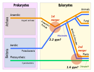
Symbiogenesis is the leading evolutionary theory of the origin of eukaryotic cells from prokaryotic organisms. The theory holds that mitochondria, plastids such as chloroplasts, and possibly other organelles of eukaryotic cells are descended from formerly free-living prokaryotes taken one inside the other in endosymbiosis. Mitochondria appear to be phylogenetically related to Rickettsiales bacteria, while chloroplasts are thought to be related to cyanobacteria.

<span class="mw-page-title-main">Excavata</span> Supergroup of unicellular organisms belonging to the domain Eukaryota

Excavata is an extensive and diverse but paraphyletic group of unicellular Eukaryota. The group was first suggested by Simpson and Patterson in 1999 and the name latinized and assigned a rank by Thomas Cavalier-Smith in 2002. It contains a variety of free-living and symbiotic protists, and includes some important parasites of humans such as Giardia and Trichomonas. Excavates were formerly considered to be included in the now obsolete Protista kingdom. They were distinguished from other lineages based on electron-microscopic information about how the cells are arranged. They are considered to be a basal flagellate lineage.

<i>Giardia duodenalis</i> Parasitic microorganism that causes giardiasis

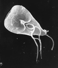
Giardia duodenalis, also known as Giardia intestinalis and Giardia lamblia, is a flagellated parasitic protozoan microorganism of the genus Giardia that colonizes the small intestine, causing a diarrheal condition known as giardiasis. The parasite attaches to the intestinal epithelium by an adhesive disc or sucker, and reproduces via binary fission. Giardiasis does not spread to other parts of the gastrointestinal tract, but remains confined to the lumen of the small intestine. The microorganism has an outer membrane that makes it possible to survive even when outside of its host, and which can render it tolerant to certain disinfectants. Giardia trophozoites are anaerobic, and absorb their nutrients from the intestinal lumen. If the organism is stained, its characteristic pattern resembles the familiar "smiley face" symbol.

<span class="mw-page-title-main">Giardiasis</span> Parasitic disease that results in diarrhea

Giardiasis is a parasitic disease caused by Giardia duodenalis. Infected individuals who experience symptoms may have diarrhoea, abdominal pain, and weight loss. Less common symptoms include vomiting and blood in the stool. Symptoms usually begin one to three weeks after exposure and, without treatment, may last two to six weeks or longer.

<i>Trichomonas vaginalis</i> Species of parasite that causes sexually transmitted infections

Trichomonas vaginalis is an anaerobic, flagellated protozoan parasite and the causative agent of a sexually transmitted disease called trichomoniasis. It is the most common pathogenic protozoan that infects humans in industrialized countries. Infection rates in men and women are similar but women are usually symptomatic, while infections in men are usually asymptomatic. Transmission usually occurs via direct, skin-to-skin contact with an infected individual, most often through vaginal intercourse. The WHO has estimated that 160 million cases of infection are acquired annually worldwide. The estimates for North America alone are between 5 and 8 million new infections each year, with an estimated rate of asymptomatic cases as high as 50%. Usually treatment consists of metronidazole and tinidazole.

<span class="mw-page-title-main">Diplomonad</span> Group of mostly parasitic flagellates

The diplomonads are a group of flagellates, most of which are parasitic. They include Giardia duodenalis, which causes giardiasis in humans. They are placed among the metamonads, and appear to be particularly close relatives of the retortamonads.

<span class="mw-page-title-main">Metamonad</span> Phylum of excavate protists

The metamonads are a large group of flagellate amitochondriate microscopic eukaryotes. Their composition is not entirely settled, but they include the retortamonads, diplomonads, and possibly the parabasalids and oxymonads as well. These four groups are all anaerobic, occurring mostly as symbiotes or parasites of animals, as is the case with Giardia lamblia which causes diarrhea in mammals.

<span class="mw-page-title-main">Microsporidia</span> Phylum of fungi

Microsporidia are a group of spore-forming unicellular parasites. These spores contain an extrusion apparatus that has a coiled polar tube ending in an anchoring disc at the apical part of the spore. They were once considered protozoans or protists, but are now known to be fungi, or a sister group to fungi. These fungal microbes are obligate eukaryotic parasites that use a unique mechanism to infect host cells. They have recently been discovered in a 2017 Cornell study to infect Coleoptera on a large scale. So far, about 1500 of the probably more than one million species are named. Microsporidia are restricted to animal hosts, and all major groups of animals host microsporidia. Most infect insects, but they are also responsible for common diseases of crustaceans and fish. The named species of microsporidia usually infect one host species or a group of closely related taxa. Approximately 10 percent of the species are parasites of vertebrates —several species, most of which are opportunistic, can infect humans, in whom they can cause microsporidiosis.

<span class="mw-page-title-main">Hydrogenosome</span> Mitochondrion-derived organelle

A hydrogenosome is a membrane-enclosed organelle found in some anaerobic ciliates, flagellates, and fungi. Hydrogenosomes are highly variable organelles that have presumably evolved from protomitochondria to produce molecular hydrogen and ATP in anaerobic conditions.

A mitosome is a mitochondrion-related organelle (MRO) found in a variety of parasitic unicellular eukaryotes, such as members of the supergroup Excavata. The mitosome was first discovered in 1999 in Entamoeba histolytica, an intestinal parasite of humans, and mitosomes have also been identified in several species of Microsporidia and in Giardia intestinalis.

<i>Hexamita</i> Genus of parasitic diplomonads

Hexamita is a genus of parasitic diplomonads. It is related to Giardia. H. columbae and H. meleagridis live in the intestines of birds. H. muris and H. pitheci live in the intestines of mammals. H. salmonis and H. truttae live in the intestines of fish. Species in the Hexamita family are most commonly spread through fecal matter.

<i>Henneguya zschokkei</i> Species of Myxosporea

Henneguya zschokkei or Henneguya salminicola is a species of a myxosporean endoparasite. It afflicts several salmon in the genera Oncorhynchus and Salmo. It causes milky flesh or tapioca disease. H. zschokkei is notable for its lack of mitochondria, mitochondrial DNA, aerobic respiration and its reliance on an exclusively anaerobic metabolism.

<i>Blastocystis</i> Genus of single-celled organisms

Blastocystis is a genus of single-celled parasites belonging to the Stramenopiles that includes algae, diatoms, and water molds. There are several species, living in the gastrointestinal tracts of species as diverse as humans, farm animals, birds, rodents, reptiles, amphibians, fish, and cockroaches. Blastocystis has low host specificity, and many different species of Blastocystis can infect humans, and by current convention, any of these species would be identified as Blastocystis hominis.

The discovery of disease-causing pathogens is an important activity in the field of medical science. Many viruses, bacteria, protozoa, fungi, helminths, and prions are identified as a confirmed or potential pathogen. In the United States, a Centers for Disease Control and Prevention program, begun in 1995, identified over a hundred patients with life-threatening illnesses that were considered to be of an infectious cause but that could not be linked to a known pathogen. The association of pathogens with disease can be a complex and controversial process, in some cases requiring decades or even centuries to achieve.

<span class="mw-page-title-main">Protozoan infection</span> Parasitic disease caused by a protozoan

Protozoan infections are parasitic diseases caused by organisms formerly classified in the kingdom Protozoa. These organisms are now classified in the supergroups Excavata, Amoebozoa, Harosa, and Archaeplastida. They are usually contracted by either an insect vector or by contact with an infected substance or surface.

Spironucleus salmonicida is a species of fish parasite. It is a flagellate adapted to micro-aerobic environments that causes systemic infections in salmonid fish. The species creates foul-smelling, pus-filled abscesses in muscles and internal organs of aquarium fish. In the late 1980s when the disease was first reported, it was believed to be caused by Spironucleus barkhanus. Anders Jørgensen was the person that found out what species really caused the disease.

Spironucleus is a diplomonad genus that is bilaterally symmetrical and can be found in various animal hosts. This genus is a binucleate flagellate, which is able to live in the anaerobic conditions of animal intestinal tracts. A characteristic of Spironucleus that is common to all metamonads is that it does not have aerobic mitochondria, but instead rely on hydrogenosomes to produce energy. Spironucleus has six anterior and two posterior flagella. The life cycle of Spironucleus involves one active trophozoite stage and one inactive cyst stage. Spironucleus undergoes asexual reproduction via longitudinal binary fission. Spironucleusvortens can cause lateral line erosion in freshwater anglefish. Spironucleuscolumbae is found to cause hexamitiasis in pigeons. Finally, Spironucleusmuris is found to cause illnesses of the digestive system in mice, rats, and hamsters. The genome of Spironucleus has been studied to exhibit the role of lateral gene transfer from prokaryotes in allowing for anaerobic metabolic processes in diplomonads.

<i>Encephalitozoon cuniculi</i> Microsporidial pathogen

Encephalitozoon cuniculi is a microsporidial parasite of mammals with world-wide distribution. An important cause of neurologic and renal disease in rabbits, E. cuniculi can also cause disease in immunocompromised people.

<i>Dientamoeba fragilis</i> Parasite of humans, pigs and gorillas

Dientamoeba fragilis is a species of single-celled excavates found in the gastrointestinal tract of some humans, pigs and gorillas. It causes gastrointestinal upset in some people, but not in others. It is an important cause of travellers diarrhoea, chronic diarrhoea, fatigue and, in children, failure to thrive. Despite this, its role as a "commensal, pathobiont, or pathogen" is still debated. D. fragilis is one of the smaller parasites that are able to live in the human intestine. Dientamoeba fragilis cells are able to survive and move in fresh feces but are sensitive to aerobic environments. They dissociate when in contact or placed in saline, tap water or distilled water.

<span class="mw-page-title-main">Mark van der Giezen</span>

Mark van der Giezen is Professor of Biological Chemistry, Centre for Organelle Research, University of Stavanger, Norway. He holds Dutch nationality and is married with three children.

References

  1. Künstler, J. (1882). "Sur cinq protozoaires parasites nouveaux". C. R. Acad. Sci. Paris. 95: 347–349.
  2. Blanchard, R. (1888). "Remarques sur le megastome intestinal". Bull. Soc. Zool. Fr. 30: 18–19.
  3. Stanley L. Erlandsen, Ernest A. Meyer (1 March 1984). Giardia and Giardiasis: Biology, Pathogenesis, and Epidemiology. Springer. pp. 131–. ISBN   978-0-306-41539-5.
  4. 1 2 Adam RD (July 2001). "Biology of Giardia lamblia". Clin. Microbiol. Rev. 14 (3): 447–75. doi:10.1128/CMR.14.3.447-475.2001. PMC   88984 . PMID   11432808.
  5. Tovar J, León-Avila G, Sánchez L, Sutak R, Tachezy J, van der Giezen M, Hernández M, Müller M, Lucocq J (2003). "Mitochondrial remnant organelles of Giardia function in iron-sulphur protein maturation". Nature. 426 (6963): 172–176. Bibcode:2003Natur.426..172T. doi:10.1038/nature01945. PMID   14614504. S2CID   4402808.
  6. Anna Karnkowska, et al. (May 2016). "A Eukaryote without a Mitochondrial Organelle". Current Biology. 26 (10): 1274–1284. Bibcode:2016CBio...26.1274K. doi: 10.1016/j.cub.2016.03.053 . PMID   27185558.
  7. Soltys BJ, Falah M, Gupta RS (July 1996). "Identification of endoplasmic reticulum in the primitive eukaryote Giardia lamblia using cryoelectron microscopy and antibody to Bip". J. Cell Sci. 109 (Pt 7): 1909–17. doi:10.1242/jcs.109.7.1909. PMID   8832413.
  8. Dolezal P, Smíd O, Rada P, et al. (August 2005). "Giardia mitosomes and trichomonad hydrogenosomes share a common mode of protein targeting". Proc. Natl. Acad. Sci. U.S.A. 102 (31): 10924–9. Bibcode:2005PNAS..10210924D. doi: 10.1073/pnas.0500349102 . PMC   1182405 . PMID   16040811.
  9. Tovar J, et al. (2003). "Mitochondrial remnant organelles of Giardia function in iron-sulphur protein maturation". Nature. 426 (6963): 172–6. Bibcode:2003Natur.426..172T. doi:10.1038/nature01945. PMID   14614504. S2CID   4402808.
  10. Cepicka I (September 2008). "Fornicata". Tree of Life Web Project.
  11. Meyer E.A., Radulescu S. (1979). "Giardia and Giardiasis". Advances in Parasitology. 17: 1–47. doi:10.1016/S0065-308X(08)60548-5. ISBN   978-0-12-031717-2. PMID   395833.
  12. Brusca R, Brusca G (2003). Invertebrates (2 ed.). Sinauer Associates. ISBN   0-87893-097-3.
  13. "Giardia Kunstler". Tree of Life Web Project. September 2008.
  14. Mahbubani 1992
  15. Thompson RC, Monis PT (2004). "Variation in Giardia: implications for taxonomy and epidemiology". Advances in Parasitology. 58: 69–137. doi:10.1016/S0065-308X(04)58002-8. ISBN   978-0-12-031758-5. PMID   15603762.
  16. Ye Q, Tian H, Chen B, Shao J, Qin Y, Wen J (25 August 2017). "Giardia's primitive GPL biosynthesis pathways with parasitic adaptation 'patches': implications for Giardia's evolutionary history and for finding targets against Giardiasis". Scientific Reports. 7 (1): 9507. Bibcode:2017NatSR...7.9507Y. doi:10.1038/s41598-017-10054-1. ISSN   2045-2322. PMC   5573378 . PMID   28842650.
  17. Lloyd D, Harris JC (1 March 2002). "Giardia: highly evolved parasite or early branching eukaryote?". Trends in Microbiology. 10 (3): 122–127. doi:10.1016/S0966-842X(02)02306-5. ISSN   0966-842X. PMID   11864821.
  18. Andersson, JO, et al. (2010). "The Genome of Giardia and Other Diplomonads". Anaerobic Parasitic Protozoa: Genomics and Molecular Biology. Caister Academic Press. ISBN   978-1-904455-61-5.
  19. Jerlström-Hultqvist J, Ankarklev J, Svärd SG (2010). "Is human giardiasis caused by two different Giardia species?". Gut Microbes. 1 (6): 379–82. doi:10.4161/gmic.1.6.13608. PMC   3056102 . PMID   21468219.
  20. Filice, F.P. (1952). "Studies on the cytology and life history of a Giardia from the laboratory rat". U. C. Publications in Zoology. 5sex7 (2). Berkeley CA: University of California Press.
  21. LaCour 2003
  22. Hörman A, Korpela H, Wedel H, Sutinen J, Hanninen M (2004). "Meta-analysis in assessment of the prevalence and annual incidence of Giardia spp. and Cryptosporidium spp. infections in humnas in the Nordic countries". Int J Parasitol. 34 (12): 1337–1346. doi:10.1016/j.ijpara.2004.08.009. PMID   15542094.
  23. Savioli L, Smith H, Thompson A (2006). "Giardia and Cryptosporidium join the "Neglected Diseases Initiative"". Trends Parasitol. 22 (5): 160–167. doi:10.1016/j.pt.2006.02.015. PMID   16545611.