Sappinia pedata

Last updated

Contents

Sappinia pedata
Scientific classification
Domain:
Phylum:
Class:
Subclass:
Family:
Genus:
Species:
S. pedata
Binomial name
Sappinia pedata

Sappinia pedata is a free living amoeboid organism, first described by Pierre Augustin Dangeard in 1896. It belongs to the genus Sappinia within the Thecamoebida clade of Amoebozoa and is characterized by its unique monopodial locomotion and cell surface morphology. S. pedata has been found in various habitats worldwide, mostly on herbivore dung, decaying plant matter, and soil. The species has gained attention due to its potential medical relevance and has been the subject of most recent and emerging studies in Protistology and Eukaryotic Microbiology as a whole.

Etyomology

The name “Sappinia” was named after the late Mister Sappin-Trouffy, a mycologist at the Paris Academy of Sciences while the Latin word “pedata” means foot-like.

Phylogeny

Sappinia pedata is a discosean amoebozoan that forms a highly supported clade with its sister species Sappinia diploidea , and the newly described species Sappinia platani, all of which are closely related to two Thecamoeba spp. within the Thecamoebida clade. [1] [2] [3] The genus Sappinia, to which S. pedata belongs, is notable for the suspected presence of sexual reproduction, setting it apart from other flabellinid genera for which the sexual status has not been explicitly established. [2]

Recent phylogenetic studies have suggested that the particular “brain-eating” strain identified in a case of amoebic encephalitis, may represent a new species more closely related to S. pedata than S. diploidea and S. platani. [4] [3] However, further research is needed to confirm the taxonomic status of this strain.

The description of S. platani as a novel species within the genus Sappinia further highlights the genetic diversity and evolutionary significance of this group of organisms and provides insight into the diversity and evolution of this major eukaryotic lineage. [3]

History of Knowledge

The species S. pedata was originally established by Pierre Augustin Dangeard in 1896, who described it as a free-living, mostly binucleate amoeba with a dense glycocalyx after isolating the species from old cultures of horse dung. [4]

Dangeard observed many remarkable things about S. pedata including cysts he described as pedicelled and a type of nuclear division that seemed to result in the formation of two closely apposed nuclei, and in some cases, four nuclei. These observations were later confirmed by Matthew Brown and colleagues well over a century later, after establishing a neotype for the same species (ATCC PRA-232) in 2007. However, no stalked cysts were found this time. [4]

The term “Standing amoeba” was coined by Brown et al (2007) after thorough observation of the lack of a cell wall in S. pedata, concluding that the cells were neither encysted nor formed spores as described in earlier publications. [1]

Habitat and Ecology

In their natural habitats, S. pedata can be found on herbivore dung, decaying plant matter, as well as soil and dog dung. In terms of geography, it has been isolated from across Europe, America and Oceania and is currently considered a worldwide species with isolates yet to be obtained from Africa. [4]

S. pedata has been decribed to be preyed upon by the dung-inhabiting fungus Stylophage anomala in nature. This close association with S. anomala suggests that the amoebae may also be dispersed phoretically by the same mites that carry the fungus due to their ability to adhere to a wide range of surfaces when in its standing form. [5]

Sappinia pedata photographed at the Brown Lab, Department of Biological Sciences, Mississippi State University, MS, USA. Showing its standing amoeba on a piece of cow dung. Photo credits: Lucia Garcia and Tristan Henderson. Sappinia pedata.jpg
Sappinia pedata photographed at the Brown Lab, Department of Biological Sciences, Mississippi State University, MS, USA. Showing its standing amoeba on a piece of cow dung. Photo credits: Lucia Garcia and Tristan Henderson.

Morphology and Life Cycle

Sappinia pedata exhibits two distinct life cycle stages: a trophozoite stage and a cyst stage.

The trophozoites are characterized by their unique erect stage, which resembles the fruiting bodies found in protostelid slime molds. The standing amoeba of S. pedata have a club-shaped appearance that is easily recognized by the nearly colorless to a very pale yellow and slightly opaque texture. Unlike the Thecamoeba spp., the amoeba form of S. pedata does not possess the characteristic longitudinal dorsal ridges. However, irregular folds can occasionally be observed along the edges of the cell.

In terms of size, the trophozoites measure approximately 45 – 65 µm in length and 18 –35 µm in width. On the other hand, the cysts are generally smaller, with a diameter ranging from 18 – 25 µm. [4] The cysts of S. pedata typically consists of a single cell, or two cells covered by a common wall with an outer mucilaginous layer. [1]

It is worth noting that certain strains of S. pedata have been shown by Wylezich et al., (2009), to have no standing form, suggesting some degree of intraspecific variation in morphology. [6]

Medical Relevance

Sappinia pedata gained clinical significance in 2003 when it was identified as the causative agent of amoebic encephalitis in a previously healthy young man. [7] This case marked the first reported instance of a Sappinia species being implicated in human disease.  

The patient, a non-immunosuppressed individual, was presented with symptoms including headache, nausea, vomiting, blurred vision, and seizures. Neuroimaging revealed a solitary mass in the patient’s brain, which was initially thought to be a tumor. However, upon surgical removal and subsequent histopathological examination, amoebic trophozoites were discovered in the brain tissue. [7]

At the time of initial diagnosis, the causative agent was provisionally identified as S. diploidea based on morphological characteristics. However, further molecular investigations performed by Qvarnstrom et al. In 2009 revealed that the amoebae isolated from the patient’s brain tissue were genetically more closely related to S. pedata. [7] This groundbreaking case highlighted the potential of Sappinia species to cause serious human infections, particularly in the central nervous system. Since this initial report, no additional cases of human infection caused by S. pedata or other Sappinia species have been documented and the patient is known to have survived the treatment procedures without any long-term consequences.

Related Research Articles

<i>Entamoeba</i> Genus of internal parasites

Entamoeba is a genus of Amoebozoa found as internal parasites or commensals of animals. In 1875, Fedor Lösch described the first proven case of amoebic dysentery in St. Petersburg, Russia. He referred to the amoeba he observed microscopically as Amoeba coli; however, it is not clear whether he was using this as a descriptive term or intended it as a formal taxonomic name. The genus Entamoeba was defined by Casagrandi and Barbagallo for the species Entamoeba coli, which is known to be a commensal organism. Lösch's organism was renamed Entamoeba histolytica by Fritz Schaudinn in 1903; he later died, in 1906, from a self-inflicted infection when studying this amoeba. For a time during the first half of the 20th century the entire genus Entamoeba was transferred to Endamoeba, a genus of amoebas infecting invertebrates about which little is known. This move was reversed by the International Commission on Zoological Nomenclature in the late 1950s, and Entamoeba has stayed 'stable' ever since.

<span class="mw-page-title-main">Vampyrellidae</span> Family of single-celled organisms

The family Vampyrellidae is a subgroup of the order Vampyrellida within the supergroup Rhizaria. Based on molecular sequence data, the family currently comprises the genus Vampyrella, and maybe several other vampyrellid amoebae. The cells are naked and characterised by radiating, filose pseudopodia and an orange colouration of the main cell body.

<i>Acanthamoeba</i> Genus of protozoans

Acanthamoeba is a genus of amoebae that are commonly recovered from soil, fresh water, and other habitats. The genus Acanthamoeba has two stages in its life cycle, the metabolically active trophozoite stage and a dormant, stress-resistant cyst stage. In nature, Acanthamoeba species are generally free-living bacterivores. However, they are also opportunistic pathogens able to cause serious and sometimes fatal infections in humans and other animals.

Free-living amoebae are a group of protozoa that are important causes of infectious disease in humans and animals.

<span class="mw-page-title-main">Amoebozoa</span> Phylum of protozoans

Amoebozoa is a major taxonomic group containing about 2,400 described species of amoeboid protists, often possessing blunt, fingerlike, lobose pseudopods and tubular mitochondrial cristae. In traditional classification schemes, Amoebozoa is usually ranked as a phylum within either the kingdom Protista or the kingdom Protozoa. In the classification favored by the International Society of Protistologists, it is retained as an unranked "supergroup" within Eukaryota. Molecular genetic analysis supports Amoebozoa as a monophyletic clade. Modern studies of eukaryotic phylogenetic trees identify it as the sister group to Opisthokonta, another major clade which contains both fungi and animals as well as several other clades comprising some 300 species of unicellular eukaryotes. Amoebozoa and Opisthokonta are sometimes grouped together in a high-level taxon, variously named Unikonta, Amorphea or Opimoda.

<span class="mw-page-title-main">Naegleriasis</span> Rare and usually fatal brain infection by a protist

Naegleriasis, also known as primary amoebic meningoencephalitis (PAM), is an almost invariably fatal infection of the brain by the free-living unicellular eukaryote Naegleria fowleri. Symptoms are meningitis-like and include headache, fever, nausea, vomiting, a stiff neck, confusion, hallucinations and seizures. Symptoms progress rapidly over around five days, and death usually results within one to two weeks of symptoms.

<i>Naegleria</i> Genus of protists

Naegleria is a free living amoebae protist genus consisting of 47 described species often found in warm aquatic environments as well as soil habitats worldwide. It has three life cycle forms: the amoeboid stage, the cyst stage, and the flagellated stage, and has been routinely studied for its ease in change from amoeboid to flagellated stages. The Naegleria genera became famous when Naegleria fowleri, a human pathogenic strain and the causative agent of primary amoebic meningoencephalitis (PAM), was discovered in 1965. Most species in the genus, however, are nonpathogenic, meaning they do not cause disease.

<i>Balamuthia mandrillaris</i> Species of pathogenic Amoebozoa

Balamuthia mandrillaris is a free-living amoeba that causes the rare but deadly neurological condition granulomatous amoebic encephalitis (GAE). B. mandrillaris is a soil-dwelling amoeba and was first discovered in 1986 in the brain of a mandrill that died in the San Diego Wild Animal Park.

<span class="mw-page-title-main">Granulomatous amoebic encephalitis</span> Rare and usually fatal brain infection by certain amoebae

Granulomatous amoebic encephalitis (GAE) is a rare, often fatal, subacute-to-chronic central nervous system disease caused by certain species of free-living amoebae of the genera Acanthamoeba, Balamuthia and Sappinia. The term is most commonly used with Acanthamoeba. In more modern references, the term "balamuthia amoebic encephalitis" (BAE) is commonly used when Balamuthia mandrillaris is the cause.

<span class="mw-page-title-main">Amoebiasis</span> Human disease caused by amoeba protists

Amoebiasis, or amoebic dysentery, is an infection of the intestines caused by a parasitic amoeba Entamoeba histolytica. Amoebiasis can be present with no, mild, or severe symptoms. Symptoms may include lethargy, loss of weight, colonic ulcerations, abdominal pain, diarrhea, or bloody diarrhea. Complications can include inflammation and ulceration of the colon with tissue death or perforation, which may result in peritonitis. Anemia may develop due to prolonged gastric bleeding.

Sappinia diploidea is a free-living amoeba species.

Sappinia is a genus of heterotrophic, lobose amoebae within the family Thecamoebidae. A defining feature of Sappinia, which separates it from its sister genus Thecamoeba, is the presence of two closely apposed nuclei with a central, flattened connection. Sappinia species have two life cycle stages: a trophozoite and a cyst. Up until 2015, only two species had been discovered, Sappinia pedata and Sappinia diploidea. Sequencing of the small subunit rRNA of a particular isolate from a sycamore tree revealed a new species, Sappinia platani.Sappinia species were once thought to be coprozoic, as the first strains were isolated from animal dung. More research has shown that they are typical free-living amoebae, and can be found worldwide in soil, plant litter, and standing decaying plants, as well as freshwater ponds. In 2001, the first and only case of human pathogenesis in Sappinia was confirmed. The patient was a non-immunocompromised 38-year-old male who presented signs of amoebic encephalitis and who patient made a full recovery after treatment with several antimicrobials. The CDC initially classified the causative agent as S. diploidea based on morphological characteristics, but in 2009, Qvarnstrom et al. used molecular data to confirm that the true causative agent was S. pedata.

<span class="mw-page-title-main">Acanthamoebidae</span> Family of protozoans

Acanthamoebidae is a family of single-celled eukaryotes within the group Amoebozoa.

Hartmannella is a genus of Amoebozoa.

<i>Trichosphaerium</i> Genus of amoebae

Trichosphaerium is a genus of amoebozoan protists that present extraordinary morphological transformations, both in size and shape, during their life cycle. They can present a test that may or may not be covered in spicules. They are related to the family Microcoryciidae, which contains other amoebae with tests, within the clade Corycidia of the phylum Amoebozoa.

Sappinia amoebic encephalitis (SAE) is the name for amoebic encephalitis caused by species of Sappinia.

<span class="mw-page-title-main">Vampyrellida</span> Order of single-celled organisms

The vampyrellids, colloquially known as vampire amoebae, are a group of free-living predatory amoebae classified as part of the lineage Endomyxa. They are distinguished from other groups of amoebae by their irregular cell shape with propensity to fuse and split like plasmodial organisms, and their life cycle with a digestive cyst stage that digests the gathered food. They appear worldwide in marine, brackish, freshwater and soil habitats. They are important predators of an enormous variety of microscopic organisms, from algae to fungi and animals. They are also known as aconchulinid amoebae.

<span class="mw-page-title-main">Amoeba</span> Cellular body type

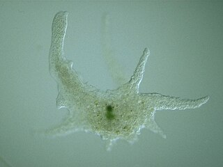
An amoeba, often called an amoeboid, is a type of cell or unicellular organism with the ability to alter its shape, primarily by extending and retracting pseudopods. Amoebae do not form a single taxonomic group; instead, they are found in every major lineage of eukaryotic organisms. Amoeboid cells occur not only among the protozoa, but also in fungi, algae, and animals.

<i>Naegleria fowleri</i> Species of free-living excavate form of protist

Naegleria fowleri, also known as the brain-eating amoeba, is a species of the genus Naegleria. It belongs to the phylum Percolozoa and is technically classified as an amoeboflagellate excavate, rather than a true amoeba. This free-living microorganism primarily feeds on bacteria but can become pathogenic in humans, causing an extremely rare, sudden, severe, and usually fatal brain infection known as naegleriasis or primary amoebic meningoencephalitis (PAM).

<i>Vampyrella</i> Genus of single-celled organisms

Vampyrella is a genus of amoebae belonging to the vampyrellid cercozoans usually ranging from 30-60 µm. Members of the genus alternate between two life stages: a free-living trophozoite stage and a cyst stage in which mitosis occurs. This taxon has received a great deal of attention due to their peculiar feeding behaviour of perforating the cell wall of algal cells and drawing out the contents for nourishment.

References

PD-icon.svg This article incorporates public domain material from websites or documents of the Centers for Disease Control and Prevention .

  1. 1 2 3 Brown, Matthew W.; Spiegel, Frederick W.; Silberman, Jeffrey D. (2007). "Amoeba at Attention: Phylogenetic Affinity of Sappinia pedata". Journal of Eukaryotic Microbiology. 54 (6): 511–519. doi:10.1111/j.1550-7408.2007.00292.x. ISSN   1066-5234. PMID   18070328.
  2. 1 2 Kang, Seungho; Tice, Alexander K; Spiegel, Frederick W; Silberman, Jeffrey D; Pánek, Tomáš; Čepička, Ivan; Kostka, Martin; Kosakyan, Anush; Alcântara, Daniel M C; Roger, Andrew J; Shadwick, Lora L; Smirnov, Alexey; Kudryavtsev, Alexander; Lahr, Daniel J G; Brown, Matthew W (2017). "Between a Pod and a Hard Test: The Deep Evolution of Amoebae". Molecular Biology and Evolution. 34 (9): 2258–2270. doi:10.1093/molbev/msx162. ISSN   0737-4038. PMC   5850466 . PMID   28505375.
  3. 1 2 3 Wylezich, C; Walochnik, J; Corsaro, D; Michel, R; Kudryavtsev, A (2015). "Electron Microscopical Investigations of a New Species of the Genus Sappinia (Thecamoebidae, Amoebozoa), Sappinia platani sp. nov., Reveal a Dictyosome in this Genus". Acta Protozoologica. 54 (1): 45–51. doi:10.4467/16890027AP.15.004.2191.
  4. 1 2 3 4 5 Walochnik, Julia; Wylezich, Claudia; Michel, Rolf (2010). "The genus Sappinia: History, phylogeny and medical relevance". Experimental Parasitology. 126 (1): 4–13. doi:10.1016/j.exppara.2009.11.017. ISSN   0014-4894. PMID   20004196.
  5. Blackwell, Meredith; Malloch, David (1991). "Life History and Arthropod Dispersal of a Coprophilous Stylopage". Mycologia. 83 (3): 360. doi:10.2307/3759996. JSTOR   3759996 via JSTOR.
  6. Wylezich, Claudia; Walochnik, Julia; Michel, Rolf (2009). "High genetic diversity of Sappinia-like strains (Amoebozoa, Thecamoebidae) revealed by SSU rRNA investigations". Parasitology Research. 105 (3): 869–873. doi:10.1007/s00436-009-1482-1. ISSN   1432-1955. PMID   19495795.
  7. 1 2 3 Qvarnstrom, Y.; Da Silva, A.J.; Schuster, F.L.; Gelman, B.B.; Visvesvara, G.S. (2009). "Molecular Confirmation of Sappinia pedata as a Causative Agent of Amoebic Encephalitis". The Journal of Infectious Diseases. 199 (8): 1139–1142. doi:10.1086/597473. ISSN   0022-1899. PMID   19302010.