Green sulfur bacteria

Last updated

Green sulfur bacteria
Green d winogradsky.jpg
Green sulfur bacteria in a Winogradsky column
Scientific classification OOjs UI icon edit-ltr.svg
Domain: Bacteria
(unranked): Bacteroidota–Chlorobiota group
Phylum: Chlorobiota
Iino et al. 2021 [1]
Class: "Chlorobia"
Garrity and Holt 2001 [2]
Order: Chlorobiales
Gibbons and Murray 1978 (Approved Lists 1980) [3]
Families and Genera
Synonyms
  • Chlorobiota:
    • Chlorobi Iino et al. 2010
    • "Chlorobi" Garrity and Holt 2001
    • "Chlorobaeota" Oren et al. 2015
    • "Chlorobiota" Whitman et al. 2018
  • Chlorobiota:
    • "Chlorobia" Whitman et al. 2018
    • Chlorobea Cavalier-Smith 2002
    • "Chlorobiia" Cavalier-Smith 2020
  • Chlorobiales:
    • "Chlorobiales" Garrity and Holt 2001

The green sulfur bacteria are a phylum, Chlorobiota, [4] of obligately anaerobic photoautotrophic bacteria that metabolize sulfur. [5]

Contents

Green sulfur bacteria are nonmotile (except Chloroherpeton thalassium, which may glide) and capable of anoxygenic photosynthesis. [5] [6] They live in anaerobic aquatic environments. [7] In contrast to plants, green sulfur bacteria mainly use sulfide ions as electron donors. [8] They are autotrophs that utilize the reverse tricarboxylic acid cycle to perform carbon fixation. [9] They are also mixotrophs and reduce nitrogen. [10] [11]

Characteristics

Green sulfur bacteria are gram-negative rod or spherical shaped bacteria. Some types of green sulfur bacteria have gas vacuoles that allow for movement. They are photolithoautotrophs, and use light energy and reduced sulfur compounds as the electron source. [12] Electron donors include H2, H2S, S. The major photosynthetic pigment in these bacteria is Bacteriochlorophylls c or d in green species and e in brown species, and is located in the chlorosomes and plasma membranes. [7] Chlorosomes are a unique feature that allow them to capture light in low-light conditions. [13]

Habitat

The majority of green sulfur bacteria are mesophilic, preferring moderate temperatures, and all live in aquatic environments. They require anaerobic conditions and reduced sulfur; they are usually found in the top millimeters of sediment. They are capable of photosynthesis in low light conditions. [7]

The Black Sea, an extremely anoxic environment, was found to house a large population of green sulfur bacteria at about 100 m depth. Due to the lack of light available in this region of the sea, most bacteria were photosynthetically inactive. The photosynthetic activity detected in the sulfide chemocline suggests that the bacteria need very little energy for cellular maintenance. [14]

A species of green sulfur bacteria has been found living near a black smoker off the coast of Mexico at a depth of 2,500 m in the Pacific Ocean. At this depth, the bacterium, designated GSB1, lives off the dim glow of the thermal vent since no sunlight can penetrate to that depth. [15]

Green sulfur bacteria has also been found living on coral reef colonies in Taiwan, they make up the majority of a "green layer" on these colonies. They likely play a role in the coral system, and there could be a symbiotic relationship between the bacteria and the coral host. [16] The coral could provide an anaerobic environment and  a source of carbon for the bacteria. The bacteria can provide nutrients and detoxify the coral by oxidizing sulfide. [17]

One type of green sulfur bacteria, Chlorobaculum tepidum , has been found in sulfur springs. These organisms are thermophilic, unlike most other green sulfur bacteria. [7]

Phylogeny

16S rRNA based LTP_08_2023 [18] [19] [20] 120 marker proteins based GTDB 08-RS214 [21] [22] [23]
Chlorobiaceae

Chloroherpeton thalassium

Prosthecochloris

P. aestuarii

P. marina

P. vibrioformis

Chlorobaculum

Chlorobaculum tepidum

C. thiosulfatiphilum

Chlorobium

C. luteolum

C. limicola

C. phaeovibrioides

C. clathratiforme

. phaeobacteroides

Chlorobiales
"Thermochlorobacteraceae"

Chloroherpeton thalassium Gibson et al. 1985

"Ca. Thermochlorobacter aerophilum" Liu et al. 2012b

Chlorobiaceae
Prosthecochloris

P. marinaBryantseva et al. 2020

P. vibrioformis(Pelsh 1936) Imhoff 2003

P. aestuariiGorlenko 1970 (type sp.)

P. ethylicaShaposhnikov, Kondrateva & Federov 1959 ex Kyndt, Van Beeumen & Meyer 2020

Chlorobaculum

C. parvumImhoff 2003

C. tepidum (Wahlund et al. 1996) Imhoff 2003 (type sp.)

C. limnaeumImhoff 2003

C. thiosulfatiphilumImhoff 2022

Chlorobium

C. limicola Nadson 1906 emend. Imhoff 2003 (type sp.)

C. phaeobacteroidesPfennig 1968

C. luteolum(Schmidle 1901) emend. Imhoff 2003

C. phaeovibrioidesPfennig 1968

" C. chlorochromatii " Meschner 1957 ex Vogl et al. 2006

C. clathratiforme(Szafer 1911) Imhoff 2003

"C. ferrooxidans" Heising et al. 1998

"Ca. C. masyuteum" Lambrecht et al. 2021

Taxonomy

Specific characteristics of genera

Green sulfur bacteria are family Chlorobiaceae. There are four genera; Chloroherpeton, Prosthecochloris, Chlorobium and Chlorobaculum. Characteristics used to distinguish between these genera include some metabolic properties, pigments, cell morphology and absorption spectra. However, it is difficult to distinguish these properties and therefore the taxonomic division is sometimes unclear. [24]

Generally, Chlorobium are rod or vibroid shaped and some species contain gas vesicles. They can develop as single or aggregate cells. They can be green or dark brown. The green strains use photosynthetic pigments Bchl c or d with chlorobactene carotenoids and the brown strains use photosynthetic pigment  Bchl e with isorenieratene carotenoids. Low amounts of salt are required for growth. [24]

Prosthecochloris are made up of vibroid, ovid or rod shaped cells. They start as single cells that form appendages that do not branch, referred to as non-branching prosthecae. They can also form gas vesicles. The photosynthetic pigments present include Bchl c, d or e. Furthermore, salt is necessary for growth. [24]

Chlorobaculum develop as single cells and are generally vibroid or rod-shaped. Some of these can form gas vesicles. The photosynthetic pigments in this genus are Bchl c, d or e. Some species require NaCl (sodium chloride) for growth. Members of this genus used to be a part of the genus Chlorobium, but have formed a separate lineage. [24]

The genus Chloroherpeton is unique because members of this genus are motile. They are flexing long rods, and can move by gliding. They are green in color and contain the photosynthetic pigment Bchl c as well as γ-carotene. Salt is required for growth. [24]

Metabolism

Photosynthesis

The green sulfur bacteria use a Type I reaction center for photosynthesis. Type I reaction centers are the bacterial homologue of photosystem I (PSI) in plants and cyanobacteria. The GSB reaction centers contain bacteriochlorophyll a and are known as P840 reaction centers due to the excitation wavelength of 840 nm that powers the flow of electrons. In green sulfur bacteria the reaction center is associated with a large antena complex called the chlorosome that captures and funnels light energy to the reaction center. The chlorosomes have a peak absorption in the far red region of the spectrum between 720 and 750 nm because they contain bacteriochlorophyll c, d and e. [25] A protein complex called the Fenna-Matthews-Olson complex (FMO) is physically located between the chlorosomes and the P840 RC. The FMO complex helps efficiently transfer the energy absorbed by the antena to the reaction center.

PSI and Type I reaction centers are able to reduce ferredoxin (Fd), a strong reductant that can be used to fix CO
2
and reduce NAD+. Once the reaction center (RC) has given an electron to Fd it becomes an oxidizing agent (P840+) with a reduction potential of around +300 mV. While this is not positive enough to strip electrons from water to synthesize O
2
(E
0
= +820 mV), it can accept electrons from other sources like H
2
S
, thiosulphate or Fe2+
ions. [26] This transport of electrons from donors like H
2
S
to the acceptor Fd is called linear electron flow or linear electron transport. The oxidation of sulfide ions leads to the production of sulfur as a waste product that accumulates as globules on the extracellular side of the membrane. These globules of sulfur give green sulfur bacteria their name. When sulfide is depleted, the sulfur globules are consumed and further oxidized to sulfate. However, the pathway of sulfur oxidation is not well-understood. [8]

Instead of passing the electrons onto Fd, the Fe-S clusters in the P840 reaction center can transfer the electrons to menaquinone (MQ:MQH
2
) which returns the electrons to the P840+ via an electron transport chain (ETC). On the way back to the RC the electrons from MQH2 pass through a cytochrome bc1 complex (similar to the complex III of mitochondria) that pumps H+
ions across the membrane. The electrochemical potential of the protons across the membrane is used to synthesize ATP by the FoF1 ATP synthase. This cyclic electron transport is responsible for converting light energy into cellular energy in the form of ATP. [25]

Sulfur metabolism

Green sulfur bacteria oxidize inorganic sulfur compounds to use as electron donors for anaerobic photosynthesis, specifically in carbon dioxide fixation. They usually prefer to utilize sulfide over other sulfur compounds as an electron donor, however they can utilize thiosulfate or H2. [27] The intermediate is usually sulfur, which is deposited outside of the cell, [28] and the end product is sulfate. The sulfur, which is deposited extracellularly, is in the form of sulfur globules, which can be later oxidized completely. [27]

The mechanisms of sulfur oxidation in green sulfur bacteria are not well characterized. Some enzymes thought to be involved in sulfide oxidation include flavocytochrome c, sulfide:quinone oxidoreductase and the SO
x
system. Flavocytochrome can catalyze the transfer of electrons to cytochromes from sulfide, and these cytochromes could then move the electrons to the photosynthetic reaction center. However, not all green sulfur bacteria produce this enzyme, demonstrating that it is not needed for the oxidation of sulfide. Sulfide:quinone oxidoreductase (SQR) also helps with electron transport, but, when alone, has been found to produce decreased rates of sulfide oxidation in green sulfur bacteria, suggesting that there is a different, more effective mechanism. [27] However, most green sulfur bacteria contain a homolog of the SQR gene. [29] The oxidation of thiosulfate to sulfate could be catalyzed by the enzymes in the SO
x
system. [27]

It is thought that the enzymes and genes related to sulfur metabolism were obtained via horizontal gene transfer during the evolution of green sulfur bacteria. [29]

Carbon fixation

Green sulfur bacteria are photoautotrophs: they not only get energy from light, they can grow using carbon dioxide as their sole source of carbon. They fix carbon dioxide using the reverse tricarboxylic acid cycle (rTCA) cycle [9] where energy is consumed to reduce carbon dioxide, rather than oxidize as seen in the forward TCA cycle, [9] in order to synthesize pyruvate and acetate. These molecules are used as the raw materials to synthesize all the building blocks a cell needs to generate macromolecules. The rTCA cycle is highly energy efficient enabling the bacteria to grow under low light conditions. [30] However it has several oxygen sensitive enzymes that limits its efficiency in aerobic conditions. [30]

Reductive TCA Cycle Diagram Reductive TCA cycle.png
Reductive TCA Cycle Diagram

The reactions of reversal of the oxidative tricarboxylic acid cycle are catalyzed by four enzymes: [9]

  1. pyruvate:ferredoxin (Fd) oxidoreductase:
    acetyl-CoA + CO2 + 2Fdred + 2H+ ⇌ pyruvate + CoA + 2Fdox
  2. ATP citrate lyase:
    ACL, acetyl-CoA + oxaloacetate + ADP + Pi ⇌ citrate + CoA + ATP
  3. α-keto-glutarate:ferredoxin oxidoreductase:
    succinyl-CoA + CO2 + 2Fdred + 2H+ ⇌ α-ketoglutarate + CoA + 2Fdox
  4. fumarare reductase
    succinate + acceptor ⇌ fumarate + reduced acceptor

However, the oxidative TCA cycle (OTCA) still is present in green sulfur bacteria. The OTCA can assimilate acetate, however the OTCA appears to be incomplete in green sulfur bacteria due to the location and down regulation of the gene during phototrophic growth. [9]

Mixotrophy

Green sulfur bacteria are often referred to as obligate photoautotrophs as they cannot grow in the absence of light even if they are provided with organic matter. [9] [26] However they exhibit a form of mixotrophy where they can consume simple organic compounds in the presence of light and CO2. [9] In the presence of CO2 or HCO3, some green sulfur bacteria can utilize acetate or pyruvate. [9]

Mixotrophy in green sulfur bacteria is best modeled by the representative green sulfur bacterium Chlorobaculum tepidum. [31] Mixotrophy occurs during amino acid biosynthesis/carbon utilization and energy metabolism. [32] The bacterium uses electrons, generated from the oxidation of sulfur, and the energy it captures from light to run the rTCA. C. tepidum also exhibits use of both pyruvate and acetate as an organic carbon source. [32]

An example of mixotrophy in C. tepidum that combines autotrophy and heterotrophy is in its synthesis of acetyl-CoA. C. tepidum can autotrophically generate acetyl-CoA through the rTCA cycle, or it can heterotrophically generate it from the uptake of acetate. Similar mixotrophic activity occurs when pyruvate is used for amino acid biosynthesis, but mixotrophic growth using acetate yields higher growth rates. [31] [32]

In energy metabolism, C. tepidum relies on light reactions to produce energy (NADPH and NADH) because the pathways typically responsible for energy production (oxidative pentose phosphate pathway and normal TCA cycle) are only partly functional. [32] Photons absorbed from the light are used to produce NADPH and NADH, the cofactors of energy metabolism. C. tepidum also generates energy in the form of ATP using the proton motive force derived from sulfide oxidation. [31] Energy production from both sulfide oxidation and photon absorption via bacteriochlorophylls. [32]

Nitrogen fixation

The majority of green sulfur bacteria are diazotrophs: they can reduce nitrogen to ammonia which is then used to synthesize amino acids. [33] Nitrogen fixation among green sulfur bacteria is generally typical of an anoxygenic phototroph, and requires the presence of light. Green sulfur bacteria exhibit activity from a Type-1 secretion system and a ferredoxin-NADP+ oxidoreductase to generate reduced iron, a trait that evolved to support nitrogen fixation. [34] Like purple sulfur bacteria, they can regulate the activity of nitrogenase post-translationally in response to ammonia concentrations. Their possession of nif genes, even though evolutionarily distinct, may suggest their nitrogen fixation abilities arose in two different events or through a shared very distant ancestor. [35]

Examples of green sulfur bacteria capable of nitrogen fixation include the genus Chlorobium and Pelodictyon, excluding P. phaeoclathratiforme. Prosthecochloris aestuarii and Chloroherpeton thalassium also fall into this category. [35] Their N2 fixation is widespread and plays an important role in overall nitrogen availability for ecosystems. Green sulfur bacteria living in coral reefs, such as Prosthecochloris, are crucial in generating available nitrogen in the already nutrient-limited environment. [36]

See also

Related Research Articles

<span class="mw-page-title-main">Photosynthesis</span> Biological process to convert light into chemical energy

Photosynthesis is a system of biological processes by which photosynthetic organisms, such as most plants, algae, and cyanobacteria, convert light energy, typically from sunlight, into the chemical energy necessary to fuel their activities. Photosynthetic organisms use intracellular organic compounds to store the chemical energy they produce in photosynthesis. Photosynthesis is usually used to refer to oxygenic photosynthesis, a form of photosynthesis where the photosynthetic processes produce oxygen as a byproduct and synthesize carbohydrate molecules like sugars, starches, glycogen, and cellulose to store the chemical energy. To use the chemical energy stored in these organic compounds, the organisms' cells metabolize the organic compounds through another process called cellular respiration. Photosynthesis is largely responsible for producing and maintaining the oxygen content of the Earth's atmosphere, and it supplies most of the biological energy necessary for complex life on Earth.

<i>Chloroflexus aurantiacus</i> Species of bacterium

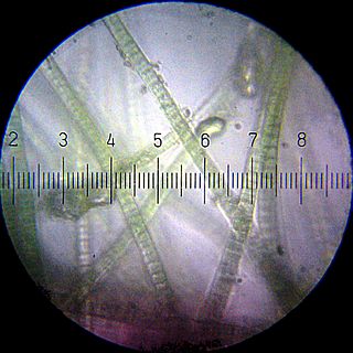
Chloroflexus aurantiacus is a photosynthetic bacterium isolated from hot springs, belonging to the green non-sulfur bacteria. This organism is thermophilic and can grow at temperatures from 35 °C to 70 °C. Chloroflexus aurantiacus can survive in the dark if oxygen is available. When grown in the dark, Chloroflexus aurantiacus has a dark orange color. When grown in sunlight it is dark green. The individual bacteria tend to form filamentous colonies enclosed in sheaths, which are known as trichomes.

<span class="mw-page-title-main">Biological carbon fixation</span> Series of interconnected biochemical reactions

Biological carbon fixation or сarbon assimilation is the process by which inorganic carbon is converted to organic compounds by living organisms. The compounds are then used to store energy and as structure for other biomolecules. Carbon is primarily fixed through photosynthesis, but some organisms use a process called chemosynthesis in the absence of sunlight.

Chlorophyll <i>a</i> Chemical compound

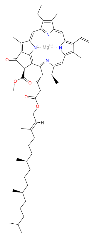
Chlorophyll a is a specific form of chlorophyll used in oxygenic photosynthesis. It absorbs most energy from wavelengths of violet-blue and orange-red light, and it is a poor absorber of green and near-green portions of the spectrum. Chlorophyll does not reflect light but chlorophyll-containing tissues appear green because green light is diffusively reflected by structures like cell walls. This photosynthetic pigment is essential for photosynthesis in eukaryotes, cyanobacteria and prochlorophytes because of its role as primary electron donor in the electron transport chain. Chlorophyll a also transfers resonance energy in the antenna complex, ending in the reaction center where specific chlorophylls P680 and P700 are located.

<span class="mw-page-title-main">Purple bacteria</span> Group of phototrophic bacteria

Purple bacteria or purple photosynthetic bacteria are Gram-negative proteobacteria that are phototrophic, capable of producing their own food via photosynthesis. They are pigmented with bacteriochlorophyll a or b, together with various carotenoids, which give them colours ranging between purple, red, brown, and orange. They may be divided into two groups – purple sulfur bacteria and purple non-sulfur bacteria. Purple bacteria are anoxygenic phototrophs widely spread in nature, but especially in aquatic environments, where there are anoxic conditions that favor the synthesis of their pigments.

<span class="mw-page-title-main">Chromatiaceae</span> Family of purple sulfur bacteria

The Chromatiaceae are one of the two families of purple sulfur bacteria, together with the Ectothiorhodospiraceae. They belong to the order Chromatiales of the class Gammaproteobacteria, which is composed by unicellular Gram-negative organisms. Most of the species are photolithoautotrophs and conduct an anoxygenic photosynthesis, but there are also representatives capable of growing under dark and/or microaerobic conditions as either chemolithoautotrophs or chemoorganoheterotrophs.

<span class="mw-page-title-main">Phototroph</span> Organism using energy from light in metabolic processes

Phototrophs are organisms that carry out photon capture to produce complex organic compounds and acquire energy. They use the energy from light to carry out various cellular metabolic processes. It is a common misconception that phototrophs are obligatorily photosynthetic. Many, but not all, phototrophs often photosynthesize: they anabolically convert carbon dioxide into organic material to be utilized structurally, functionally, or as a source for later catabolic processes. All phototrophs either use electron transport chains or direct proton pumping to establish an electrochemical gradient which is utilized by ATP synthase, to provide the molecular energy currency for the cell. Phototrophs can be either autotrophs or heterotrophs. If their electron and hydrogen donors are inorganic compounds they can be also called lithotrophs, and so, some photoautotrophs are also called photolithoautotrophs. Examples of phototroph organisms are Rhodobacter capsulatus, Chromatium, and Chlorobium.

Photoheterotrophs are heterotrophic phototrophs—that is, they are organisms that use light for energy, but cannot use carbon dioxide as their sole carbon source. Consequently, they use organic compounds from the environment to satisfy their carbon requirements; these compounds include carbohydrates, fatty acids, and alcohols. Examples of photoheterotrophic organisms include purple non-sulfur bacteria, green non-sulfur bacteria, and heliobacteria. These microorganisms are ubiquitous in aquatic habitats, occupy unique niche-spaces, and contribute to global biogeochemical cycling. Recent research has also indicated that the oriental hornet and some aphids may be able to use light to supplement their energy supply.

<span class="mw-page-title-main">Chlorosome</span>

A chlorosome is a photosynthetic antenna complex found in green sulfur bacteria (GSB) and many green non-sulfur bacteria (GNsB), together known as green bacteria. They differ from other antenna complexes by their large size and lack of protein matrix supporting the photosynthetic pigments. Green sulfur bacteria are a group of organisms that generally live in extremely low-light environments, such as at depths of 100 metres in the Black Sea. The ability to capture light energy and rapidly deliver it to where it needs to go is essential to these bacteria, some of which see only a few photons of light per chlorophyll per day. To achieve this, the bacteria contain chlorosome structures, which contain up to 250,000 chlorophyll molecules. Chlorosomes are ellipsoidal bodies, in GSB their length varies from 100 to 200 nm, width of 50-100 nm and height of 15 – 30 nm, in GNsB the chlorosomes are somewhat smaller.

Microbial metabolism is the means by which a microbe obtains the energy and nutrients it needs to live and reproduce. Microbes use many different types of metabolic strategies and species can often be differentiated from each other based on metabolic characteristics. The specific metabolic properties of a microbe are the major factors in determining that microbe's ecological niche, and often allow for that microbe to be useful in industrial processes or responsible for biogeochemical cycles.

<span class="mw-page-title-main">Fenna–Matthews–Olson complex</span>

The Fenna–Matthews–Olson (FMO) complex is a water-soluble complex and was the first pigment-protein complex (PPC) to be structure analyzed by x-ray spectroscopy. It appears in green sulfur bacteria and mediates the excitation energy transfer from light-harvesting chlorosomes to the membrane-embedded bacterial reaction center (bRC). Its structure is trimeric (C3-symmetry). Each of the three monomers contains eight bacteriochlorophyll a molecules. They are bound to the protein scaffold via chelation of their central magnesium atom either to amino acids of the protein or water-bridged oxygen atoms.

Sulfur is metabolized by all organisms, from bacteria and archaea to plants and animals. Sulfur can have an oxidation state from -2 to +6 and is reduced or oxidized by a diverse range of organisms. The element is present in proteins, sulfate esters of polysaccharides, steroids, phenols, and sulfur-containing coenzymes.

<span class="mw-page-title-main">Light-dependent reactions</span> Photosynthetic reactions

Light-dependent reactions are certain photochemical reactions involved in photosynthesis, the main process by which plants acquire energy. There are two light dependent reactions: the first occurs at photosystem II (PSII) and the second occurs at photosystem I (PSI).

<span class="mw-page-title-main">Anoxygenic photosynthesis</span> Process used by obligate anaerobes

Anoxygenic photosynthesis is a special form of photosynthesis used by some bacteria and archaea, which differs from the better known oxygenic photosynthesis in plants in the reductant used and the byproduct generated.

<span class="mw-page-title-main">Isorenieratene</span> Chemical compound

Isorenieratene /ˌaɪsoʊrəˈnɪərətiːn/ is a carotenoid light harvesting pigment produced exclusively by the genus Chlorobium. Chlorobium are the brown-colored strains of the family of green sulfur bacteria (Chlorobiaceae). Green sulfur bacteria are anaerobic photoautotrophic organisms meaning they perform photosynthesis in the absence of oxygen using hydrogen sulfide in the following reaction:

Rhodovulum sulfidophilum is a gram-negative purple nonsulfur bacteria. The cells are rod-shaped, and range in size from 0.6 to 0.9 μm wide and 0.9 to 2.0 μm long, and have a polar flagella. These cells reproduce asexually by binary fission. This bacterium can grow anaerobically when light is present, or aerobically (chemoheterotrophic) under dark conditions. It contains the photosynthetic pigments bacteriochlorophyll a and of carotenoids.

Chlorobaculum tepidum, previously known as Chlorobium tepidum, is an anaerobic, thermophilic green sulfur bacteria first isolated from New Zealand. Its cells are gram-negative and non-motile rods of variable length. They contain chlorosomes and bacteriochlorophyll a and c.

Chlorobium chlorochromatii, originally known as Chlorobium aggregatum, is a symbiotic green sulfur bacteria that performs anoxygenic photosynthesis and functions as an obligate photoautotroph using reduced sulfur species as electron donors. Chlorobium chlorochromatii can be found in stratified freshwater lakes.

Okenane, the diagenetic end product of okenone, is a biomarker for Chromatiaceae, the purple sulfur bacteria. These anoxygenic phototrophs use light for energy and sulfide as their electron donor and sulfur source. Discovery of okenane in marine sediments implies a past euxinic environment, where water columns were anoxic and sulfidic. This is potentially tremendously important for reconstructing past oceanic conditions, but so far okenane has only been identified in one Paleoproterozoic rock sample from Northern Australia.

<span class="mw-page-title-main">Microbial oxidation of sulfur</span>

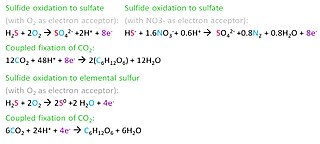
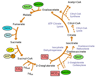
Microbial oxidation of sulfur is the oxidation of sulfur by microorganisms to build their structural components. The oxidation of inorganic compounds is the strategy primarily used by chemolithotrophic microorganisms to obtain energy to survive, grow and reproduce. Some inorganic forms of reduced sulfur, mainly sulfide (H2S/HS) and elemental sulfur (S0), can be oxidized by chemolithotrophic sulfur-oxidizing prokaryotes, usually coupled to the reduction of oxygen (O2) or nitrate (NO3). Anaerobic sulfur oxidizers include photolithoautotrophs that obtain their energy from sunlight, hydrogen from sulfide, and carbon from carbon dioxide (CO2).

References

  1. Oren A, Garrity GM (2021). "Valid publication of the names of forty-two phyla of prokaryotes". Int J Syst Evol Microbiol. 71 (10): 5056. doi: 10.1099/ijsem.0.005056 . PMID   34694987. S2CID   239887308.
  2. Garrity GM, Holt JG. (2001). "Phylum BXI. Chlorobi phy. nov.". In Boone DR, Castenholz RW, Garrity GM. (eds.). Bergey's Manual of Systematic Bacteriology. Vol. 1 (The Archaea and the deeply branching and phototrophic Bacteria) (2nd ed.). New York, NY: Springer–Verlag. pp. 601–623.
  3. Gibbons NE, Murray RGE. (1978). "Proposals Concerning the Higher Taxa of Bacteria". International Journal of Systematic Bacteriology. 28: 1–6. doi: 10.1099/00207713-28-1-1 .
  4. "Phylum Chlorobiota". List of Prokaryotic Names with Standing in Nomenclature. 25907. Retrieved 22 August 2023.
  5. 1 2 Bryant DA, Frigaard NU (November 2006). "Prokaryotic photosynthesis and phototrophy illuminated". Trends in Microbiology. 14 (11): 488–96. doi:10.1016/j.tim.2006.09.001. PMID   16997562.
  6. Green BR (2003). Light-Harvesting Antennas in Photosynthesis. p. 8. ISBN   0792363353.
  7. 1 2 3 4 Kushkevych, Ivan; Procházka, Jiří; Gajdács, Márió; Rittmann, Simon K.-M. R.; Vítězová, Monika (2021-06-15). "Molecular Physiology of Anaerobic Phototrophic Purple and Green Sulfur Bacteria". International Journal of Molecular Sciences. 22 (12): 6398. doi: 10.3390/ijms22126398 . ISSN   1422-0067. PMC   8232776 . PMID   34203823.
  8. 1 2 Sakurai H, Ogawa T, Shiga M, Inoue K (June 2010). "Inorganic sulfur oxidizing system in green sulfur bacteria". Photosynthesis Research. 104 (2–3): 163–76. doi:10.1007/s11120-010-9531-2. PMID   20143161. S2CID   1091791.
  9. 1 2 3 4 5 6 7 8 Tang KH, Blankenship RE (November 2010). "Both forward and reverse TCA cycles operate in green sulfur bacteria". The Journal of Biological Chemistry. 285 (46): 35848–54. doi: 10.1074/jbc.M110.157834 . PMC   2975208 . PMID   20650900.
  10. Wahlund, Thomas (1993). "Nitrogen Fixation by the Thermophilic Green Sulfur Bacterium Chlorobium tepidum". Journal of Bacteriology. 175 (2): 474–478. doi:10.1128/jb.175.2.474-478.1993. PMC   196162 . PMID   8093448.
  11. Feng, Xueyang; Tang, Kuo-Hsiang; Blankenship, Robert E.; Tang, Yinjie J. (2010-12-10). "Metabolic Flux Analysis of the Mixotrophic Metabolisms in the Green Sulfur Bacterium Chlorobaculum tepidum*". Journal of Biological Chemistry. 285 (50): 39544–39550. doi: 10.1074/jbc.M110.162958 . ISSN   0021-9258. PMC   2998096 . PMID   20937805.
  12. "Green Sulfur Bacteria - an overview | ScienceDirect Topics". www.sciencedirect.com. Retrieved 2022-04-22.
  13. John Wiley & Sons, Ltd, ed. (2001-05-30). eLS (1 ed.). Wiley. doi:10.1002/9780470015902.a0000458.pub2. ISBN   978-0-470-01617-6. S2CID   82067054.
  14. Marschall E, Jogler M, Hessge U, Overmann J (May 2010). "Large-scale distribution and activity patterns of an extremely low-light-adapted population of green sulfur bacteria in the Black Sea". Environmental Microbiology. 12 (5): 1348–62. doi:10.1111/j.1462-2920.2010.02178.x. PMID   20236170.
  15. Beatty JT, Overmann J, Lince MT, Manske AK, Lang AS, Blankenship RE, Van Dover CL, Martinson TA, Plumley FG (June 2005). "An obligately photosynthetic bacterial anaerobe from a deep-sea hydrothermal vent". Proceedings of the National Academy of Sciences of the United States of America. 102 (26): 9306–10. Bibcode:2005PNAS..102.9306B. doi: 10.1073/pnas.0503674102 . PMC   1166624 . PMID   15967984.
  16. Yang, Shan-Hua; Lee, Sonny T. M.; Huang, Chang-Rung; Tseng, Ching-Hung; Chiang, Pei-Wen; Chen, Chung-Pin; Chen, Hsing-Ju; Tang, Sen-Lin (2016-02-26). "Prevalence of potential nitrogen-fixing, green sulfur bacteria in the skeleton of reef-building coral Isopora palifera". Limnology and Oceanography. 61 (3): 1078–1086. Bibcode:2016LimOc..61.1078Y. doi: 10.1002/lno.10277 . ISSN   0024-3590. S2CID   87463811.
  17. Cai, Lin; Zhou, Guowei; Tian, Ren-Mao; Tong, Haoya; Zhang, Weipeng; Sun, Jin; Ding, Wei; Wong, Yue Him; Xie, James Y.; Qiu, Jian-Wen; Liu, Sheng (2017-08-24). "Metagenomic analysis reveals a green sulfur bacterium as a potential coral symbiont". Scientific Reports. 7 (1): 9320. Bibcode:2017NatSR...7.9320C. doi:10.1038/s41598-017-09032-4. ISSN   2045-2322. PMC   5571212 . PMID   28839161.
  18. "The LTP" . Retrieved 20 November 2023.
  19. "LTP_all tree in newick format" . Retrieved 20 November 2023.
  20. "LTP_08_2023 Release Notes" (PDF). Retrieved 20 November 2023.
  21. "GTDB release 08-RS214". Genome Taxonomy Database . Retrieved 10 May 2023.
  22. "bac120_r214.sp_label". Genome Taxonomy Database . Retrieved 10 May 2023.
  23. "Taxon History". Genome Taxonomy Database . Retrieved 10 May 2023.
  24. 1 2 3 4 5 Bryantseva, Irina A.; Tarasov, Alexey L.; Kostrikina, Nadezhda A.; Gaisin, Vasil A.; Grouzdev, Denis S.; Gorlenko, Vladimir M. (2019-12-01). "Prosthecochloris marina sp. nov., a new green sulfur bacterium from the coastal zone of the South China Sea". Archives of Microbiology. 201 (10): 1399–1404. doi:10.1007/s00203-019-01707-y. ISSN   1432-072X. PMID   31338544. S2CID   198190182.
  25. 1 2 Hauska G, Schoedl T, Remigy H, Tsiotis G (October 2001). "The reaction center of green sulfur bacteria(1)". Biochimica et Biophysica Acta. 1507 (1–3): 260–77. doi: 10.1016/S0005-2728(01)00200-6 . PMID   11687219.
  26. 1 2 Ligrone, Roberto (2019). "Moving to the Light: The Evolution of Photosynthesis". In Roberto Ligrone (ed.). Biological Innovations that Built the World: A Four-billion-year Journey through Life and Earth History. Cham: Springer International Publishing. pp. 99–127. doi:10.1007/978-3-030-16057-9_4. ISBN   978-3-030-16057-9. S2CID   189992218 . Retrieved 2021-01-29.
  27. 1 2 3 4 Frigaard, Niels-Ulrik; Dahl, Christiane (2008-01-01), Poole, Robert K. (ed.), "Sulfur Metabolism in Phototrophic Sulfur Bacteria", Advances in Microbial Physiology, vol. 54, Academic Press, pp. 103–200, retrieved 2022-04-22
  28. van Gemerden, Hans (1986-10-01). "Production of elemental sulfur by green and purple sulfur bacteria". Archives of Microbiology. 146 (1): 52–56. doi:10.1007/BF00690158. ISSN   1432-072X. S2CID   30812886.
  29. 1 2 Gregersen, Lea; Bryant, Donald; Frigaard, Niels-Ulrik (2011). "Mechanisms and Evolution of Oxidative Sulfur Metabolism in Green Sulfur Bacteria". Frontiers in Microbiology. 2: 116. doi: 10.3389/fmicb.2011.00116 . ISSN   1664-302X. PMC   3153061 . PMID   21833341.
  30. 1 2 Bar-Even, Arren; Noor, Elad; Milo, Ron (2012). "A survey of carbon fixation pathways through a quantitative lens". Journal of Experimental Botany. 63 (6): 2325–2342. doi: 10.1093/jxb/err417 . ISSN   1460-2431. PMID   22200662.
  31. 1 2 3 Frigaard, Niels-Ulrik; Chew, Aline Gomez Maqueo; Li, Hui; Maresca, Julia A.; Bryant, Donald A. (2003). "Chlorobium Tepidum : Insights into the Structure, Physiology, and Metabolism of a Green Sulfur Bacterium Derived from the Complete Genome Sequence". Photosynthesis Research. 78 (2): 93–117. doi:10.1023/B:PRES.0000004310.96189.b4. ISSN   0166-8595. PMID   16245042. S2CID   30218833.
  32. 1 2 3 4 5 Feng, Xueyang; Tang, Kuo-Hsiang; Blankenship, Robert E.; Tang, Yinjie J. (2010-12-10). "Metabolic Flux Analysis of the Mixotrophic Metabolisms in the Green Sulfur Bacterium Chlorobaculum tepidum*". Journal of Biological Chemistry. 285 (50): 39544–39550. doi: 10.1074/jbc.M110.162958 . ISSN   0021-9258. PMC   2998096 . PMID   20937805.
  33. Madigan, Michael T. (1995). "Microbiology of Nitrogen Fixation by Anoxygenic Photosynthetic Bacteria". In Robert E. Blankenship; Michael T. Madigan; Carl E. Bauer (eds.). Anoxygenic Photosynthetic Bacteria. Advances in Photosynthesis and Respiration. Vol. 2. Dordrecht: Springer Netherlands. pp. 915–928. doi:10.1007/0-306-47954-0_42. ISBN   978-0-306-47954-0.
  34. Mus, Florence; Colman, Daniel R.; Peters, John W.; Boyd, Eric S. (2019-08-20). "Geobiological feedbacks, oxygen, and the evolution of nitrogenase". Free Radical Biology and Medicine. Early Life on Earth and Oxidative Stress. 140: 250–259. doi: 10.1016/j.freeradbiomed.2019.01.050 . ISSN   0891-5849. PMID   30735835. S2CID   73433517.
  35. 1 2 Madigan, Michael T. (1995), Blankenship, Robert E.; Madigan, Michael T.; Bauer, Carl E. (eds.), "Microbiology of Nitrogen Fixation by Anoxygenic Photosynthetic Bacteria", Anoxygenic Photosynthetic Bacteria, Advances in Photosynthesis and Respiration, vol. 2, Dordrecht: Springer Netherlands, pp. 915–928, doi:10.1007/0-306-47954-0_42, ISBN   978-0-306-47954-0 , retrieved 2022-05-01
  36. Yang, Shan-Hua; Lee, Sonny T. M.; Huang, Chang-Rung; Tseng, Ching-Hung; Chiang, Pei-Wen; Chen, Chung-Pin; Chen, Hsing-Ju; Tang, Sen-Lin (May 2016). "Prevalence of potential nitrogen-fixing, green sulfur bacteria in the skeleton of reef-building coral Isopora palifera: Endolithic bacteria in coral skeletons". Limnology and Oceanography. 61 (3): 1078–1086. Bibcode:2016LimOc..61.1078Y. doi: 10.1002/lno.10277 . S2CID   87463811.