Chlorosome

Last updated
Schematic of the chlorosome (rod hypothesis) Chlorosome by Hartmann.PNG
Schematic of the chlorosome (rod hypothesis)
Bacteriochlorophyll c-binding protein
Identifiers
SymbolBac_chlorC
Pfam PF02043
InterPro IPR001470
CATH 2k37
Available protein structures:
Pfam   structures / ECOD  
PDB RCSB PDB; PDBe; PDBj
PDBsum structure summary

A chlorosome is a photosynthetic antenna complex found in green sulfur bacteria (GSB) and many green non-sulfur bacteria (GNsB), together known as green bacteria. [2] They differ from other antenna complexes by their large size and lack of protein matrix supporting the photosynthetic pigments. Green sulfur bacteria are a group of organisms that generally live in extremely low-light environments, such as at depths of 100 metres in the Black Sea. The ability to capture light energy and rapidly deliver it to where it needs to go is essential to these bacteria, some of which see only a few photons of light per chlorophyll per day. To achieve this, the bacteria contain chlorosome structures, which contain up to 250,000 chlorophyll molecules. Chlorosomes are ellipsoidal bodies, in GSB their length varies from 100 to 200 nm, width of 50-100 nm and height of 15 – 30 nm, [3] in GNsB the chlorosomes are somewhat smaller.

Contents

Chlorosomes are a type of chromatophores that are found in photosynthetic bacteria (e.g. purple bacteria).

Structure

Chlorosome shape can vary between species, with some species containing ellipsoidal shaped chlorosomes and others containing conical or irregular shaped chlorosomes. [4] Inside green sulfur bacteria, the chlorosomes are attached to type-I reaction centers in the cell membrane via FMO-proteins and a chlorosome baseplate composed of CsmA proteins. [5] Filamentous anoxygenic phototrophs of the phylum Chloroflexota lack the FMO complex, but instead use a protein complex called B808-866. Unlike the FMO proteins in green sulfur bacteria, B808-866 proteins are embedded in the cytoplasmic membrane and surround type-II reaction centers, providing the link between the reaction centers and the baseplate. [6]

The composition of the chlorosomes is mostly bacteriochlorophyll (BChl) with small amounts of carotenoids and quinones surrounded by a galactolipid monolayer. [5] In Chlorobi, chlorosome monolayers can contain up to eleven different proteins. The proteins of Chlorobi are the ones currently best understood in terms of structure and function. These proteins are named CsmA through CsmF, CsmH through CsmK, and CsmX. Other Csm proteins with different letter suffixes can be found in Chloroflexota and Ca. "Chloracidobacterium". [5]

Within the chlorosome, the thousands of BChl pigment molecules have the ability to self assemble with each other, meaning they do not interact with protein scaffolding complexes for assembly. [5] These pigments self assemble in lamellar structures about 10-30 nm wide. [4]

Organization of the light harvesting pigments

Bacteriochlorophyll and carotenoids are two molecules responsible for harvesting light energy. Current models of the organization of bacteriochlorophyll and carotenoids (the main constituents) inside the chlorosomes have put them in a lamellar organization, where the long farnesol tails of the bacteriochlorophyll intermix with carotenoids and each other, forming a structure resembling a lipid multilayer. [7]

Recently, another study has determined the organization of the bacteriochlorophyll molecules in green sulfur bacteria. [8] Because they have been so difficult to study, the chlorosomes in green sulfur bacteria are the last class of light-harvesting complexes to be characterized structurally by scientists. Each individual chlorosome has a unique organization and this variability in composition had prevented scientists from using X-ray crystallography to characterize the internal structure. To get around this problem, the team used a combination of different experimental approaches. Genetic techniques to create a mutant bacterium with a more regular internal structure, cryo-electron microscopy to identify the larger distance constraints for the chlorosome, solid-state nuclear magnetic resonance (NMR) spectroscopy to determine the structure of the chlorosome's component chlorophyll molecules, and modeling to bring together all of the pieces and create a final picture of the chlorosome.

To create the mutant, three genes were inactivated that green sulfur bacteria acquired late in their evolution. In this way it was possible to go backward in evolutionary time to an intermediate state with much less variable and better ordered chlorosome organelles than the wild-type. The chlorosomes were isolated from the mutant and the wild-type forms of the bacteria. Cryo-electron microscopy was used to take pictures of the chlorosomes. The images reveal that the chlorophyll molecules inside chlorosomes have a nanotube shape. The team then used MAS NMR spectroscopy to resolve the microscopic arrangement of chlorophyll inside the chlorosome. With distance constraints and DFT ring current analyses, the organization was found to consist of unique syn-anti monomer stacking. The combination of NMR, cryo-electron microscopy and modeling enabled the scientists to determine that the chlorophyll molecules in green sulfur bacteria are arranged in helices. In the mutant bacteria, the chlorophyll molecules are positioned at a nearly 90-degree angle in relation to the long axis of the nanotubes, whereas the angle is less steep in the wild-type organism. The structural framework can accommodate disorder to improve the biological light harvesting function, which implies that a less ordered structure has a better performance.

An alternative energy source

The interactions that lead to the assembly of the chlorophylls in chlorosomes are rather simple and the results may one day be used to build artificial photosynthetic systems that convert solar energy to electricity or biofuel.

List of bacterial taxa containing chlorosomes

List adapted from, [9] Figure 1.

Related Research Articles

<span class="mw-page-title-main">Photosynthesis</span> Biological process to convert light into chemical energy

Photosynthesis is a biological process used by many organisms to convert light energy into chemical energy, which is stored in organic compounds that can later be metabolized through cellular respiration to fuel the organism's activities. The term usually refers to oxygenic photosynthesis, where oxygen is produced as a byproduct and some of the chemical energy produced is stored in carbohydrate molecules such as sugars, starch, glycogen, and cellulose, which are synthesized from an endergonic reaction of carbon dioxide with water. Organisms that perform photosynthesis are called photoautotrophs; most plants, algae, and cyanobacteria are photoautotrophs. Photosynthesis is largely responsible for producing and maintaining the oxygen content of the Earth's atmosphere, and it supplies most of the biological energy necessary for complex life on Earth.

<span class="mw-page-title-main">Green sulfur bacteria</span> Family of bacteria

The green sulfur bacteria are a phylum, Chlorobiota, of obligately anaerobic photoautotrophic bacteria that metabolize sulfur.

<i>Chloroflexus aurantiacus</i> Species of bacterium

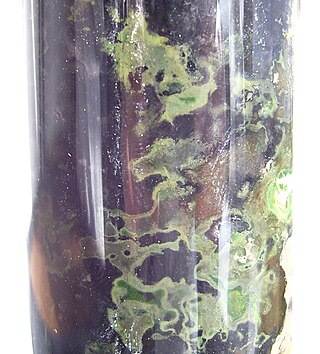
Chloroflexus aurantiacus is a photosynthetic bacterium isolated from hot springs, belonging to the green non-sulfur bacteria. This organism is thermophilic and can grow at temperatures from 35 °C to 70 °C. Chloroflexus aurantiacus can survive in the dark if oxygen is available. When grown in the dark, Chloroflexus aurantiacus has a dark orange color. When grown in sunlight it is dark green. The individual bacteria tend to form filamentous colonies enclosed in sheaths, which are known as trichomes.

<span class="mw-page-title-main">Bacteriochlorophyll</span> Chemical compound

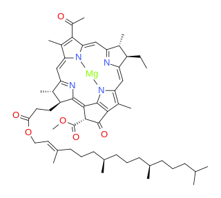
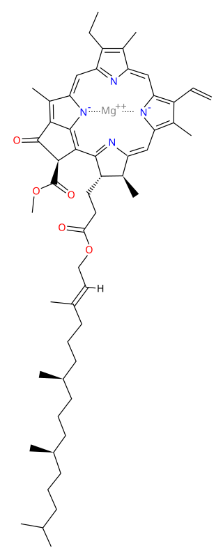
Bacteriochlorophylls (BChl) are photosynthetic pigments that occur in various phototrophic bacteria. They were discovered by C. B. van Niel in 1932. They are related to chlorophylls, which are the primary pigments in plants, algae, and cyanobacteria. Organisms that contain bacteriochlorophyll conduct photosynthesis to sustain their energy requirements, but the process is anoxygenic and does not produce oxygen as a byproduct. They use wavelengths of light not absorbed by plants or cyanobacteria. Replacement of Mg2+ with protons gives bacteriophaeophytin (BPh), the phaeophytin form.

<span class="mw-page-title-main">Photosystem</span> Structural units of protein involved in photosynthesis

Photosystems are functional and structural units of protein complexes involved in photosynthesis. Together they carry out the primary photochemistry of photosynthesis: the absorption of light and the transfer of energy and electrons. Photosystems are found in the thylakoid membranes of plants, algae, and cyanobacteria. These membranes are located inside the chloroplasts of plants and algae, and in the cytoplasmic membrane of photosynthetic bacteria. There are two kinds of photosystems: PSI and PSII.

Chlorophyll <i>a</i> Chemical compound

Chlorophyll a is a specific form of chlorophyll used in oxygenic photosynthesis. It absorbs most energy from wavelengths of violet-blue and orange-red light, and it is a poor absorber of green and near-green portions of the spectrum. Chlorophyll does not reflect light but chlorophyll-containing tissues appear green because green light is diffusively reflected by structures like cell walls. This photosynthetic pigment is essential for photosynthesis in eukaryotes, cyanobacteria and prochlorophytes because of its role as primary electron donor in the electron transport chain. Chlorophyll a also transfers resonance energy in the antenna complex, ending in the reaction center where specific chlorophylls P680 and P700 are located.

<span class="mw-page-title-main">Purple bacteria</span> Group of phototrophic bacteria

Purple bacteria or purple photosynthetic bacteria are Gram-negative proteobacteria that are phototrophic, capable of producing their own food via photosynthesis. They are pigmented with bacteriochlorophyll a or b, together with various carotenoids, which give them colours ranging between purple, red, brown, and orange. They may be divided into two groups – purple sulfur bacteria and purple non-sulfur bacteria. Purple bacteria are anoxygenic phototrophs widely spread in nature, but especially in aquatic environments, where there are anoxic conditions that favor the synthesis of their pigments.

Photoheterotrophs are heterotrophic phototrophs—that is, they are organisms that use light for energy, but cannot use carbon dioxide as their sole carbon source. Consequently, they use organic compounds from the environment to satisfy their carbon requirements; these compounds include carbohydrates, fatty acids, and alcohols. Examples of photoheterotrophic organisms include purple non-sulfur bacteria, green non-sulfur bacteria, and heliobacteria. These microorganisms are ubiquitous in aquatic habitats, occupy unique niche-spaces, and contribute to global biogeochemical cycling. Recent research has also indicated that the oriental hornet and some aphids may be able to use light to supplement their energy supply.

<i>Chlorobium</i> Genus of bacteria

Chlorobium is a genus of green sulfur bacteria. They are photolithotrophic oxidizers of sulfur and most notably utilise a noncyclic electron transport chain to reduce NAD+. Photosynthesis is achieved using a Type 1 Reaction Centre using bacteriochlorophyll (BChl) a. Two photosynthetic antenna complexes aid in light absorption: the Fenna-Matthews-Olson complex, and the chlorosomes which employ mostly BChl c, d, or e. Hydrogen sulfide is used as an electron source and carbon dioxide its carbon source.

A light-harvesting complex consists of a number of chromophores which are complex subunit proteins that may be part of a larger super complex of a photosystem, the functional unit in photosynthesis. It is used by plants and photosynthetic bacteria to collect more of the incoming light than would be captured by the photosynthetic reaction center alone. The light which is captured by the chromophores is capable of exciting molecules from their ground state to a higher energy state, known as the excited state. This excited state does not last very long and is known to be short-lived.

<span class="mw-page-title-main">Fenna–Matthews–Olson complex</span>

The Fenna–Matthews–Olson (FMO) complex is a water-soluble complex and was the first pigment-protein complex (PPC) to be structure analyzed by x-ray spectroscopy. It appears in green sulfur bacteria and mediates the excitation energy transfer from light-harvesting chlorosomes to the membrane-embedded bacterial reaction center (bRC). Its structure is trimeric (C3-symmetry). Each of the three monomers contains eight bacteriochlorophyll a molecules. They are bound to the protein scaffold via chelation of their central magnesium atom either to amino acids of the protein or water-bridged oxygen atoms.

<span class="mw-page-title-main">Antenna complex in purple bacteria</span>

The antenna complex in purple photosynthetic bacteria are protein complexes responsible for the transfer of solar energy to the photosynthetic reaction centre. Purple bacteria, particularly Rhodopseudomonas acidophila of purple non-sulfur bacteria, have been one of the main groups of organisms used to study bacterial antenna complexes so much is known about this group's photosynthetic components. It is one of the many independent types of light-harvesting complex used by various photosynthetic organisms.

<span class="mw-page-title-main">Light-dependent reactions</span> Photosynthetic reactions

Light-dependent reactions refers to certain photochemical reactions that are involved in photosynthesis, the main process by which plants acquire energy. There are two light dependent reactions, the first occurs at photosystem II (PSII) and the second occurs at photosystem I (PSI).

<span class="mw-page-title-main">Anoxygenic photosynthesis</span> Process used by obligate anaerobes

Anoxygenic photosynthesis is a special form of photosynthesis used by some bacteria and archaea, which differs from the better known oxygenic photosynthesis in plants in the reductant used and the byproduct generated.

<span class="mw-page-title-main">Isorenieratene</span> Chemical compound

Isorenieratene /ˌaɪsoʊrəˈnɪərətiːn/ is a carotenoid light harvesting pigment produced exclusively by the genus Chlorobium. Chlorobium are the brown-colored strains of the family of green sulfur bacteria (Chlorobiaceae). Green sulfur bacteria are anaerobic photoautotrophic organisms meaning they perform photosynthesis in the absence of oxygen using hydrogen sulfide in the following reaction:

Chlorobaculum tepidum, previously known as Chlorobium tepidum, is an anaerobic, thermophilic green sulfur bacteria first isolated from New Zealand. Its cells are gram-negative and non-motile rods of variable length. They contain chlorosomes and bacteriochlorophyll a and c.

Roseiflexus castenholzii is a heterotrophic, thermophilic, filamentous anoxygenetic phototroph (FAP) bacterium. This species is in one of two genera of FAPs that lack chlorosomes. R. castenholzii was first isolated from red-colored bacterial mats located Nakabusa hot springs in Japan. Because this organism is a phototroph, it utilizes photosynthesis to fix carbon dioxide and build biomolecules. R. castenholzii has three photosynthetic complexes: light-harvesting only, reaction center only, and light-harvesting with reaction center.

In some forms of photosynthetic bacteria, a chromatophore is a pigmented(coloured), membrane-associated vesicle used to perform photosynthesis. They contain different coloured pigments.

John M. Olson was an American biophysicist and pioneer researcher in photosynthesis, especially light harvesting complex of green sulfur bacteria.

<span class="mw-page-title-main">Photoautotrophism</span> Organisms that use light and inorganic carbon to produce organic materials

Photoautotrophs are organisms that can utilize light energy from sunlight and elements from inorganic compounds to produce organic materials needed to sustain their own metabolism. This biological activity is known as photosynthesis, and examples of such photosynthetic organisms include plants, algae and cyanobacteria.

References

  1. Bryant, D.A. et al. Molecular Contacts for Chlorosome Envelope Proteins Revealed by Cross-Linking Studies with Chlorosomes from Chlorobium tepidum. Biochemistry 45, pp. 9095-9103 (2006)
  2. Shively, J.M.; Cannon, G.C.; Heinhorst, S.; Fuerst, J.A.; Bryant, D.A.; Gantt, E.; Maupin-Furlow, J.A.; Schüler, D.; Pfeifer, F.; Docampo, R.; Dahl, C.; Preiss, J.; Steinbüchel, A.; Federici, B.A. (2009). "Intracellular Structures of Prokaryotes: Inclusions, Compartments and Assemblages". Encyclopedia of Microbiology. pp. 404–424. doi:10.1016/B978-012373944-5.00048-1. ISBN   9780123739445. Chlorosomes are the light-harvesting organelles of green bacteria, which include all known members of the phylum Chlorobi (green sulfur bacteria) and most filamentous anoxygenic phototrophs belonging to the Chloroflexi.
  3. Martinez-Planells A, Arellano JB, Borrego CM, López-Iglesias C, Gich F, Garcia-Gil J (2002). "Determination of the topography and biometry of chlorosomes by atomic force microscopy". Photosynthesis Research. 71 (1–2): 83–90. doi:10.1023/A:1014955614757. PMID   16228503. S2CID   20689329.
  4. 1 2 Oostergetel GT, van Amerongen H, Boekema EJ (June 2010). "The chlorosome: a prototype for efficient light harvesting in photosynthesis". Photosynthesis Research. 104 (2–3): 245–55. doi:10.1007/s11120-010-9533-0. PMC   2882566 . PMID   20130996.
  5. 1 2 3 4 Orf GS, Blankenship RE (October 2013). "Chlorosome antenna complexes from green photosynthetic bacteria". Photosynthesis Research. 116 (2–3): 315–31. doi:10.1007/s11120-013-9869-3. PMID   23761131. S2CID   8613450.
  6. Linnanto JM, Korppi-Tommola JE (September 2013). "Exciton description of chlorosome to baseplate excitation energy transfer in filamentous anoxygenic phototrophs and green sulfur bacteria". The Journal of Physical Chemistry B. 117 (38): 11144–61. doi:10.1021/jp4011394. PMID   23848459.
  7. Psencík J, Ikonen TP, Laurinmäki P, Merckel MC, Butcher SJ, Serimaa RE, Tuma R (August 2004). "Lamellar organization of pigments in chlorosomes, the light harvesting complexes of green photosynthetic bacteria". Biophysical Journal. 87 (2): 1165–72. Bibcode:2004BpJ....87.1165P. doi:10.1529/biophysj.104.040956. PMC   1304455 . PMID   15298919.
  8. Ganapathy S, Oostergetel GT, Wawrzyniak PK, Reus M, Gomez Maqueo Chew A, Buda F, Boekema EJ, Bryant DA, Holzwarth AR, de Groot HJ (May 2009). "Alternating syn-anti bacteriochlorophylls form concentric helical nanotubes in chlorosomes". Proceedings of the National Academy of Sciences of the United States of America. 106 (21): 8525–30. Bibcode:2009PNAS..106.8525G. doi: 10.1073/pnas.0903534106 . PMC   2680731 . PMID   19435848.
  9. Thweatt, Jennifer L.; Canniffe, Daniel P.; Bryant, Donald A. (2019). "Biosynthesis of chlorophylls and bacteriochlorophylls in green bacteria". Advances in Botanical Research. 90: 35–89. doi:10.1016/bs.abr.2019.03.002. ISBN   9780081027523. S2CID   109529231.
  10. Bryant DA, Costas AM, Maresca JA, Chew AG, Klatt CG, Bateson MM, Tallon LJ, Hostetler J, Nelson WC, Heidelberg JF, Ward DM (July 2007). "Candidatus Chloracidobacterium thermophilum: an aerobic phototrophic Acidobacterium". Science. 317 (5837): 523–6. Bibcode:2007Sci...317..523B. doi:10.1126/science.1143236. PMID   17656724. S2CID   20419870.
  11. Saini, MK; Sebastian, A; Shirotori, Y; Soulier, NT; Garcia Costas, AM; Drautz-Moses, DI; Schuster, SC; Albert, I; Haruta, S; Hanada, S; Thiel, V; Tank, M; Bryant, DA (2021). "Genomic and Phenotypic Characterization of Chloracidobacterium Isolates Provides Evidence for Multiple Species". Frontiers in Microbiology. 12: 704168. doi: 10.3389/fmicb.2021.704168 . PMC   8245765 . PMID   34220789.