Related Research Articles

Entamoeba is a genus of Amoebozoa found as internal parasites or commensals of animals. In 1875, Fedor Lösch described the first proven case of amoebic dysentery in St. Petersburg, Russia. He referred to the amoeba he observed microscopically as Amoeba coli; however, it is not clear whether he was using this as a descriptive term or intended it as a formal taxonomic name. The genus Entamoeba was defined by Casagrandi and Barbagallo for the species Entamoeba coli, which is known to be a commensal organism. Lösch's organism was renamed Entamoeba histolytica by Fritz Schaudinn in 1903; he later died, in 1906, from a self-inflicted infection when studying this amoeba. For a time during the first half of the 20th century the entire genus Entamoeba was transferred to Endamoeba, a genus of amoebas infecting invertebrates about which little is known. This move was reversed by the International Commission on Zoological Nomenclature in the late 1950s, and Entamoeba has stayed 'stable' ever since.
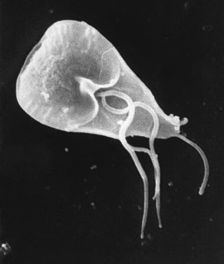
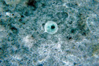
Giardia duodenalis, also known as Giardia intestinalis and Giardia lamblia, is a flagellated parasitic protozoan microorganism of the genus Giardia that colonizes the small intestine, causing a diarrheal condition known as giardiasis. The parasite attaches to the intestinal epithelium by an adhesive disc or sucker, and reproduces via binary fission. Giardiasis does not spread to other parts of the gastrointestinal tract, but remains confined to the lumen of the small intestine. The microorganism has an outer membrane that makes it possible to survive even when outside of its host, and which can render it tolerant to certain disinfectants. Giardia trophozoites are anaerobic, and absorb their nutrients from the intestinal lumen. If the organism is stained, its characteristic pattern resembles the familiar "smiley face" symbol.

Entamoeba histolytica is an anaerobic parasitic amoebozoan, part of the genus Entamoeba. Predominantly infecting humans and other primates causing amoebiasis, E. histolytica is estimated to infect about 35-50 million people worldwide. E. histolytica infection is estimated to kill more than 55,000 people each year. Previously, it was thought that 10% of the world population was infected, but these figures predate the recognition that at least 90% of these infections were due to a second species, E. dispar. Mammals such as dogs and cats can become infected transiently, but are not thought to contribute significantly to transmission.
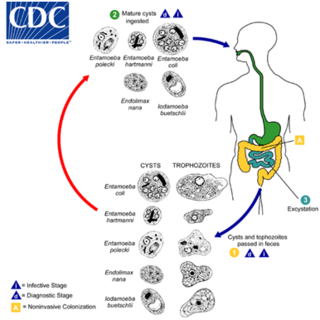
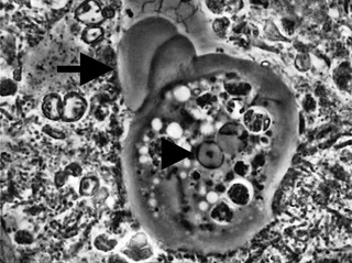
Entamoeba coli is a non-pathogenic species of Entamoeba that frequently exists as a commensal parasite in the human gastrointestinal tract. E. coli is important in medicine because it can be confused during microscopic examination of stained stool specimens with the pathogenic Entamoeba histolytica. This amoeba does not move much by the use of its pseudopod, and creates a "sur place (non-progressive) movement" inside the large intestine. Usually, the amoeba is immobile, and keeps its round shape. This amoeba, in its trophozoite stage, is only visible in fresh, unfixed stool specimens. Sometimes the Entamoeba coli have parasites as well. One is the fungus Sphaerita spp. This fungus lives in the cytoplasm of the E. coli. While this differentiation is typically done by visual examination of the parasitic cysts via light microscopy, new methods using molecular biology techniques have been developed. The scientific name of the amoeba, E. coli, is often mistaken for the bacterium, Escherichia coli. Unlike the bacterium, the amoeba is mostly harmless, and does not cause as many intestinal problems as some strains of the E. coli bacterium. To make the naming of these organisms less confusing, "alternate contractions" are used to name the species for the purpose making the naming easier; for example, using Esch. coli and Ent. coli for the bacterium and amoeba, instead of using E. coli for both.
Amoebozoa is a major taxonomic group containing about 2,400 described species of amoeboid protists, often possessing blunt, fingerlike, lobose pseudopods and tubular mitochondrial cristae. In traditional classification schemes, Amoebozoa is usually ranked as a phylum within either the kingdom Protista or the kingdom Protozoa. In the classification favored by the International Society of Protistologists, it is retained as an unranked "supergroup" within Eukaryota. Molecular genetic analysis supports Amoebozoa as a monophyletic clade. Modern studies of eukaryotic phylogenetic trees identify it as the sister group to Opisthokonta, another major clade which contains both fungi and animals as well as several other clades comprising some 300 species of unicellular eukaryotes. Amoebozoa and Opisthokonta are sometimes grouped together in a high-level taxon, variously named Unikonta, Amorphea or Opimoda.
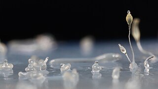
A trophozoite is the activated, feeding stage in the life cycle of certain protozoa such as malaria-causing Plasmodium falciparum and those of the Giardia group. The complementary form of the trophozoite state is the thick-walled cyst form. They are often different from the cyst stage, which is a protective, dormant form of the protozoa. Trophozoites are often found in the host's body fluids and tissues and in many cases, they are the form of the protozoan that causes disease in the host. In the protozoan, Entamoeba histolytica it invades the intestinal mucosa of its host, causing dysentery, which aid in the trophozoites traveling to the liver and leading to the production of hepatic abscesses.

Retortamonas is a genus of flagellated excavates. It is one of only two genera belonging to the family Retortamonadidae along with the genus Chilomastix. The genus parasitizes a large range of hosts including humans. Species within this genus are considered harmless commensals which reside in the intestine of their host. The wide host diversity is a useful factor given that species are distinguished based on their host rather than morphology. This is because all species share similar morphology, which would present challenges when trying to make classifications based on structural anatomy. Although Retortamonas currently includes over 25 known species, it is possible that some defined species are synonymous, given that such overlapping species have been discovered in the past. Further efforts into learning about this genus must be done such as cross-transmission testing as well as biochemical and genetic studies. One of the most well-known species within this genus is Retortamonas intestinalis, a human parasite that lives in the large intestine of humans.
A mitosome is a mitochondrion-related organelle (MRO) found in a variety of parasitic unicellular eukaryotes, such as members of the supergroup Excavata. The mitosome was first discovered in 1999 in Entamoeba histolytica, an intestinal parasite of humans, and mitosomes have also been identified in several species of Microsporidia and in Giardia intestinalis.
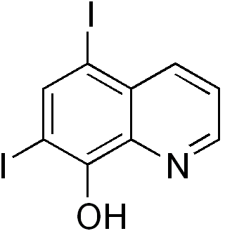
The quinoline derivative diiodohydroxyquinoline (INN), or iodoquinol (USAN), brand name Diodoquin, can be used in the treatment of amoebiasis.
A microbial cyst is a resting or dormant stage of a microorganism, that can be thought of as a state of suspended animation in which the metabolic processes of the cell are slowed and the cell ceases all activities like feeding and locomotion. Many groups of single-celled, microscopic organisms, or microbes, possess the ability to enter this dormant state.
Entamoeba gingivalis is an opportunistic Amoebozoa and is the first amoeba in humans to be described.

Amoebiasis, or amoebic dysentery, is an infection of the intestines caused by a parasitic amoeba Entamoeba histolytica. Amoebiasis can be present with no, mild, or severe symptoms. Symptoms may include lethargy, loss of weight, colonic ulcerations, abdominal pain, diarrhea, or bloody diarrhea. Complications can include inflammation and ulceration of the colon with tissue death or perforation, which may result in peritonitis. Anemia may develop due to prolonged gastric bleeding.

Amoebic brain abscess is an affliction caused by the anaerobic parasitic protist Entamoeba histolytica. It is extremely rare; the first case being reported in 1849. Brain abscesses resulting from Entamoeba histolytica are difficult to diagnose and very few case reports suggest complete recovery even after the administration of appropriate treatment regimen.
Entamoeba polecki is an intestinal parasite of the genus Entamoeba. E. polecki is found primarily in pigs and monkeys and is largely considered non-pathogenic in humans, although there have been some reports regarding symptomatic infections of humans. Prevalence is concentrated in New Guinea, with distribution also recorded in areas of southeast Asia, France, and the United States.
Conosa is a grouping of Amoebozoa. It is subdivided into three groups: Archamoeba, Variosea and Mycetozoa.
Apicomplexans, a group of intracellular parasites, have life cycle stages that allow them to survive the wide variety of environments they are exposed to during their complex life cycle. Each stage in the life cycle of an apicomplexan organism is typified by a cellular variety with a distinct morphology and biochemistry.
Entamoeba moshkovskii is part of the genus Entamoeba. It is found in areas with polluted water sources, and is prevalent in places such as Malaysia, India, and Bangladesh, but more recently has made its way to Turkey, Australia, and North America. This amoeba is said to rarely infect humans, but recently this has changed. It is in question as to whether it is pathogenic or not. Despite some sources stating this is a free living amoeba, various studies worldwide have shown it contains the ability to infect humans, with some cases of pathogenic potential being reported. Some of the symptoms that often occur are diarrhea, weight loss, bloody stool, and abdominal pain. The first known human infection also known as the "Laredo strain" of Entamoebic mushkovskii was in Laredo, Texas in 1991, although it was first described by a man named Tshalaia in 1941 in Moscow, Russia. It is known to affect people of all ages and genders.

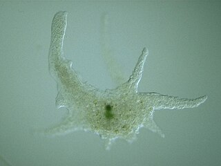
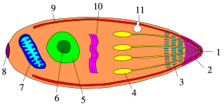
An amoeba, often called an amoeboid, is a type of cell or unicellular organism with the ability to alter its shape, primarily by extending and retracting pseudopods. Amoebae do not form a single taxonomic group; instead, they are found in every major lineage of eukaryotic organisms. Amoeboid cells occur not only among the protozoa, but also in fungi, algae, and animals.
Alok Bhattacharya is an Indian parasitologist, academic and a professor at the School of Life Sciences of the Jawaharlal Nehru University. He chairs the Biotechnology Information System Network (BITSNET) as well as the Life Sciences Expert Committee of FIST program of the Department of Science and Technology (DST). He is an elected fellow of the Indian Academy of Sciences and the Indian National Science Academy and is known for his studies on Entamoeba histolytica and species-specific calcium binding protein and its gene.

Chilomastix is a genus of pyriform excavates within the family Retortamonadidae All species within this genus are flagellated, structured with three flagella pointing anteriorly and a fourth contained within the feeding groove. Chilomastix also lacks Golgi apparatus and mitochondria but does possess a single nucleus. The genus parasitizes a wide range of vertebrate hosts, but is known to be typically non-pathogenic, and is therefore classified as harmless. The life cycle of Chilomastix lacks an intermediate host or vector. Chilomastix has a resistant cyst stage responsible for transmission and a trophozoite stage, which is recognized as the feeding stage. Chilomastix mesnili is one of the more studied species in this genus due to the fact it is a human parasite. Therefore, much of the information on this genus is based on what is known about this one species.
References
- 1 2 3 4 Ehrenkaufer; et al. (26 July 2013). "The genome and transcriptome of the enteric parasite Entamoeba invadens, a model for encystation". Genome Biology. 14 (7): R77. doi: 10.1186/gb-2013-14-7-r77 . PMC 4053983 . PMID 23889909.
- ↑ Sanchez; et al. (September 1994). "Identification of a developmentally regulated transcript expressed during encystation of Entamoeba invadens". Molecular and Biochemical Parasitology. 67 (1): 125–135. doi:10.1016/0166-6851(94)90102-3. PMID 7838173.
- 1 2 Segovia-Gamboa, Norma Cristina (2010). "Entabmoeba Invadens, encystation process and enolase". Experimental Parasitology. 125 (2): 63–69. doi:10.1016/j.exppara.2009.12.019. PMID 20045689.
- 1 2 3 Rivas, A. (2014). "Pathology In Practice". JAVMA. 245 (4): 501–503. doi:10.2460/javma.245.4.389. PMID 25075821.
- ↑ Brewer, Laurie A. (2017). "Analysis of Commercial Entamoeba histolytica Elisa Kits for the Detection of Entamoeba invadens in Reptiles". Journal of Zoo and Wildlife Medicine. 39 (3): 493–495. doi:10.1638/2007-0182.1. JSTOR 20460506. PMID 18817019. S2CID 20666660.
- 1 2 3 4 Welter, Brenda (2017). "Flow cytometric characterization of encystation in Entamoeba invadens". Molecular and Biochemical Parasitology. 218 (October): 23–27. doi:10.1016/j.molbiopara.2017.10.002. PMC 5682210 . PMID 29037797.
- 1 2 Chia; et al. (July 2009). "Entamoeba invadens Myositis in a Common Water Monitor Lizard (Varanus salvator)". Veterinary Pathology. 46 (4): 673–676. doi:10.1354/vp.08-VP-0224-P-CR. PMID 19276058. S2CID 36602674.
- 1 2 Eichinger, Daniel (2001). "Encystation in Parasitic Protozoa". Current Opinion in Microbiology. 4 (4): 421–426. doi:10.1016/S1369-5274(00)00229-0. PMID 11495805.
- ↑ Chatterjee, Anirban (2009). "Evidence for a "wattle and daub" model of the cyst wall of Entamoeba". PLOS Pathogens. 5 (7): e1000498. doi: 10.1371/journal.ppat.1000498 . PMC 2698119 . PMID 19578434.
- ↑ Vázquezdelara-Cisneros, Luis (1984). "Induction of Encystation of Entamoeba invadens by Removal of Glucose from the Culture Medium". The Journal of Parasitology. 70 (5): 629–633. doi:10.2307/3281741. JSTOR 3281741. PMID 6512629.
- ↑ Mitra, B. (2010). "Compounds of the upper gastrointestinal tract induce rapid and efficient excystation of Entamoeba invadens". International Journal for Parasitology. 40 (6): 751–760. doi:10.1016/j.ijpara.2009.11.012. PMC 2881592 . PMID 20018192.
- ↑ Herrera-Martínez, M. (2013). "Actin, RhoA, and Rab11 participation during encystment in entamoeba invadens". BioMed Research International. 2013: 919345. doi: 10.1155/2013/919345 . PMC 3794519 . PMID 24175308.
- ↑ A, Rawat; P, Singh; A, Jyoti; S, Kaushik; Vk, Srivastava (2020-04-30). "Averting Transmission: A Pivotal Target to Manage Amoebiasis". Chemical Biology & Drug Design. 96 (2): 731–744. doi:10.1111/cbdd.13699. PMID 32356312.
- 1 2 Singh N, Bhattacharya A, Bhattacharya S (2013). "Homologous recombination occurs in Entamoeba and is enhanced during growth stress and stage conversion". PLOS ONE. 8 (9): e74465. Bibcode:2013PLoSO...874465S. doi: 10.1371/journal.pone.0074465 . PMC 3787063 . PMID 24098652.
- ↑ Wang, Z. (2003). "Gene discovery in the Entamoeba invadens genome". Molecular and Biochemical Parasitology. 129 (1): 23–31. doi:10.1016/S0166-6851(03)00073-2. PMID 12798503.