Protease

Last updated
Ribbon diagram of a protease (TEV protease) complexed with its peptide substrate in black with catalytic residues in red.(PDB: 1LVB ) TEV protease summary.png
Ribbon diagram of a protease (TEV protease) complexed with its peptide substrate in black with catalytic residues in red.( PDB: 1LVB )

A protease (also called a peptidase, proteinase, or proteolytic enzyme) [1] is an enzyme that catalyzes proteolysis, breaking down proteins into smaller polypeptides or single amino acids, and spurring the formation of new protein products. [2] They do this by cleaving the peptide bonds within proteins by hydrolysis, a reaction where water breaks bonds. Proteases are involved in numerous biological pathways, including digestion of ingested proteins, protein catabolism (breakdown of old proteins), [3] [4] and cell signaling.

Contents

In the absence of functional accelerants, proteolysis would be very slow, taking hundreds of years. [5] Proteases can be found in all forms of life and viruses. They have independently evolved multiple times, and different classes of protease can perform the same reaction by completely different catalytic mechanisms.

Classification

Based on catalytic residue

Proteases can be classified into seven broad groups: [6]

Proteases were first grouped into 84 families according to their evolutionary relationship in 1993, and classified under four catalytic types: serine, cysteine, aspartic, and metallo proteases. [7] The threonine and glutamic proteases were not described until 1995 and 2004 respectively. The mechanism used to cleave a peptide bond involves making an amino acid residue that has the cysteine and threonine (proteases) or a water molecule (aspartic, glutamic and metalloproteases) nucleophilic so that it can attack the peptide carbonyl group. One way to make a nucleophile is by a catalytic triad, where a histidine residue is used to activate serine, cysteine, or threonine as a nucleophile. This is not an evolutionary grouping, however, as the nucleophile types have evolved convergently in different superfamilies, and some superfamilies show divergent evolution to multiple different nucleophiles. Metalloproteases, aspartic, and glutamic proteases utilize their active site residues to activate a water molecule, which then attacks the scissile bond. [8]

Peptide lyases

A seventh catalytic type of proteolytic enzymes, asparagine peptide lyase, was described in 2011. Its proteolytic mechanism is unusual since, rather than hydrolysis, it performs an elimination reaction. [9] During this reaction, the catalytic asparagine forms a cyclic chemical structure that cleaves itself at asparagine residues in proteins under the right conditions. Given its fundamentally different mechanism, its inclusion as a peptidase may be debatable. [9]

Based on evolutionary phylogeny

An up-to-date classification of protease evolutionary superfamilies is found in the MEROPS database. [10] In this database, proteases are classified firstly by 'clan' (superfamily) based on structure, mechanism and catalytic residue order (e.g. the PA clan where P indicates a mixture of nucleophile families). Within each 'clan', proteases are classified into families based on sequence similarity (e.g. the S1 and C3 families within the PA clan). Each family may contain many hundreds of related proteases (e.g. trypsin, elastase, thrombin and streptogrisin within the S1 family).

Currently more than 50 clans are known, each indicating an independent evolutionary origin of proteolysis. [10]

Based on optimal pH

Alternatively, proteases may be classified by the optimal pH in which they are active:

Enzymatic function and mechanism

A comparison of the two hydrolytic mechanisms used for proteolysis. Enzyme is shown in black, substrate protein in red and water in blue. The top panel shows 1-step hydrolysis where the enzyme uses an acid to polarise water, which then hydrolyses the substrate. The bottom panel shows 2-step hydrolysis where a residue within the enzyme is activated to act as a nucleophile (Nu) and attack the substrate. This forms an intermediate where the enzyme is covalently linked to the N-terminal half of the substrate. In a second step, water is activated to hydrolyse this intermediate and complete catalysis. Other enzyme residues (not shown) donate and accept hydrogens and electrostatically stabilise charge build-up along the reaction mechanism. Protease mechanism summary.svg
A comparison of the two hydrolytic mechanisms used for proteolysis. Enzyme is shown in black, substrate protein in red and water in blue. The top panel shows 1-step hydrolysis where the enzyme uses an acid to polarise water, which then hydrolyses the substrate. The bottom panel shows 2-step hydrolysis where a residue within the enzyme is activated to act as a nucleophile (Nu) and attack the substrate. This forms an intermediate where the enzyme is covalently linked to the N-terminal half of the substrate. In a second step, water is activated to hydrolyse this intermediate and complete catalysis. Other enzyme residues (not shown) donate and accept hydrogens and electrostatically stabilise charge build-up along the reaction mechanism.

Proteases are involved in digesting long protein chains into shorter fragments by splitting the peptide bonds that link amino acid residues. Some detach the terminal amino acids from the protein chain (exopeptidases, such as aminopeptidases, carboxypeptidase A); others attack internal peptide bonds of a protein (endopeptidases, such as trypsin, chymotrypsin, pepsin, papain, elastase).

Catalysis

Catalysis is achieved by one of two mechanisms:

Specificity

Proteolysis can be highly promiscuous such that a wide range of protein substrates are hydrolyzed. This is the case for digestive enzymes such as trypsin, which have to be able to cleave the array of proteins ingested into smaller peptide fragments. Promiscuous proteases typically bind to a single amino acid on the substrate and so only have specificity for that residue. For example, trypsin is specific for the sequences ...K\... or ...R\... ('\'=cleavage site). [12]

Conversely some proteases are highly specific and only cleave substrates with a certain sequence. Blood clotting (such as thrombin) and viral polyprotein processing (such as TEV protease) requires this level of specificity in order to achieve precise cleavage events. This is achieved by proteases having a long binding cleft or tunnel with several pockets that bind to specified residues. For example, TEV protease is specific for the sequence ...ENLYFQ\S... ('\'=cleavage site). [13]

Degradation and autolysis

Proteases, being themselves proteins, are cleaved by other protease molecules, sometimes of the same variety. This acts as a method of regulation of protease activity. Some proteases are less active after autolysis (e.g. TEV protease) whilst others are more active (e.g. trypsinogen).

Biodiversity of proteases

Proteases occur in all organisms, from prokaryotes to eukaryotes to viruses. These enzymes are involved in a multitude of physiological reactions from simple digestion of food proteins to highly regulated cascades (e.g., the blood-clotting cascade, the complement system, apoptosis pathways, and the invertebrate prophenoloxidase-activating cascade). Proteases can either break specific peptide bonds (limited proteolysis), depending on the amino acid sequence of a protein, or completely break down a peptide to amino acids (unlimited proteolysis). The activity can be a destructive change (abolishing a protein's function or digesting it to its principal components), it can be an activation of a function, or it can be a signal in a signalling pathway.

Plants

Plant genomes encode hundreds of proteases, largely of unknown function. Those with known function are largely involved in developmental regulation. [14] Plant proteases also play a role in regulation of photosynthesis. [15]

Animals

Proteases are used throughout an organism for various metabolic processes. Acid proteases secreted into the stomach (such as pepsin) and serine proteases present in the duodenum (trypsin and chymotrypsin) enable us to digest the protein in food. Proteases present in blood serum (thrombin, plasmin, Hageman factor, etc.) play an important role in blood-clotting, as well as lysis of the clots, and the correct action of the immune system. Other proteases are present in leukocytes (elastase, cathepsin G) and play several different roles in metabolic control. Some snake venoms are also proteases, such as pit viper haemotoxin and interfere with the victim's blood clotting cascade. Proteases determine the lifetime of other proteins playing important physiological roles like hormones, antibodies, or other enzymes. This is one of the fastest "switching on" and "switching off" regulatory mechanisms in the physiology of an organism.

By a complex cooperative action, proteases can catalyze cascade reactions, which result in rapid and efficient amplification of an organism's response to a physiological signal.

Bacteria

Bacteria secrete proteases to hydrolyse the peptide bonds in proteins and therefore break the proteins down into their constituent amino acids. Bacterial and fungal proteases are particularly important to the global carbon and nitrogen cycles in the recycling of proteins, and such activity tends to be regulated by nutritional signals in these organisms. [16] The net impact of nutritional regulation of protease activity among the thousands of species present in soil can be observed at the overall microbial community level as proteins are broken down in response to carbon, nitrogen, or sulfur limitation. [17]

Bacteria contain proteases responsible for general protein quality control (e.g. the AAA+ proteasome) by degrading unfolded or misfolded proteins.

A secreted bacterial protease may also act as an exotoxin, and be an example of a virulence factor in bacterial pathogenesis (for example, exfoliative toxin). Bacterial exotoxic proteases destroy extracellular structures.

Viruses

The genomes of some viruses encode one massive polyprotein, which needs a protease to cleave this into functional units (e.g. the hepatitis C virus and the picornaviruses). [18] These proteases (e.g. TEV protease) have high specificity and only cleave a very restricted set of substrate sequences. They are therefore a common target for protease inhibitors. [19] [20]

Archaea

Archaea use proteases to regulate various cellular processes from cell-signaling, metabolism, secretion and protein quality control. [21] [22] Only two ATP-dependent proteases are found in archaea: the membrane associated LonB protease and a soluble 20S proteosome complex . [21]

Uses

The field of protease research is enormous. Since 2004, approximately 8000 papers related to this field were published each year. [23] Proteases are used in industry, medicine and as a basic biological research tool. [24] [25]

Digestive proteases are part of many laundry detergents and are also used extensively in the bread industry in bread improver. A variety of proteases are used medically both for their native function (e.g. controlling blood clotting) or for completely artificial functions (e.g. for the targeted degradation of pathogenic proteins). Highly specific proteases such as TEV protease and thrombin are commonly used to cleave fusion proteins and affinity tags in a controlled fashion. Protease-containing plant-solutions called vegetarian rennet have been in use for hundreds of years in Europe and the Middle East for making kosher and halal Cheeses. Vegetarian rennet from Withania coagulans has been in use for thousands of years as a Ayurvedic remedy for digestion and diabetes in the Indian subcontinent. It is also used to make Paneer.

Inhibitors

The activity of proteases is inhibited by protease inhibitors. [26] One example of protease inhibitors is the serpin superfamily. It includes alpha 1-antitrypsin (which protects the body from excessive effects of its own inflammatory proteases), alpha 1-antichymotrypsin (which does likewise), C1-inhibitor (which protects the body from excessive protease-triggered activation of its own complement system), antithrombin (which protects the body from excessive coagulation), plasminogen activator inhibitor-1 (which protects the body from inadequate coagulation by blocking protease-triggered fibrinolysis), and neuroserpin. [27]

Natural protease inhibitors include the family of lipocalin proteins, which play a role in cell regulation and differentiation. Lipophilic ligands, attached to lipocalin proteins, have been found to possess tumor protease inhibiting properties. The natural protease inhibitors are not to be confused with the protease inhibitors used in antiretroviral therapy. Some viruses, with HIV/AIDS among them, depend on proteases in their reproductive cycle. Thus, protease inhibitors are developed as antiviral therapeutic agents.

Other natural protease inhibitors are used as defense mechanisms. Common examples are the trypsin inhibitors found in the seeds of some plants, most notable for humans being soybeans, a major food crop, where they act to discourage predators. Raw soybeans are toxic to many animals, including humans, until the protease inhibitors they contain have been denatured.

See also

Related Research Articles

<span class="mw-page-title-main">Chymotrypsin</span> Digestive enzyme

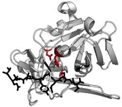
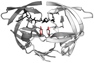
Chymotrypsin (EC 3.4.21.1, chymotrypsins A and B, alpha-chymar ophth, avazyme, chymar, chymotest, enzeon, quimar, quimotrase, alpha-chymar, alpha-chymotrypsin A, alpha-chymotrypsin) is a digestive enzyme component of pancreatic juice acting in the duodenum, where it performs proteolysis, the breakdown of proteins and polypeptides. Chymotrypsin preferentially cleaves peptide amide bonds where the side chain of the amino acid N-terminal to the scissile amide bond (the P1 position) is a large hydrophobic amino acid (tyrosine, tryptophan, and phenylalanine). These amino acids contain an aromatic ring in their side chain that fits into a hydrophobic pocket (the S1 position) of the enzyme. It is activated in the presence of trypsin. The hydrophobic and shape complementarity between the peptide substrate P1 side chain and the enzyme S1 binding cavity accounts for the substrate specificity of this enzyme. Chymotrypsin also hydrolyzes other amide bonds in peptides at slower rates, particularly those containing leucine at the P1 position.

<span class="mw-page-title-main">Proteolysis</span> Breakdown of proteins into smaller polypeptides or amino acids

Proteolysis is the breakdown of proteins into smaller polypeptides or amino acids. Uncatalysed, the hydrolysis of peptide bonds is extremely slow, taking hundreds of years. Proteolysis is typically catalysed by cellular enzymes called proteases, but may also occur by intra-molecular digestion.

<span class="mw-page-title-main">Trypsin</span> Family of digestive enzymes

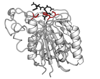
Trypsin is an enzyme in the first section of the small intestine that starts the digestion of protein molecules by cutting long chains of amino acids into smaller pieces. It is a serine protease from the PA clan superfamily, found in the digestive system of many vertebrates, where it hydrolyzes proteins. Trypsin is formed in the small intestine when its proenzyme form, the trypsinogen produced by the pancreas, is activated. Trypsin cuts peptide chains mainly at the carboxyl side of the amino acids lysine or arginine. It is used for numerous biotechnological processes. The process is commonly referred to as trypsinogen proteolysis or trypsinization, and proteins that have been digested/treated with trypsin are said to have been trypsinized. Trypsin was discovered in 1876 by Wilhelm Kühne and was named from the Ancient Greek word for rubbing since it was first isolated by rubbing the pancreas with glycerin.

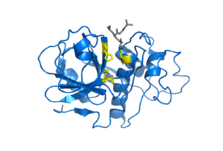
In biology and biochemistry, protease inhibitors, or antiproteases, are molecules that inhibit the function of proteases. Many naturally occurring protease inhibitors are proteins.

<span class="mw-page-title-main">Serine protease</span> Class of enzymes

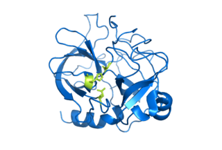
Serine proteases are enzymes that cleave peptide bonds in proteins. Serine serves as the nucleophilic amino acid at the (enzyme's) active site. They are found ubiquitously in both eukaryotes and prokaryotes. Serine proteases fall into two broad categories based on their structure: chymotrypsin-like (trypsin-like) or subtilisin-like.

<span class="mw-page-title-main">Metalloproteinase</span> Type of enzyme

A metalloproteinase, or metalloprotease, is any protease enzyme whose catalytic mechanism involves a metal. An example is ADAM12 which plays a significant role in the fusion of muscle cells during embryo development, in a process known as myogenesis.

<span class="mw-page-title-main">Papain</span> Widely used enzyme extracted from papayas

Papain, also known as papaya proteinase I, is a cysteine protease enzyme present in papaya and mountain papaya. It is the namesake member of the papain-like protease family.

<span class="mw-page-title-main">Catalytic triad</span> Set of three coordinated amino acids

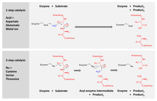
A catalytic triad is a set of three coordinated amino acids that can be found in the active site of some enzymes. Catalytic triads are most commonly found in hydrolase and transferase enzymes. An acid-base-nucleophile triad is a common motif for generating a nucleophilic residue for covalent catalysis. The residues form a charge-relay network to polarise and activate the nucleophile, which attacks the substrate, forming a covalent intermediate which is then hydrolysed to release the product and regenerate free enzyme. The nucleophile is most commonly a serine or cysteine amino acid, but occasionally threonine or even selenocysteine. The 3D structure of the enzyme brings together the triad residues in a precise orientation, even though they may be far apart in the sequence.

<span class="mw-page-title-main">Cysteine protease</span> Class of enzymes

Cysteine proteases, also known as thiol proteases, are hydrolase enzymes that degrade proteins. These proteases share a common catalytic mechanism that involves a nucleophilic cysteine thiol in a catalytic triad or dyad.

In molecular biology, the Signal Peptide Peptidase (SPP) is a type of protein that specifically cleaves parts of other proteins. It is an intramembrane aspartyl protease with the conserved active site motifs 'YD' and 'GxGD' in adjacent transmembrane domains (TMDs). Its sequences is highly conserved in different vertebrate species. SPP cleaves remnant signal peptides left behind in membrane by the action of signal peptidase and also plays key roles in immune surveillance and the maturation of certain viral proteins.

Protein metabolism denotes the various biochemical processes responsible for the synthesis of proteins and amino acids (anabolism), and the breakdown of proteins by catabolism.

<span class="mw-page-title-main">Aspartic protease</span>

Aspartic proteases are a catalytic type of protease enzymes that use an activated water molecule bound to one or more aspartate residues for catalysis of their peptide substrates. In general, they have two highly conserved aspartates in the active site and are optimally active at acidic pH. Nearly all known aspartyl proteases are inhibited by pepstatin.

<span class="mw-page-title-main">Subtilase</span>

Subtilases are a family of subtilisin-like serine proteases. They appear to have independently and convergently evolved an Asp/Ser/His catalytic triad, like in the trypsin serine proteases. The structure of proteins in this family shows that they have an alpha/beta fold containing a 7-stranded parallel beta sheet.

<span class="mw-page-title-main">Threonine protease</span> Class of enzymes

Threonine proteases are a family of proteolytic enzymes harbouring a threonine (Thr) residue within the active site. The prototype members of this class of enzymes are the catalytic subunits of the proteasome, however, the acyltransferases convergently evolved the same active site geometry and mechanism.

<span class="mw-page-title-main">Scytalidopepsin B</span>

Scytalidocarboxyl peptidase B, also known as Scytalidoglutamic peptidase and Scytalidopepsin B is a proteolytic enzyme. It was previously thought to be an aspartic protease, but determination of its molecular structure showed it to belong a novel group of proteases, glutamic protease.

<span class="mw-page-title-main">PA clan of proteases</span>

The PA clan is the largest group of proteases with common ancestry as identified by structural homology. Members have a chymotrypsin-like fold and similar proteolysis mechanisms but can have identity of <10%. The clan contains both cysteine and serine proteases. PA clan proteases can be found in plants, animals, fungi, eubacteria, archaea and viruses.

<span class="mw-page-title-main">Glutamic protease</span>

Glutamic proteases are a group of proteolytic enzymes containing a glutamic acid residue within the active site. This type of protease was first described in 2004 and became the sixth catalytic type of protease. Members of this group of protease had been previously assumed to be an aspartate protease, but structural determination showed it to belong to a novel protease family. The first structure of this group of protease was scytalidoglutamic peptidase, the active site of which contains a catalytic dyad, glutamic acid (E) and glutamine (Q), which give rise to the name eqolisin. This group of proteases are found primarily in pathogenic fungi affecting plant and human.

Asparagine peptide lyase are one of the seven groups in which proteases, also termed proteolytic enzymes, peptidases, or proteinases, are classified according to their catalytic residue. The catalytic mechanism of the asparagine peptide lyases involves an asparagine residue acting as nucleophile to perform a nucleophilic elimination reaction, rather than hydrolysis, to catalyse the breaking of a peptide bond.

<span class="mw-page-title-main">Asparagine endopeptidase</span> Class of enzymes

Asparagine endopeptidase is a proteolytic enzyme from C13 peptidase family which hydrolyses a peptide bond using the thiol group of a cysteine residue as a nucleophile. It is also known as asparaginyl endopeptidase, citvac, proteinase B, hemoglobinase, PRSC1 gene product or LGMN, vicilin peptidohydrolase and bean endopeptidase. In humans it is encoded by the LGMN gene.

<span class="mw-page-title-main">Papain-like protease</span> Protein family of cysteine protease enzymes

Papain-like proteases are a large protein family of cysteine protease enzymes that share structural and enzymatic properties with the group's namesake member, papain. They are found in all domains of life. In animals, the group is often known as cysteine cathepsins or, in older literature, lysosomal peptidases. In the MEROPS protease enzyme classification system, papain-like proteases form Clan CA. Papain-like proteases share a common catalytic dyad active site featuring a cysteine amino acid residue that acts as a nucleophile.

References

  1. "Proteolytic enzyme | Description, Types, & Functions | Britannica".
  2. López-Otín C, Bond JS (November 2008). "Proteases: multifunctional enzymes in life and disease". The Journal of Biological Chemistry. 283 (45): 30433–30437. doi: 10.1074/jbc.R800035200 . PMC   2576539 . PMID   18650443.
  3. 1 2 King JV, Liang WG, Scherpelz KP, Schilling AB, Meredith SC, Tang WJ (July 2014). "Molecular basis of substrate recognition and degradation by human presequence protease". Structure. 22 (7): 996–1007. doi:10.1016/j.str.2014.05.003. PMC   4128088 . PMID   24931469.
  4. 1 2 Shen Y, Joachimiak A, Rosner MR, Tang WJ (October 2006). "Structures of human insulin-degrading enzyme reveal a new substrate recognition mechanism". Nature. 443 (7113): 870–874. Bibcode:2006Natur.443..870S. doi:10.1038/nature05143. PMC   3366509 . PMID   17051221.
  5. Radzicka A, Wolfenden R (July 1996). "Rates of Uncatalyzed Peptide Bond Hydrolysis in Neutral Solution and the Transition State Affinities of Proteases". Journal of the American Chemical Society. 118 (26): 6105–6109. doi:10.1021/ja954077c. To assess the relative proficiencies of enzymes that catalyze the hydrolysis of internal and C-terminal peptide bonds [...]
  6. Oda K (January 2012). "New families of carboxyl peptidases: serine-carboxyl peptidases and glutamic peptidases". Journal of Biochemistry. 151 (1): 13–25. doi: 10.1093/jb/mvr129 . PMID   22016395.
  7. Rawlings ND, Barrett AJ (February 1993). "Evolutionary families of peptidases". The Biochemical Journal. 290 (Pt 1): 205–218. doi:10.1042/bj2900205. PMC   1132403 . PMID   8439290.
  8. Sanman, Laura E. (June 2014). "Activity-Based Profiling of Proteases". Annual Review of Biochemistry. 83: 249–273. doi:10.1146/annurev-biochem-060713-035352. PMID   24905783.
  9. 1 2 Rawlings ND, Barrett AJ, Bateman A (November 2011). "Asparagine peptide lyases: a seventh catalytic type of proteolytic enzymes". The Journal of Biological Chemistry. 286 (44): 38321–38328. doi: 10.1074/jbc.M111.260026 . PMC   3207474 . PMID   21832066.
  10. 1 2 Rawlings ND, Barrett AJ, Bateman A (January 2010). "MEROPS: the peptidase database". Nucleic Acids Research. 38 (Database issue): D227–D233. doi:10.1093/nar/gkp971. PMC   2808883 . PMID   19892822.
  11. Mitchell RS, Kumar V, Abbas AK, Fausto N (2007). Robbins Basic Pathology (8th ed.). Philadelphia: Saunders. p. 122. ISBN   978-1-4160-2973-1.
  12. Rodriguez J, Gupta N, Smith RD, Pevzner PA (January 2008). "Does trypsin cut before proline?". Journal of Proteome Research. 7 (1): 300–305. doi:10.1021/pr0705035. PMID   18067249.
  13. Renicke C, Spadaccini R, Taxis C (2013-06-24). "A tobacco etch virus protease with increased substrate tolerance at the P1' position". PLOS ONE. 8 (6): e67915. Bibcode:2013PLoSO...867915R. doi: 10.1371/journal.pone.0067915 . PMC   3691164 . PMID   23826349.
  14. van der Hoorn RA (2008). "Plant proteases: from phenotypes to molecular mechanisms". Annual Review of Plant Biology. 59: 191–223. doi:10.1146/annurev.arplant.59.032607.092835. hdl: 11858/00-001M-0000-0012-37C7-9 . PMID   18257708.
  15. Zelisko A, Jackowski G (October 2004). "Senescence-dependent degradation of Lhcb3 is mediated by a thylakoid membrane-bound protease". Journal of Plant Physiology. 161 (10): 1157–1170. doi:10.1016/j.jplph.2004.01.006. PMID   15535125.
  16. Sims GK (2006). "Nitrogen Starvation Promotes Biodegradation of N-Heterocyclic Compounds in Soil". Soil Biology & Biochemistry. 38 (8): 2478–2480. doi:10.1016/j.soilbio.2006.01.006. Archived from the original on 2021-04-28. Retrieved 2018-12-29.
  17. Sims GK, Wander MM (2002). "Proteolytic activity under nitrogen or sulfur limitation". Appl. Soil Ecol. 568 (3): 1–5. Bibcode:2002AppSE..19..217S. doi:10.1016/S0929-1393(01)00192-5.
  18. Tong L (December 2002). "Viral proteases". Chemical Reviews. 102 (12): 4609–4626. doi:10.1021/cr010184f. PMID   12475203.
  19. Skoreński M, Sieńczyk M (2013). "Viral proteases as targets for drug design". Current Pharmaceutical Design. 19 (6): 1126–1153. doi:10.2174/13816128130613. PMID   23016690.
  20. Kurt Yilmaz N, Swanstrom R, Schiffer CA (July 2016). "Improving Viral Protease Inhibitors to Counter Drug Resistance". Trends in Microbiology. 24 (7): 547–557. doi:10.1016/j.tim.2016.03.010. PMC   4912444 . PMID   27090931.
  21. 1 2 Giménez MI, Cerletti M, De Castro RE (2015). "Archaeal membrane-associated proteases: insights on Haloferax volcanii and other haloarchaea". Frontiers in Microbiology. 6: 39. doi: 10.3389/fmicb.2015.00039 . PMC   4343526 . PMID   25774151.
  22. Maupin-Furlow JA (December 2018). Robinson NP (ed.). "Proteolytic systems of archaea: slicing, dicing, and mincing in the extreme". Emerging Topics in Life Sciences. 2 (4): 561–580. doi:10.1042/ETLS20180025. PMC   7497159 . PMID   32953999.
  23. Barrett AJ, Rawlings ND, Woessnerd JF (2004). Handbook of proteolytic enzymes (2nd ed.). London, UK: Elsevier Academic Press. ISBN   978-0-12-079610-6.
  24. Hooper NM, ed. (2002). Proteases in biology and medicine. London: Portland Press. ISBN   978-1-85578-147-4.
  25. Feijoo-Siota L, Villa TG (28 September 2010). "Native and Biotechnologically Engineered Plant Proteases with Industrial Applications". Food and Bioprocess Technology. 4 (6): 1066–1088. doi:10.1007/s11947-010-0431-4. S2CID   84748291.
  26. Southan C (July 2001). "A genomic perspective on human proteases as drug targets". Drug Discovery Today. 6 (13): 681–688. doi:10.1016/s1359-6446(01)01793-7. PMID   11427378.
  27. Puente XS, López-Otín C (April 2004). "A genomic analysis of rat proteases and protease inhibitors". Genome Research. 14 (4): 609–622. doi:10.1101/gr.1946304. PMC   383305 . PMID   15060002.