Lipocalin

Last updated
1kt6 opm.png
Retinol-binding protein in a calculated membrane-bound state of the protein 1kt6
Identifiers
SymbolLipocalin
Pfam PF00061
Pfam clan CL0116
InterPro IPR000566
PROSITE PDOC00187
SCOP2 1hms / SCOPe / SUPFAM
OPM superfamily 50
OPM protein 1kt6
Available protein structures:
Pfam   structures / ECOD  
PDB RCSB PDB; PDBe; PDBj
PDBsum structure summary
Lipocalin-like domain
PDB 1qwd EBI.jpg
Structure of the Escherichia coli lipocalin. [1]
Identifiers
SymbolLipocalin_2
Pfam PF08212
Pfam clan CL0116
InterPro IPR013208
Available protein structures:
Pfam   structures / ECOD  
PDB RCSB PDB; PDBe; PDBj
PDBsum structure summary

The lipocalins are a family of proteins which transport small hydrophobic molecules such as steroids, bilins, retinoids, and lipids, and most lipocalins are also able to bind to complexed iron (via siderophores [2] or flavonoids [3] ) as well as heme. [4] They share limited regions of sequence homology and a common tertiary structure architecture. [5] [6] [7] [8] [9] This is an eight stranded antiparallel beta barrel with a repeated + 1 topology enclosing an internal ligand binding site. [7] [8]

Contents

These proteins are found in gram negative bacteria, vertebrate cells, and invertebrate cells, and in plants. Lipocalins have been associated with many biological processes, among them immune response, pheromone transport, biological prostaglandin synthesis, retinoid binding, and cancer cell interactions. [10]

Function

Immune response

Lipocalin proteins are important key players of nutritional immunity by withholding and sequestering micronutrients. [11] They are thereby able to regulate inflammatory and detoxification processes caused by immune system activation in mammals. They are known respiratory allergens of mice, cats, dogs, horses, and other animals. Examples of lipocalin proteins involved in immune system responses include alpha-1-microglobulin, alpha-1-acid glycoprotein, and C8gamma. Structural information for many immune system influencing lipocalin proteins is available, while their exact role in biological systems is still somewhat unclear. Lipocalin allergens have been shown to evoke a Th2-deviated immune response, important for allergic sensitization, when applied in their apo-form (with an empty calyx devoid of ligands), whereas the holo-form seemed to exert immune-suppressive properties in vitro. [12]

Pheromone transport

The lipocalin family has been connected with the transport of mammalian pheromones due to easily observable protein-pheromone interactions. Lipocalins are comparatively small in size, and are thus less complicated to study as opposed to large, bulky proteins. They can also bind to various ligands for different biological purposes. Lipocalins have been detected as carrier proteins of important pheromones in the nasal mucus of rodents. Major urinary proteins, a lipocalin subfamily, are found in mouse and rat urine and may act as protein pheromones themselves. [13]

Prostaglandin synthesis

This family of proteins plays a part in the biological system of terminal prostaglandin synthesis.

Retinoid binding

Retinol, (vitamin A), is an important micronutrient that affects eyesight, cell differentiation, immune system function, bone growth, and tumor suppression. Retinol absorption and metabolism depends on lipocalins that act as binding proteins. Retinyl esters (present in meats) and beta-carotene (present in plants) are the two main sources of retinoids in the diet. After intake, they are converted to retinol, successively metabolized, and finally bound to retinol binding proteins (lipocalins) in the blood plasma.

Cancer cell interactions

Because lipocalins are extracellular proteins, their intracellular effects are not obvious, and demand further study. However, lipophilic ligands, present as substituents to the lipocalins, have the ability to enter the cell, where they can act as tumor protease inhibitors. This research suggests another possible route of protein-tumor investigations.

Allergens

Some of the proteins in this family are allergens. Allergies are hypersensitivity reactions of the immune system to specific substances called allergens (such as pollen, stings, drugs, or food) that, in most people, result in no symptoms. A nomenclature system has been established for antigens (allergens) that cause IgE-mediated atopic allergies in humans. [14] This nomenclature system is defined by a designation that is composed of the first three letters of the genus; a space; the first letter of the species name; a space and an Arabic number. In the event that two species names have identical designations, they are discriminated from one another by adding one or more letters (as necessary) to each species designation.

The allergens in this family include allergens with the following designations: Bla g 4, Bos d 2, Bos d 5, Can f 1, Can f 2, Fel d 4, Equ c 1 and Equ c 2.[ citation needed ]

Hormone

LCN2 (Lipocalin 2) acts as bone-derived hormone which crosses the BBB and acts on PVN paraventricular nucleus of hypothalamus in the brain.[ citation needed ]

Structure

Although lipocalins are a broad family of greatly varied proteins, their three-dimensional structure is a unifying characteristic. Lipocalins have an eight-stranded, antiparallel, symmetrical β-barrel fold, which is, in essence, a beta sheet which has been rolled into a cylindrical shape. Inside this barrel is located a ligand binding site, which plays an important role in the lipocalin classification as a transport protein. If lipocalins are genetically engineered in the attempt to modify their binding properties, they are called anticalins.

Family members

The name "lipocalin" has been proposed [5] for this protein family, but cytosolic fatty acid binding proteins are also included. The sequences of most members of the family, the core or kernel lipocalins, are characterised by three short conserved stretches of residues, while others, the outlier lipocalin group, share only one or two of these. [8] [15] Proteins known to belong to this family include alpha-1-microglobulin (protein HC); major urinary proteins; alpha-1-acid glycoprotein (orosomucoid); [16] aphrodisin; apolipoprotein D; beta-lactoglobulin; complement component C8 gamma chain; [17] crustacyanin; [18] epididymal-retinoic acid binding protein (E-RABP); [19] insectacyanin; odorant binding protein (OBP); human pregnancy-associated endometrial alpha-2 globulin (PAEP); probasin (PB), a prostatic protein; prostaglandin D synthase; [20] purpurin; Von Ebner's gland protein (VEGP); [21] and lizard epididymal secretory protein IV (LESP IV). [22]

Human proteins that contain lipocalin domain include:

See also

Related Research Articles

<span class="mw-page-title-main">Siderophore</span> Iron compounds secreted by microorganisms

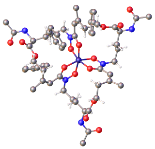
Siderophores (Greek: "iron carrier") are small, high-affinity iron-chelating compounds that are secreted by microorganisms such as bacteria and fungi. They help the organism accumulate iron. Although a widening range of siderophore functions is now being appreciated, siderophores are among the strongest (highest affinity) Fe3+ binding agents known. Phytosiderophores are siderophores produced by plants.

<span class="mw-page-title-main">CD36</span> Mammalian protein found in Homo sapiens

CD36, also known as platelet glycoprotein 4, fatty acid translocase (FAT), scavenger receptor class B member 3 (SCARB3), and glycoproteins 88 (GP88), IIIb (GPIIIB), or IV (GPIV) is a protein that in humans is encoded by the CD36 gene. The CD36 antigen is an integral membrane protein found on the surface of many cell types in vertebrate animals. It imports fatty acids inside cells and is a member of the class B scavenger receptor family of cell surface proteins. CD36 binds many ligands including collagen, thrombospondin, erythrocytes parasitized with Plasmodium falciparum, oxidized low density lipoprotein, native lipoproteins, oxidized phospholipids, and long-chain fatty acids.

<span class="mw-page-title-main">Beta-lactoglobulin</span>

β-Lactoglobulin (BLG) is the major whey protein of cow and sheep's milk, and is also present in many other mammalian species; a notable exception being humans. Its structure, properties and biological role have been reviewed many times. BLG is considered to be a milk allergen.

<span class="mw-page-title-main">Protein contact map</span>

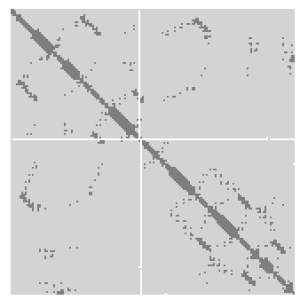
A protein contact map represents the distance between all possible amino acid residue pairs of a three-dimensional protein structure using a binary two-dimensional matrix. For two residues and , the element of the matrix is 1 if the two residues are closer than a predetermined threshold, and 0 otherwise. Various contact definitions have been proposed: The distance between the Cα-Cα atom with threshold 6-12 Å; distance between Cβ-Cβ atoms with threshold 6-12 Å ; and distance between the side-chain centers of mass.

<span class="mw-page-title-main">CD14</span> Mammalian protein found in Homo sapiens

CD14 is a human protein made mostly by macrophages as part of the innate immune system. It helps to detect bacteria in the body by binding lipopolysaccharide (LPS), a pathogen-associated molecular pattern (PAMP).

<span class="mw-page-title-main">Beta barrel</span>

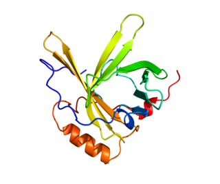
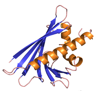
In protein structures, a beta barrel is a beta sheet composed of tandem repeats that twists and coils to form a closed toroidal structure in which the first strand is bonded to the last strand. Beta-strands in many beta-barrels are arranged in an antiparallel fashion. Beta barrel structures are named for resemblance to the barrels used to contain liquids. Most of them are water-soluble proteins and frequently bind hydrophobic ligands in the barrel center, as in lipocalins. Others span cell membranes and are commonly found in porins. Porin-like barrel structures are encoded by as many as 2–3% of the genes in Gram-negative bacteria. It has been shown that more than 600 proteins with various function contain the beta barrel structure.

<span class="mw-page-title-main">Fel d 1</span> Secretoglobin protein

Fel d 1 is a secretoglobin protein complex that, in cats, is encoded by the CH1 and CH2 genes.

<span class="mw-page-title-main">Bilin (biochemistry)</span> Class of chemical compound

Bilins, bilanes or bile pigments are biological pigments formed in many organisms as a metabolic product of certain porphyrins. Bilin was named as a bile pigment of mammals, but can also be found in lower vertebrates, invertebrates, as well as red algae, green plants and cyanobacteria. Bilins can range in color from red, orange, yellow or brown to blue or green.

<span class="mw-page-title-main">Apolipoprotein D</span> Protein-coding gene in the species Homo sapiens

Apolipoprotein D(ApoD) is a protein that in humans is encoded by the APOD gene. Unlike other lipoproteins, which are mainly produced in the liver, apolipoprotein D is mainly produced in the brain and testes. It is a 29 kDa glycoprotein discovered in 1963 as a component of the high-density lipoprotein (HDL) fraction of human plasma. It is the major component of human mammary cyst fluid. The human gene encoding it was cloned in 1986 and the deduced protein sequence revealed that ApoD is a member of the lipocalin family, small hydrophobic molecule transporters. ApoD is 169 amino acids long, including a secretion peptide signal of 20 amino acids. It contains two glycosylation sites (aspargines 45 and 78) and the molecular weight of the mature protein varies from 20 to 32 kDa (see figure 1).

<span class="mw-page-title-main">Retinol binding protein 4</span> Protein-coding gene in the species Homo sapiens

Retinol binding protein 4, also known as RBP4, is a transporter protein for retinol. RBP4 has a molecular weight of approximately 21 kDa and is encoded by the RBP4 gene in humans. It is mainly, though not exclusively, synthesized in the liver and circulates in the bloodstream as a hepatokine bound to retinol in a complex with transthyretin. RBP4 has been a drug target for ophthalmology research due to its role in vision. RBP4 may also be involved in metabolic diseases as suggested by recent studies.

<span class="mw-page-title-main">RAR-related orphan receptor alpha</span> Protein-coding gene in the species Homo sapiens

RAR-related orphan receptor alpha (RORα), also known as NR1F1 is a nuclear receptor that in humans is encoded by the RORA gene. RORα participates in the transcriptional regulation of some genes involved in circadian rhythm. In mice, RORα is essential for development of cerebellum through direct regulation of genes expressed in Purkinje cells. It also plays an essential role in the development of type 2 innate lymphoid cells (ILC2) and mutant animals are ILC2 deficient. In addition, although present in normal numbers, the ILC3 and Th17 cells from RORα deficient mice are defective for cytokine production.

<span class="mw-page-title-main">Retinoic acid receptor alpha</span> Protein-coding gene in the species Homo sapiens

Retinoic acid receptor alpha (RAR-α), also known as NR1B1 is a nuclear receptor that in humans is encoded by the RARA gene.

Pectate lyase is an enzyme involved in the maceration and soft rotting of plant tissue. Pectate lyase is responsible for the eliminative cleavage of pectate, yielding oligosaccharides with 4-deoxy-α-D-mann-4-enuronosyl groups at their non-reducing ends. The protein is maximally expressed late in pollen development. It has been suggested that the pollen expression of pectate lyase genes might relate to a requirement for pectin degradation during pollen tube growth.

Prostaglandin DP<sub>2</sub> receptor Protein-coding gene in the species Homo sapiens

Prostaglandin D2 receptor 2 (DP2 or CRTH2) is a human protein encoded by the PTGDR2 gene and GPR44. DP2 has also been designated as CD294 (cluster of differentiation 294). It is a member of the class of prostaglandin receptors which bind with and respond to various prostaglandins. DP2 along with Prostaglandin DP1 receptor are receptors for prostaglandin D2 (PGD2). Activation of DP2 by PGD2 or other cognate receptor ligands has been associated with certain physiological and pathological responses, particularly those associated with allergy and inflammation, in animal models and certain human diseases.

<span class="mw-page-title-main">Histidine-rich glycoprotein</span> Glycoprotein

Histidine-rich glycoprotein (HRG) is a glycoprotein that in humans is encoded by the HRG gene. The HRG protein is produced in the liver, and it could also be synthesized by monocytes, macrophages, and megakaryocytes. It possesses a multi-domain structure, which makes it capable of binding to numerous ligands and modulating various biological processes including immunity, vascularization and coagulation.

<span class="mw-page-title-main">SIGLEC8</span> Protein-coding gene in the species Homo sapiens

Sialic acid-binding Ig-like lectin 8 is a protein that in humans is encoded by the SIGLEC8 gene. This gene is located on chromosome 19q13.4, about 330 kb downstream of the SIGLEC9 gene. Within the siglec family of transmembrane proteins, Siglec-8 belongs to the CD33-related siglec subfamily, a subfamily that has undergone rapid evolution.

<span class="mw-page-title-main">Plant lipid transfer proteins</span>

Plant lipid transfer proteins, also known as plant LTPs or PLTPs, are a group of highly-conserved proteins of about 7-9kDa found in higher plant tissues. As its name implies, lipid transfer proteins facilitate the shuttling of phospholipids and other fatty acid groups between cell membranes. LTPs are divided into two structurally related subfamilies according to their molecular masses: LTP1s (9 kDa) and LTP2s (7 kDa). Various LTPs bind a wide range of ligands, including fatty acids with a C10–C18 chain length, acyl derivatives of coenzyme A, phospho- and galactolipids, prostaglandin B2, sterols, molecules of organic solvents, and some drugs.

<span class="mw-page-title-main">Major urinary proteins</span> Proteins found in the urine and other secretions of many animals

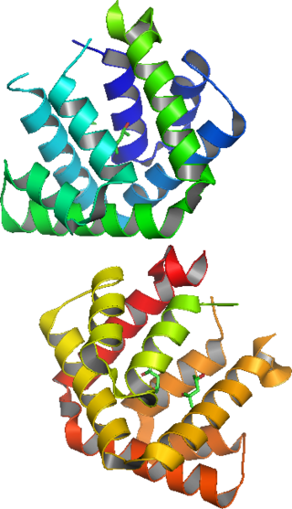
Major urinary proteins (Mups), also known as α2u-globulins, are a subfamily of proteins found in abundance in the urine and other secretions of many animals. Mups provide a small range of identifying information about the donor animal, when detected by the vomeronasal organ of the receiving animal. They belong to a larger family of proteins known as lipocalins. Mups are encoded by a cluster of genes, located adjacent to each other on a single stretch of DNA, that varies greatly in number between species: from at least 21 functional genes in mice to none in humans. Mup proteins form a characteristic glove shape, encompassing a ligand-binding pocket that accommodates specific small organic chemicals.

<span class="mw-page-title-main">Bet v I allergen</span>

Bet v I allergen is a family of protein allergens. Allergies are hypersensitivity reactions of the immune system to specific substances called allergens that, in most people, result in no symptoms.

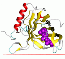
Siderocalin(Scn), lipocalin-2, NGAL, 24p3 is a mammalian lipocalin-type protein that can prevent iron acquisition by pathogenic bacteria by binding siderophores, which are iron-binding chelators made by microorganisms. Iron serves as a key nutrient in host-pathogen interactions, and pathogens can acquire iron from the host organism via synthesis and release siderophores such as enterobactin. Siderocalin is a part of the mammalian defence mechanism and acts as an antibacterial agent. Crystallographic studies of Scn demonstrated that it includes a calyx, a ligand-binding domain that is lined with polar cationic groups. Central to the siderophore/siderocalin recognition mechanism are hybrid electrostatic/cation-pi interactions. To evade the host defences, pathogens evolved to produce structurally varied siderophores that would not be recognized by siderocalin, allowing the bacteria to acquire iron.

References

  1. Campanacci V, Nurizzo D, Spinelli S, Valencia C, Tegoni M, Cambillau C (March 2004). "The crystal structure of the Escherichia coli lipocalin Blc suggests a possible role in phospholipid binding". FEBS Letters. 562 (1–3): 183–188. doi: 10.1016/S0014-5793(04)00199-1 . PMID   15044022. S2CID   26737744.
  2. Goetz DH, Holmes MA, Borregaard N, Bluhm ME, Raymond KN, Strong RK (November 2002). "The neutrophil lipocalin NGAL is a bacteriostatic agent that interferes with siderophore-mediated iron acquisition". Molecular Cell. 10 (5): 1033–1043. doi: 10.1016/s1097-2765(02)00708-6 . PMID   12453412.
  3. Roth-Walter F, Pacios LF, Bianchini R, Jensen-Jarolim E (December 2017). "Linking iron-deficiency with allergy: role of molecular allergens and the microbiome". Metallomics. 9 (12): 1676–1692. doi: 10.1039/c7mt00241f . PMID   29120476.
  4. Matz JM, Drepper B, Blum TB, van Genderen E, Burrell A, Martin P, et al. (July 2020). "A lipocalin mediates unidirectional heme biomineralization in malaria parasites". Proceedings of the National Academy of Sciences of the United States of America. 117 (28): 16546–16556. doi: 10.1073/pnas.2001153117 . PMC   7368307 . PMID   32601225.
  5. 1 2 Pervaiz S, Brew K (September 1987). "Homology and structure-function correlations between alpha 1-acid glycoprotein and serum retinol-binding protein and its relatives". FASEB Journal. 1 (3): 209–214. doi:10.1096/fasebj.1.3.3622999. PMID   3622999. S2CID   12416282.
  6. Igarashi M, Nagata A, Toh H, Urade Y, Hayaishi O (June 1992). "Structural organization of the gene for prostaglandin D synthase in the rat brain". Proceedings of the National Academy of Sciences of the United States of America. 89 (12): 5376–5380. Bibcode:1992PNAS...89.5376I. doi: 10.1073/pnas.89.12.5376 . PMC   49294 . PMID   1608945.
  7. 1 2 Cowan SW, Newcomer ME, Jones TA (1990). "Crystallographic refinement of human serum retinol binding protein at 2A resolution". Proteins. 8 (1): 44–61. doi:10.1002/prot.340080108. PMID   2217163. S2CID   21613341.
  8. 1 2 3 Flower DR, North AC, Attwood TK (May 1993). "Structure and sequence relationships in the lipocalins and related proteins". Protein Science. 2 (5): 753–761. doi:10.1002/pro.5560020507. PMC   2142497 . PMID   7684291.
  9. Godovac-Zimmermann J (February 1988). "The structural motif of beta-lactoglobulin and retinol-binding protein: a basic framework for binding and transport of small hydrophobic molecules?". Trends in Biochemical Sciences. 13 (2): 64–66. doi:10.1016/0968-0004(88)90031-X. PMID   3238752.
  10. Araos P, Amador CA (2022). "Neutrophil gelatinase-associated lipocalin as an immunomodulator in endocrine hypertension". Front Endocrinol (Lausanne). 13: 1006790. doi: 10.3389/fendo.2022.1006790 . PMC   9640732 . PMID   36387895.
  11. Roth-Walter, Franziska (2022). "Iron-Deficiency in Atopic Diseases: Innate Immune Priming by Allergens and Siderophores". Frontiers in Allergy. 3: 859922. doi: 10.3389/falgy.2022.859922 . ISSN   2673-6101. PMC   9234869 . PMID   35769558.
  12. Roth-Walter F, Pacios LF, Gomez-Casado C, Hofstetter G, Roth GA, Singer J, et al. (August 2014). "The major cow milk allergen Bos d 5 manipulates T-helper cells depending on its load with siderophore-bound iron". PLOS ONE. 9 (8): e104803. Bibcode:2014PLoSO...9j4803R. doi: 10.1371/journal.pone.0104803 . PMC   4130594 . PMID   25117976.
  13. Chamero P, Marton TF, Logan DW, Flanagan K, Cruz JR, Saghatelian A, et al. (December 2007). "Identification of protein pheromones that promote aggressive behaviour". Nature. 450 (7171): 899–902. Bibcode:2007Natur.450..899C. doi:10.1038/nature05997. PMID   18064011. S2CID   4398766.
  14. [WHO/IUIS Allergen Nomenclature Subcommittee King T.P., Hoffmann D., Loewenstein H., Marsh D.G., Platts-Mills T.A.E., Thomas W. Bull. World Health Organ. 72:797-806(1994)]
  15. Flower DR, North AC, Attwood TK (October 1991). "Mouse oncogene protein 24p3 is a member of the lipocalin protein family". Biochemical and Biophysical Research Communications. 180 (1): 69–74. doi:10.1016/S0006-291X(05)81256-2. PMID   1834059.
  16. Kremer JM, Wilting J, Janssen LH (March 1988). "Drug binding to human alpha-1-acid glycoprotein in health and disease". Pharmacological Reviews. 40 (1): 1–47. PMID   3064105.
  17. Haefliger JA, Peitsch MC, Jenne DE, Tschopp J (1991). "Structural and functional characterization of complement C8 gamma, a member of the lipocalin protein family". Molecular Immunology. 28 (1–2): 123–131. doi:10.1016/0161-5890(91)90095-2. PMID   1707134.
  18. Keen JN, Caceres I, Eliopoulos EE, Zagalsky PF, Findlay JB (April 1991). "Complete sequence and model for the A2 subunit of the carotenoid pigment complex, crustacyanin". European Journal of Biochemistry. 197 (2): 407–417. doi:10.1111/j.1432-1033.1991.tb15925.x. PMID   2026162.
  19. Newcomer ME (September 1993). "Structure of the epididymal retinoic acid binding protein at 2.1 A resolution". Structure. 1 (1): 7–18. doi: 10.1016/0969-2126(93)90004-Z . PMID   8069623.
  20. Peitsch MC, Boguski MS (October 1991). "The first lipocalin with enzymatic activity". Trends in Biochemical Sciences. 16 (10): 363. doi:10.1016/0968-0004(91)90149-P. PMID   1723819.
  21. Kock K, Ahlers C, Schmale H (May 1994). "Structural organization of the genes for rat von Ebner's gland proteins 1 and 2 reveals their close relationship to lipocalins". European Journal of Biochemistry. 221 (3): 905–916. doi: 10.1111/j.1432-1033.1994.tb18806.x . PMID   7514123.
  22. Morel L, Dufaure JP, Depeiges A (May 1993). "LESP, an androgen-regulated lizard epididymal secretory protein family identified as a new member of the lipocalin superfamily". The Journal of Biological Chemistry. 268 (14): 10274–10281. doi: 10.1016/S0021-9258(18)82200-1 . PMID   8486691.

Further reading

This article incorporates text from the public domain Pfam and InterPro: IPR000566