DD-transpeptidase

Last updated
Serine-type D-Ala-D-Ala carboxypeptidase
DD-Transpeptidase.png
Structure of the streptomyces K15 DD-transpeptidase
Identifiers
EC no. 3.4.16.4
CAS no. 9077-67-2
Databases
IntEnz IntEnz view
BRENDA BRENDA entry
ExPASy NiceZyme view
KEGG KEGG entry
MetaCyc metabolic pathway
PRIAM profile
PDB structures RCSB PDB PDBe PDBsum
Search
PMC articles
PubMed articles
NCBI proteins

DD-transpeptidase (EC 3.4.16.4, DD-peptidase, DD-transpeptidase, DD-carboxypeptidase, D-alanyl-D-alanine carboxypeptidase , D-alanyl-D-alanine-cleaving-peptidase, D-alanine carboxypeptidase, D-alanyl carboxypeptidase, and serine-type D-Ala-D-Ala carboxypeptidase. [1] ) is a bacterial enzyme that catalyzes the transfer of the R-L-αα-D-alanyl moiety of R-L-αα-D-alanyl-D-alanine carbonyl donors to the γ-OH of their active-site serine and from this to a final acceptor. [2] It is involved in bacterial cell wall biosynthesis, namely, the transpeptidation that crosslinks the peptide side chains of peptidoglycan strands. [3]

Contents

The antibiotic penicillin irreversibly binds to and inhibits the activity of the transpeptidase enzyme by forming a highly stable penicilloyl-enzyme intermediate. [4] Because of the interaction between penicillin and transpeptidase, this enzyme is also known as penicillin-binding protein (PBP).

Mechanism

DD-transpeptidase is mechanistically similar to the proteolytic reactions of the trypsin protein family. [5]

DD-transpeptidase catalytic mechanism DD-Transpeptidase mechanism fixed.png
DD-transpeptidase catalytic mechanism

Crosslinking of peptidyl moieties of adjacent glycan strands is a two-step reaction. The first step involves the cleavage of the D-alanyl-D-alanine bond of a peptide unit precursor acting as carbonyl donor, the release of the carboxyl-terminal D-alanine, and the formation of the acyl-enzyme. The second step involves the breakdown of the acyl-enzyme intermediate and the formation of a new peptide bond between the carbonyl of the D-alanyl moiety and the amino group of another peptide unit. [6]

Most discussion of DD-peptidase mechanisms revolves around the catalysts of proton transfer. During formation of the acyl-enzyme intermediate, a proton must be removed from the active site serine hydroxyl group and one must be added to the amine leaving group. A similar proton movement must be facilitated in deacylation. The identity of the general acid and base catalysts involved in these proton transfers has not yet been elucidated. [7] However, the catalytic triad tyrosine, lysine, and serine, as well as serine, lysine, serine have been proposed. [7]

Structure

Transpeptidases are members of the penicilloyl-serine transferase superfamily, which has a signature SxxK conserved motif. [8] With "x" denoting a variable amino acid residue, the transpeptidases of this superfamily show a trend in the form of three motifs: SxxK, SxN (or analogue), and KTG (or analogue). These motifs occur at equivalent places, and are roughly equally spaced, along the polypeptide chain. The folded protein brings these motifs close to each other at the catalytic center between an all-α domain and an α/β domain. [9] [10]

The structure of the streptomyces K15 DD-transpeptidase has been studied, and consists of a single polypeptide chain organized into two domains. One domain contains mainly α-helices, and the second one is of α/β-type. [6] The center of the catalytic cleft is occupied by the Ser35-Thr36-Thr37-Lys38 tetrad, which includes the nucleophilic Ser35 residue at the amino-terminal end of helix α2. One side of the cavity is defined by the Ser96-Gly97-Cys98 loop connecting helices α4 and α5. The Lys213-Thr214-Gly215 triad lies on strand β3 on the opposite side of the cavity. The backbone NH group of the essential Ser35 residue and that of Ser216 downstream from the motif Lys213-Thr214-Gly215 occupy positions that are compatible with the oxyanion hole function required for catalysis. [6]

The enzyme is classified as a DD-transpeptidase because the susceptible peptide bond of the carbonyl donor extends between two carbon atoms with the D-configuration. [6]

Biological Function

All bacteria possess at least one, most often several, monofunctional serine DD-peptidases. [2]

Disease Relevance

The structural similarity between (A) D-Ala-D-Ala terminus of peptidoglycan terminus and (B) penicillins. Transpeptidases misrecognize penicillins for the TPase catalytic reaction. Penicillin vs PG terminus structure.png
The structural similarity between (A) D-Ala-D-Ala terminus of peptidoglycan terminus and (B) penicillins. Transpeptidases misrecognize penicillins for the TPase catalytic reaction.

This enzyme is an excellent drug target because it is essential, is accessible from the periplasm, and has no equivalent in mammalian cells. DD-transpeptidase is the target protein of β-lactam antibiotics (e.g. penicillin) This is because the structure of the β-lactam closely resembles the D-ala-D-ala residue.

β-lactams exert their effect by competitively inactivating the serine DD-transpeptidase catalytic site. Penicillin is a cyclic analogue of the D-Ala-D-Ala terminated carbonyl donors, therefore in the presence of this antibiotic, the reaction stops at the level of the serine ester-linked penicilloyl enzyme. [11] Thus β-lactam antibiotics force these enzymes to behave like penicillin binding proteins. [12]

Kinetically, the interaction between the DD-peptidase and beta-lactams is a three-step reaction:

[12]

Beta-Iactams may form an adduct E-I* of high stability with DD-transpeptidase. The half life of this adduct is around hours, whereas the half-life of the normal reaction is in the order of milliseconds. [8]

The interference with the enzyme processes responsible for cell wall formation results in cellular lysis and death due to the triggering of the autolytic system in the bacteria. [13]

See also

Related Research Articles

<span class="mw-page-title-main">Beta-lactam</span> Family of chemical compounds

A beta-lactam (β-lactam) ring is a four-membered lactam. A lactam is a cyclic amide, and beta-lactams are named so because the nitrogen atom is attached to the β-carbon atom relative to the carbonyl. The simplest β-lactam possible is 2-azetidinone. β-lactams are significant structural units of medicines as manifested in many β-lactam antibiotics Up to 1970, most β-lactam research was concerned with the penicillin and cephalosporin groups, but since then, a wide variety of structures have been described.

<span class="mw-page-title-main">Beta-lactamase</span> Class of enzymes

Beta-lactamases (β-lactamases) are enzymes produced by bacteria that provide multi-resistance to beta-lactam antibiotics such as penicillins, cephalosporins, cephamycins, monobactams and carbapenems (ertapenem), although carbapenems are relatively resistant to beta-lactamase. Beta-lactamase provides antibiotic resistance by breaking the antibiotics' structure. These antibiotics all have a common element in their molecular structure: a four-atom ring known as a beta-lactam (β-lactam) ring. Through hydrolysis, the enzyme lactamase breaks the β-lactam ring open, deactivating the molecule's antibacterial properties.

<span class="mw-page-title-main">Penicillin</span> Group of antibiotics derived from Penicillium fungi

Penicillins are a group of β-lactam antibiotics originally obtained from Penicillium moulds, principally P. chrysogenum and P. rubens. Most penicillins in clinical use are synthesised by P. chrysogenum using deep tank fermentation and then purified. A number of natural penicillins have been discovered, but only two purified compounds are in clinical use: penicillin G and penicillin V. Penicillins were among the first medications to be effective against many bacterial infections caused by staphylococci and streptococci. They are still widely used today for different bacterial infections, though many types of bacteria have developed resistance following extensive use.

Peptidoglycan or murein is a unique large macromolecule, a polysaccharide, consisting of sugars and amino acids that forms a mesh-like peptidoglycan layer (sacculus) that surrounds the bacterial cytoplasmic membrane. The sugar component consists of alternating residues of β-(1,4) linked N-acetylglucosamine (NAG) and N-acetylmuramic acid (NAM). Attached to the N-acetylmuramic acid is an oligopeptide chain made of three to five amino acids. The peptide chain can be cross-linked to the peptide chain of another strand forming the 3D mesh-like layer. Peptidoglycan serves a structural role in the bacterial cell wall, giving structural strength, as well as counteracting the osmotic pressure of the cytoplasm. This repetitive linking results in a dense peptidoglycan layer which is critical for maintaining cell form and withstanding high osmotic pressures, and it is regularly replaced by peptidoglycan production. Peptidoglycan hydrolysis and synthesis are two processes that must occur in order for cells to grow and multiply, a technique carried out in three stages: clipping of current material, insertion of new material, and re-crosslinking of existing material to new material.

<span class="mw-page-title-main">Beta-lactam antibiotics</span> Class of broad-spectrum antibiotics

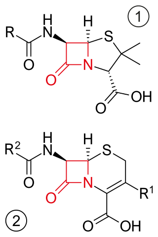
β-lactam antibiotics are antibiotics that contain a beta-lactam ring in their chemical structure. This includes penicillin derivatives (penams), cephalosporins and cephamycins (cephems), monobactams, carbapenems and carbacephems. Most β-lactam antibiotics work by inhibiting cell wall biosynthesis in the bacterial organism and are the most widely used group of antibiotics. Until 2003, when measured by sales, more than half of all commercially available antibiotics in use were β-lactam compounds. The first β-lactam antibiotic discovered, penicillin, was isolated from a strain of Penicillium rubens.

<span class="mw-page-title-main">Methicillin</span> Antibiotic medication

Methicillin (USAN), also known as meticillin (INN), is a narrow-spectrum β-lactam antibiotic of the penicillin class.

<span class="mw-page-title-main">Clavulanic acid</span> Molecule used to overcome antibiotic resistance in bacteria

Clavulanic acid is a β-lactam drug that functions as a mechanism-based β-lactamase inhibitor. While not effective by itself as an antibiotic, when combined with penicillin-group antibiotics, it can overcome antibiotic resistance in bacteria that secrete β-lactamase, which otherwise inactivates most penicillins.

<span class="mw-page-title-main">Acyl carrier protein</span> Cofactor of both fatty acid and polyketide biosynthesis

The acyl carrier protein (ACP) is a cofactor of both fatty acid and polyketide biosynthesis machinery. It is one of the most abundant proteins in cells of E. coli. In both cases, the growing chain is bound to the ACP via a thioester derived from the distal thiol of a 4'-phosphopantetheine moiety.

<span class="mw-page-title-main">Catalytic triad</span> Set of three coordinated amino acids

A catalytic triad is a set of three coordinated amino acids that can be found in the active site of some enzymes. Catalytic triads are most commonly found in hydrolase and transferase enzymes. An acid-base-nucleophile triad is a common motif for generating a nucleophilic residue for covalent catalysis. The residues form a charge-relay network to polarise and activate the nucleophile, which attacks the substrate, forming a covalent intermediate which is then hydrolysed to release the product and regenerate free enzyme. The nucleophile is most commonly a serine or cysteine amino acid, but occasionally threonine or even selenocysteine. The 3D structure of the enzyme brings together the triad residues in a precise orientation, even though they may be far apart in the sequence.

<span class="mw-page-title-main">Penicillin-binding proteins</span> Class of proteins

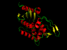
Penicillin-binding proteins (PBPs) are a group of proteins that are characterized by their affinity for and binding of penicillin. They are a normal constituent of many bacteria; the name just reflects the way by which the protein was discovered. All β-lactam antibiotics bind to PBPs, which are essential for bacterial cell wall synthesis. PBPs are members of a subgroup of enzymes called transpeptidases. Specifically, PBPs are DD-transpeptidases.

<span class="mw-page-title-main">Carboxypeptidase</span>

A carboxypeptidase is a protease enzyme that hydrolyzes (cleaves) a peptide bond at the carboxy-terminal (C-terminal) end of a protein or peptide. This is in contrast to an aminopeptidases, which cleave peptide bonds at the N-terminus of proteins. Humans, animals, bacteria and plants contain several types of carboxypeptidases that have diverse functions ranging from catabolism to protein maturation. At least two mechanisms have been discussed.

β-Lactamase inhibitor Family of enzymes

Beta-lactamases are a family of enzymes involved in bacterial resistance to beta-lactam antibiotics. In bacterial resistance to beta-lactam antibiotics, the bacteria have beta-lactamase which degrade the beta-lactam rings, rendering the antibiotic ineffective. However, with beta-lactamase inhibitors, these enzymes on the bacteria are inhibited, thus allowing the antibiotic to take effect. Strategies for combating this form of resistance have included the development of new beta-lactam antibiotics that are more resistant to cleavage and the development of the class of enzyme inhibitors called beta-lactamase inhibitors. Although β-lactamase inhibitors have little antibiotic activity of their own, they prevent bacterial degradation of beta-lactam antibiotics and thus extend the range of bacteria the drugs are effective against.

The discovery of an orally inactive peptide from snake venom established the important role of angiotensin converting enzyme (ACE) inhibitors in regulating blood pressure. This led to the development of captopril, the first ACE inhibitor. When the adverse effects of captopril became apparent new derivates were designed. Then after the discovery of two active sites of ACE: N-domain and C-domain, the development of domain-specific ACE inhibitors began.

Jack Leonard Strominger is the Higgins Professor of Biochemistry at Harvard University, specializing in the structure and function of human histocompatibility proteins and their role in disease. He won the Albert Lasker Award for Basic Medical Research in 1995.

<span class="mw-page-title-main">Peptidoglycan binding domain</span> Class of protein structural domains

Peptidoglycan binding domains have a general peptidoglycan binding function and a common core structure consisting of a closed, three-helical bundle with a left-handed twist. It is found at the N or C terminus of a variety of enzymes involved in bacterial cell wall degradation. Examples are:

Cephalosporins are a broad class of bactericidal antibiotics that include the β-lactam ring and share a structural similarity and mechanism of action with other β-lactam antibiotics. The cephalosporins have the ability to kill bacteria by inhibiting essential steps in the bacterial cell wall synthesis which in the end results in osmotic lysis and death of the bacterial cell. Cephalosporins are widely used antibiotics because of their clinical efficiency and desirable safety profile.

In molecular biology, VanY are protein domains found in enzymes named metallopeptidases. They are vital to bacterial cell wall synthesis and antibiotic resistance.

Muramoylpentapeptide carboxypeptidase is an enzyme. This enzyme catalyses the following chemical reaction.

Muramoyltetrapeptide carboxypeptidase is an enzyme. This enzyme catalyses the following chemical reaction

Zinc D-Ala-D-Ala carboxypeptidase (EC 3.4.17.14, Zn2+ G peptidase, D-alanyl-D-alanine hydrolase, D-alanyl-D-alanine-cleaving carboxypeptidase, DD-carboxypeptidase, G enzyme, DD-carboxypeptidase-transpeptidase) is an enzyme. This enzyme catalyses the following chemical reaction

References

  1. "E.C.3.4.16.4 Serine-type D-Ala-D-Ala carboxypeptidase". Enzyme Structures Database. Archived from the original on May 17, 2006. Retrieved February 26, 2006.
  2. 1 2 Grandchamps J, Nguyen-Distèche M, Damblon C, Frère JM, Ghuysen JM (1995). "Streptomyces K15 active-site serine DD-transpeptidase: specificity profile for peptide, thiol ester and ester carbonyl donors and pathways of the transfer reactions". Biochem J. 307 ( Pt 2) (2): 335–9. doi:10.1042/bj3070335. PMC   1136653 . PMID   7733866.
  3. Yocum RR, Waxman DJ, Rasmussen JR, Strominger JL (1979). "Mechanism of penicillin action: penicillin and substrate bind covalently to the same active site serine in two bacterial D-alanine carboxypeptidases". Proc Natl Acad Sci U S A. 76 (6): 2730–4. Bibcode:1979PNAS...76.2730Y. doi: 10.1073/pnas.76.6.2730 . PMC   383682 . PMID   111240.
  4. Gordon E, Mouz N, Duée E, Dideberg O (June 2000). "The crystal structure of the penicillin-binding protein 2x from Streptococcus pneumoniae and its acyl-enzyme form: implication in drug resistance". Journal of Molecular Biology. 299 (2): 477–85. doi:10.1006/jmbi.2000.3740. PMID   10860753.
  5. Goffin C, Ghuysen JM (December 2002). "Biochemistry and comparative genomics of SxxK superfamily acyltransferases offer a clue to the mycobacterial paradox: presence of penicillin-susceptible target proteins versus lack of efficiency of penicillin as therapeutic agent". Microbiology and Molecular Biology Reviews. 66 (4): 702–38, table of contents. doi:10.1128/MMBR.66.4.702-738.2002. PMC   134655 . PMID   12456788.
  6. 1 2 3 4 Fonzé E, Vermeire M, Nguyen-Distèche M, Brasseur R, Charlier P (July 1999). "The crystal structure of a penicilloyl-serine transferase of intermediate penicillin sensitivity. The DD-transpeptidase of streptomyces K15". The Journal of Biological Chemistry. 274 (31): 21853–60. doi: 10.1074/jbc.274.31.21853 . PMID   10419503.
  7. 1 2 Pratt RF (July 2008). "Substrate specificity of bacterial DD-peptidases (penicillin-binding proteins)". Cellular and Molecular Life Sciences. 65 (14): 2138–55. doi:10.1007/s00018-008-7591-7. PMID   18408890. S2CID   25147733.
  8. 1 2 Walsh C, Wencewicz T (2016). Antibiotics: Challenges, Mechanisms, Opportunities (2nd ed.). American Society for Microbiology (Verlag). ISBN   978-1-55581-930-9.
  9. Ghuysen JM (October 1994). "Molecular structures of penicillin-binding proteins and beta-lactamases". Trends in Microbiology. 2 (10): 372–80. doi:10.1016/0966-842X(94)90614-9. hdl: 2268/96404 . PMID   7850204.
  10. Kelly JA, Kuzin AP, Charlier P, Fonzé E (April 1998). "X-ray studies of enzymes that interact with penicillins". Cellular and Molecular Life Sciences (Submitted manuscript). 54 (4): 353–8. doi:10.1007/s000180050163. hdl: 2268/77968 . PMID   9614972. S2CID   21504958.
  11. Nguyen-Distèche M, Leyh-Bouille M, Ghuysen JM (October 1982). "Isolation of the membrane-bound 26 000-Mr penicillin-binding protein of Streptomyces strain K15 in the form of a penicillin-sensitive D-alanyl-D-alanine-cleaving transpeptidase". The Biochemical Journal. 207 (1): 109–15. doi:10.1042/bj2070109. PMC   1153830 . PMID   7181854.
  12. 1 2 Ghuysen JM, Frère JM, Leyh-Bouille M, Nguyen-Distèche M, Coyette J, Dusart J, Joris B, Duez C, Dideberg O, Charlier P (1984). "Bacterial wall peptidoglycan, DD-peptidases and beta-lactam antibiotics". Scandinavian Journal of Infectious Diseases. Supplementum. 42: 17–37. PMID   6597561.
  13. Spratt BG (May 1983). "Penicillin-binding proteins and the future of beta-lactam antibiotics. The Seventh Fleming Lecture". Journal of General Microbiology. 129 (5): 1247–60. doi: 10.1099/00221287-129-5-1247 . PMID   6352855.