Glycan

Last updated

The terms glycans and polysaccharides are defined by IUPAC as synonyms meaning "compounds consisting of a large number of monosaccharides linked glycosidically". [1] However, in practice the term glycan may also be used to refer to the carbohydrate portion of a glycoconjugate, such as a glycoprotein, glycolipid, or a proteoglycan, even if the carbohydrate is only an oligosaccharide. [2] Glycans usually consist solely of O-glycosidic linkages of monosaccharides. For example, cellulose is a glycan (or, to be more specific, a glucan) composed of β-1,4-linked D-glucose, and chitin is a glycan composed of β-1,4-linked N-acetyl-D-glucosamine. Glycans can be homo- or heteropolymers of monosaccharide residues, and can be linear or branched.

Contents

Glycans and proteins

Glycans can be found attached to proteins as in glycoproteins and proteoglycans. In general, they are found on the exterior surface of cells. O- and N-linked glycans are very common in eukaryotes but may also be found, although less commonly, in prokaryotes.

N-Linked glycans

Introduction

N-Linked glycans are attached in the endoplasmic reticulum to the nitrogen (N) in the side chain of asparagine (Asn) in the sequon. The sequon is an Asn-X-Ser or Asn-X-Thr sequence, where X is any amino acid except proline and the glycan may be composed of N-acetylgalactosamine, galactose, neuraminic acid, N-acetylglucosamine, fucose, mannose, and other monosaccharides.

Assembly

In eukaryotes, N-linked glycans are derived from a core 14-sugar unit assembled in the cytoplasm and endoplasmic reticulum. First, two N-acetylglucosamine residues are attached to dolichol monophosphate, a lipid, on the external side of the endoplasmic reticulum membrane. Five mannose residues are then added to this structure. At this point, the partially finished core glycan is flipped across the endoplasmic reticulum membrane, so that it is now located within the reticular lumen. Assembly then continues within the endoplasmic reticulum, with the addition of four more mannose residues. Finally, three glucose residues are added to this structure. Following full assembly, the glycan is transferred en bloc by the glycosyltransferase oligosaccharyltransferase to a nascent peptide chain, within the reticular lumen. This core structure of N-linked glycans, thus, consists of 14 residues (3 glucose, 9 mannose, and 2 N-acetylglucosamine).

Image: https://www.ncbi.nlm.nih.gov/books/bv.fcgi?rid=glyco.figgrp.469

Dark squares are N-acetylglucosamine; light circles are mannose; dark triangles are glucose.

Processing, modification, and diversity

Once transferred to the nascent peptide chain, N-linked glycans, in general, undergo extensive processing reactions, whereby the three glucose residues are removed, as well as several mannose residues, depending on the N-linked glycan in question. The removal of the glucose residues is dependent on proper protein folding. These processing reactions occur in the Golgi apparatus. Modification reactions may involve the addition of a phosphate or acetyl group onto the sugars, or the addition of new sugars, such as neuraminic acid. Processing and modification of N-linked glycans within the Golgi does not follow a linear pathway. As a result, many different variations of N-linked glycan structure are possible, depending on enzyme activity in the Golgi.

Functions and importance

N-linked glycans are extremely important in proper protein folding in eukaryotic cells. Chaperone proteins in the endoplasmic reticulum, such as calnexin and calreticulin, bind to the three glucose residues present on the core N-linked glycan. These chaperone proteins then serve to aid in the folding of the protein that the glycan is attached to. Following proper folding, the three glucose residues are removed, and the glycan moves on to further processing reactions. If the protein fails to fold properly, the three glucose residues are reattached, allowing the protein to re-associate with the chaperones. This cycle may repeat several times until a protein reaches its proper conformation. If a protein repeatedly fails to properly fold, it is excreted from the endoplasmic reticulum and degraded by cytoplasmic proteases.

N-linked glycans also contribute to protein folding by steric effects. For example, cysteine residues in the peptide may be temporarily blocked from forming disulfide bonds with other cysteine residues, due to the size of a nearby glycan. Therefore, the presence of a N-linked glycan allows the cell to control which cysteine residues will form disulfide bonds.

N-linked glycans also play an important role in cell-cell interactions. For example, tumour cells make N-linked glycans that are abnormal. These are recognized by the CD337 receptor on Natural Killer cells as a sign that the cell in question is cancerous.

Within the immune system the N-linked glycans on an immune cell's surface will help dictate that migration pattern of the cell, e.g. immune cells that migrate to the skin have specific glycosylations that favor homing to that site. [3] The glycosylation patterns on the various immunoglobulins including IgE, IgM, IgD, IgE, IgA, and IgG bestow them with unique effector functions by altering their affinities for Fc and other immune receptors. [3] Glycans may also be involved in "self" and "non self" discrimination, which may be relevant to the pathophysiology of various autoimmune diseases; [3] including rheumatoid arthritis [4] and type 1 diabetes. [5]

The targeting of degradative lysosomal enzymes is also accomplished by N-linked glycans. The modification of an N-linked glycan with a mannose-6-phosphate residue serves as a signal that the protein to which this glycan is attached should be moved to the lysosome. This recognition and trafficking of lysosomal enzymes by the presence of mannose-6-phosphate is accomplished by two proteins: CI-MPR (cation-independent mannose-6-phosphate receptor) and CD-MPR (cation-dependent mannose-6-phosphate receptor).

O-Linked glycans

Introduction

In eukaryotes, O-linked glycans are assembled one sugar at a time on a serine or threonine residue of a peptide chain in the Golgi apparatus. Unlike N-linked glycans, there is no known consensus sequence yet. However, the placement of a proline residue at either -1 or +3 relative to the serine or threonine is favourable for O-linked glycosylation.

Assembly

The first monosaccharide attached in the synthesis of O-linked glycans is N-acetyl-galactosamine. After this, several different pathways are possible. A Core 1 structure is generated by the addition of galactose. A Core 2 structure is generated by the addition of N-acetyl-glucosamine to the N-acetyl-galactosamine of the Core 1 structure. Core 3 structures are generated by the addition of a single N-acetyl-glucosamine to the original N-acetyl-galactosamine. Core 4 structures are generated by the addition of a second N-acetyl-glucosamine to the Core 3 structure. Other core structures are possible, though less common.

Images:

https://www.ncbi.nlm.nih.gov/books/bv.fcgi?rid=glyco.figgrp.561  : Core 1 and Core 2 generation. White square = N-acetyl-galactosamine; black circle = galactose; Black square = N-acetyl-glucosamine. Note: There is a mistake in this diagram. The bottom square should always be white in each image, not black.

https://www.ncbi.nlm.nih.gov/books/bv.fcgi?rid=glyco.figgrp.562  : Core 3 and Core 4 generation.

A common structural theme in O-linked glycans is the addition of polylactosamine units to the various core structures. These are formed by the repetitive addition of galactose and N-acetyl-glucosamine units. Polylactosamine chains on O-linked glycans are often capped by the addition of a sialic acid residue (similar to neuraminic acid). If a fucose residue is also added, to the next to penultimate residue, a Sialyl-Lewis X (SLex) structure is formed.

Functions and importance

Sialyl lewis x is important in ABO blood antigen determination.

SLex is also important to proper immune response. P-selectin release from Weibel-Palade bodies, on blood vessel endothelial cells, can be induced by a number of factors. One such factor is the response of the endothelial cell to certain bacterial molecules, such as peptidoglycan. P-selectin binds to the SLex structure that is present on neutrophils in the bloodstream and helps to mediate the extravasation of these cells into the surrounding tissue during infection.

O-linked glycans, in particular mucin, have been found to be important in developing normal intestinal microflora. Certain strains of intestinal bacteria bind specifically to mucin, allowing them to colonize the intestine.

Examples of O-linked glycoproteins are:

Glycosaminoglycans

Another type of cellular glycan is the glycosaminoglycans (GAGs). These comprise 2-aminosugars linked in an alternating fashion with uronic acids, and include polymers such as heparin, heparan sulfate, chondroitin, keratan and dermatan. Some glycosaminoglycans, such as heparan sulfate, are found attached to the cell surface, where they are linked through a tetrasacharide linker via a xylosyl residue to a protein (forming a glycoprotein or proteoglycan).

Glycoscience

A 2012 report from the U.S. National Research Council calls for a new focus on glycoscience, a field that explores the structures and functions of glycans and promises great advances in areas as diverse as medicine, energy generation, and materials science. [6] Until now, glycans have received little attention from the research community due to a lack of tools to probe their often complex structures and properties. [7] The report presents a roadmap for transforming glycoscience from a field dominated by specialists to a widely studied and integrated discipline.

Glycans and lipids

See glycolipids

GPI-Anchors

See glycophosphatidylinositol

Tools used for glycan research

The following are examples of the commonly used techniques in glycan analysis: [8] [9]

High-resolution mass spectrometry (MS) and high-performance liquid chromatography (HPLC)

The most commonly applied methods are MS and HPLC, in which the glycan part is cleaved either enzymatically or chemically from the target and subjected to analysis. [10] In case of glycolipids, they can be analyzed directly without separation of the lipid component.

N-glycans from glycoproteins are analyzed routinely by high-performance-liquid-chromatography (reversed phase, normal phase and ion exchange HPLC) after tagging the reducing end of the sugars with a fluorescent compound (reductive labeling). [11] A large variety of different labels were introduced in the recent years, where 2-aminobenzamide (AB), anthranilic acid (AA), 2-aminopyridin (PA), 2-aminoacridone (AMAC) and 3-(acetylamino)-6-aminoacridine (AA-Ac) are just a few of them. [12] Different labels have to be used for different ESI modes and MS systems used. [13]

O-glycans are usually analysed without any tags, due to the chemical release conditions preventing them to be labeled.

Fractionated glycans from high-performance liquid chromatography (HPLC) instruments can be further analyzed by MALDI-TOF-MS(MS) to get further information about structure and purity. Sometimes glycan pools are analyzed directly by mass spectrometry without prefractionation, although a discrimination between isobaric glycan structures is more challenging or even not always possible. Anyway, direct MALDI-TOF-MS analysis can lead to a fast and straightforward illustration of the glycan pool. [14]

In recent years, high performance liquid chromatography online coupled to mass spectrometry became very popular. By choosing porous graphitic carbon as a stationary phase for liquid chromatography, even non derivatized glycans can be analyzed. Detection is here done by mass spectrometry, but in instead of MALDI-MS, electrospray ionisation (ESI) is more frequently used. [15] [16] [17]

Multiple reaction monitoring (MRM)

Although MRM has been used extensively in metabolomics and proteomics, its high sensitivity and linear response over a wide dynamic range make it especially suited for glycan biomarker research and discovery. MRM is performed on a triple quadrupole (QqQ) instrument, which is set to detect a predetermined precursor ion in the first quadrupole, a fragmented in the collision quadrupole, and a predetermined fragment ion in the third quadrupole. It is a non-scanning technique, wherein each transition is detected individually and the detection of multiple transitions occurs concurrently in duty cycles. This technique is being used to characterize the immune glycome. [3] [18]

Table 1:Advantages and disadvantages of mass spectrometry in glycan analysis

AdvantagesDisadvantages
  • Applicable for small sample amounts (lower fmol range)
  • Useful for complex glycan mixtures (generation of a further analysis dimension).
  • Attachment sides can be analysed by tandem MS experiments (side-specific glycan analysis).
  • Glycan sequencing by tandem MS experiments.
  • Destructive method.
  • Need of a proper experimental design.

Arrays

Lectin and antibody arrays provide high-throughput screening of many samples containing glycans. This method uses either naturally occurring lectins or artificial monoclonal antibodies, where both are immobilized on a certain chip and incubated with a fluorescent glycoprotein sample.

Glycan arrays, like that offered by the Consortium for Functional Glycomics and Z Biotech LLC, contain carbohydrate compounds that can be screened with lectins or antibodies to define carbohydrate specificity and identify ligands.

Metabolic and covalent labeling of glycans

Metabolic labeling of glycans can be used as a way to detect glycan structures. A well-known strategy involves the use of azide-labeled sugars which can be reacted using the Staudinger ligation. This method has been used for in vitro and in vivo imaging of glycans.

Tools for glycoproteins

X-ray crystallography and nuclear magnetic resonance (NMR) spectroscopy for complete structural analysis of complex glycans is a difficult and complex field. However, the structure of the binding site of numerous lectins, enzymes and other carbohydrate-binding proteins have revealed a wide variety of the structural basis for glycome function. The purity of test samples have been obtained through chromatography (affinity chromatography etc.) and analytical electrophoresis (PAGE (polyacrylamide electrophoresis), capillary electrophoresis, affinity electrophoresis, etc.).

See also

Resources

Related Research Articles

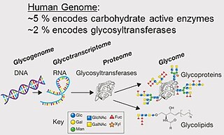
Glycomics is the comprehensive study of glycomes, including genetic, physiologic, pathologic, and other aspects. Glycomics "is the systematic study of all glycan structures of a given cell type or organism" and is a subset of glycobiology. The term glycomics is derived from the chemical prefix for sweetness or a sugar, "glyco-", and was formed to follow the omics naming convention established by genomics and proteomics.

<span class="mw-page-title-main">Glycoprotein</span> Protein with oligosaccharide modifications

Glycoproteins are proteins which contain oligosaccharide chains covalently attached to amino acid side-chains. The carbohydrate is attached to the protein in a cotranslational or posttranslational modification. This process is known as glycosylation. Secreted extracellular proteins are often glycosylated.

<span class="mw-page-title-main">Mannose</span> Chemical compound

Mannose is a sugar monomer of the aldohexose series of carbohydrates. It is a C-2 epimer of glucose. Mannose is important in human metabolism, especially in the glycosylation of certain proteins. Several congenital disorders of glycosylation are associated with mutations in enzymes involved in mannose metabolism.

<span class="mw-page-title-main">Glycome</span> Complete set of all sugars, free or bound, in an organism.

A glycome is the entire complement or complete set of all sugars, whether free or chemically bound in more complex molecules, of an organism. An alternative definition is the entirety of carbohydrates in a cell. The glycome may in fact be one of the most complex entities in nature. "Glycomics, analogous to genomics and proteomics, is the systematic study of all glycan structures of a given cell type or organism" and is a subset of glycobiology.

Defined in the narrowest sense, glycobiology is the study of the structure, biosynthesis, and biology of saccharides that are widely distributed in nature. Sugars or saccharides are essential components of all living things and aspects of the various roles they play in biology are researched in various medical, biochemical and biotechnological fields.

Glycosylation is the reaction in which a carbohydrate, i.e. a glycosyl donor, is attached to a hydroxyl or other functional group of another molecule in order to form a glycoconjugate. In biology, glycosylation usually refers to an enzyme-catalysed reaction, whereas glycation may refer to a non-enzymatic reaction.

An oligosaccharide is a saccharide polymer containing a small number of monosaccharides. Oligosaccharides can have many functions including cell recognition and cell adhesion.

<span class="mw-page-title-main">Calnexin</span> Mammalian protein found in Homo sapiens

Calnexin (CNX) is a 67kDa integral protein of the endoplasmic reticulum (ER). It consists of a large N-terminal calcium-binding lumenal domain, a single transmembrane helix and a short, acidic cytoplasmic tail. In humans, calnexin is encoded by the gene CANX.

<span class="mw-page-title-main">Oligosaccharyltransferase</span> Class of enzymes

Oligosaccharyltransferase or OST (EC 2.4.1.119) is a membrane protein complex that transfers a 14-sugar oligosaccharide from dolichol to nascent protein. It is a type of glycosyltransferase. The sugar Glc3Man9GlcNAc2 (where Glc=Glucose, Man=Mannose, and GlcNAc=N-acetylglucosamine) is attached to an asparagine (Asn) residue in the sequence Asn-X-Ser or Asn-X-Thr where X is any amino acid except proline. This sequence is called a glycosylation sequon. The reaction catalyzed by OST is the central step in the N-linked glycosylation pathway.

<span class="mw-page-title-main">Endoplasmic-reticulum-associated protein degradation</span>

Endoplasmic-reticulum-associated protein degradation (ERAD) designates a cellular pathway which targets misfolded proteins of the endoplasmic reticulum for ubiquitination and subsequent degradation by a protein-degrading complex, called the proteasome.

The mannose receptor is a C-type lectin primarily present on the surface of macrophages, immature dendritic cells and liver sinusoidal endothelial cells, but is also expressed on the surface of skin cells such as human dermal fibroblasts and keratinocytes. It is the first member of a family of endocytic receptors that includes Endo180 (CD280), M-type PLA2R, and DEC-205 (CD205).

The enzyme mannosyl-glycoprotein endo-β-N-acetylglucosaminidase (endoglycosidase H) (EC 3.2.1.96) has systematic name glycopeptide-D-mannosyl-N4-(N-acetyl-D-glucosaminyl)2-asparagine 1,4-N-acetyl-β-glucosaminohydrolase. It is a highly specific endoglycosidase which cleaves asparagine-linked mannose rich oligosaccharides, but not highly processed complex oligosaccharides from glycoproteins. It is used for research purposes to deglycosylate glycoproteins and to monitor intracellular protein trafficking through the secretory pathway.

<span class="mw-page-title-main">MAN1B1</span> Protein-coding gene in the species Homo sapiens

Endoplasmic reticulum mannosyl-oligosaccharide 1,2-alpha-mannosidase is an enzyme that in humans is encoded by the MAN1B1 gene.

Oligosaccharides and polysaccharides are an important class of polymeric carbohydrates found in virtually all living entities. Their structural features make their nomenclature challenging and their roles in living systems make their nomenclature important.

Glycopeptides are peptides that contain carbohydrate moieties (glycans) covalently attached to the side chains of the amino acid residues that constitute the peptide.

<i>N</i>-linked glycosylation Attachment of an oligosaccharide to a nitrogen atom

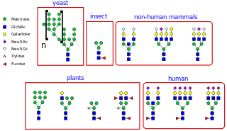
N-linked glycosylation, is the attachment of an oligosaccharide, a carbohydrate consisting of several sugar molecules, sometimes also referred to as glycan, to a nitrogen atom, in a process called N-glycosylation, studied in biochemistry. The resulting protein is called an N-linked glycan, or simply an N-glycan.

O-linked glycosylation is the attachment of a sugar molecule to the oxygen atom of serine (Ser) or threonine (Thr) residues in a protein. O-glycosylation is a post-translational modification that occurs after the protein has been synthesised. In eukaryotes, it occurs in the endoplasmic reticulum, Golgi apparatus and occasionally in the cytoplasm; in prokaryotes, it occurs in the cytoplasm. Several different sugars can be added to the serine or threonine, and they affect the protein in different ways by changing protein stability and regulating protein activity. O-glycans, which are the sugars added to the serine or threonine, have numerous functions throughout the body, including trafficking of cells in the immune system, allowing recognition of foreign material, controlling cell metabolism and providing cartilage and tendon flexibility. Because of the many functions they have, changes in O-glycosylation are important in many diseases including cancer, diabetes and Alzheimer's. O-glycosylation occurs in all domains of life, including eukaryotes, archaea and a number of pathogenic bacteria including Burkholderia cenocepacia, Neisseria gonorrhoeae and Acinetobacter baumannii.

Mannosyl-oligosaccharide glucosidase (MOGS) (EC 3.2.1.106, processing α-glucosidase I,Glc3Man9NAc2 oligosaccharide glucosidase, trimming glucosidase I, GCS1) is an enzyme with systematic name mannosyl-oligosaccharide glucohydrolase. MOGS is a transmembrane protein found in the membrane of the endoplasmic reticulum of eukaryotic cells. Biologically, it functions within the N-glycosylation pathway.

Translational glycobiology or applied glycobiology is the branch of glycobiology and glycochemistry that focuses on developing new pharmaceuticals through glycomics and glycoengineering. Although research in this field presents many difficulties, translational glycobiology presents applications with therapeutic glycoconjugates, with treating various bone diseases, and developing therapeutic cancer vaccines and other targeted therapies. Some mechanisms of action include using the glycan for drug targeting, engineering protein glycosylation for better efficacy, and glycans as drugs themselves.

Glycan nomenclature is the systematic naming of glycans, which are carbohydrate-based polymers made by all living organisms. In general glycans can be represented in (i) text formats, these include commonly used CarbBank, IUPAC name, and several other types; and (ii) symbol formats, these are consisting of Symbol Nomenclature For Glycans and Oxford Notations.

References

  1. "Glycans". IUPAC Gold Book - Glycans. 2009. doi:10.1351/goldbook.G02645. ISBN   978-0-9678550-9-7.
  2. Dwek, Raymond A. (1996). "Glycobiology: Toward Understanding the Function of Sugars". Chem. Rev. 96 (2): 683–720. doi:10.1021/cr940283b. PMID   11848770.
  3. 1 2 3 4 Maverakis E, Kim K, Shimoda M, Gershwin M, Patel F, Wilken R, Raychaudhuri S, Ruhaak LR, Lebrilla CB (2015). "Glycans in the immune system and The Altered Glycan Theory of Autoimmunity". J Autoimmun. 57 (6): 1–13. doi:10.1016/j.jaut.2014.12.002. PMC   4340844 . PMID   25578468.
  4. Nakagawa, S; Hato, M; Takegawa, Y; Deguchi, K; Ito, H; Takahata, M; Iwasaki, N; Minami, A; Nishimura, S-I (2007). "Detection of altered N-glycan profiles in whole serum from rheumatoid arthritis patients". J. Chromatogr. B. 853 (1–2): 133–137. doi:10.1016/j.jchromb.2007.03.003. hdl: 2115/28276 . PMID   17392038.
  5. Bermingham, ML; Colombo, M; McGurnaghan, SJ; Blackbourn, LAK; Vučković, F; Pučić Baković, M; Trbojević-Akmačić, I; Lauc, G; Agakov, F; Agakova, AS; Hayward, C; Klarić, L; Palmer, CNA; Petrie, JR; Chalmers, J; Collier, A; Green, F; Lindsay, RS; Macrury, S; McKnight, JA; Patrick, AW; Thekkepat, S; Gornik, O; McKeigue, PM; Colhoun, HM (2018). "N-Glycan Profile and Kidney Disease in Type 1 Diabetes". Diabetes Care. 41 (1): 79–87. doi: 10.2337/dc17-1042 . hdl: 20.500.11820/413dce5a-e852-4787-aac9-62c2c6d4389f . PMID   29146600.
  6. "U.S. National Research Council Report, Transforming Glycoscience: A Roadmap for the Future".
  7. "U.S. National Research Council Report-in-Brief, Transforming Glycoscience: A Roadmap for the Future".
  8. Essentials of Glycobiology (2nd ed.). Cold Spring Harbor Laboratory Press. 2009. ISBN   978-087969770-9.
  9. Aizpurua-Olaizola, O.; Toraño, J. Sastre; Falcon-Perez, J.M.; Williams, C.; Reichardt, N.; Boons, G.-J. (2018). "Mass spectrometry for glycan biomarker discovery". TrAC Trends in Analytical Chemistry. 100: 7–14. doi:10.1016/j.trac.2017.12.015.
  10. Wada Y, Azadi P, Costello CE, et al. (April 2007). "Comparison of the methods for profiling glycoprotein glycans—HUPO Human Disease Glycomics/Proteome Initiative multi-institutional study". Glycobiology. 17 (4): 411–22. doi: 10.1093/glycob/cwl086 . PMID   17223647.
  11. Hase S, Ikenaka T, Matsushima Y (November 1978). "Structure analyses of oligosaccharides by tagging of the reducing end sugars with a fluorescent compound". Biochem. Biophys. Res. Commun. 85 (1): 257–63. doi:10.1016/S0006-291X(78)80037-0. PMID   743278.
  12. Pabst M, Kolarich D, Pöltl G, et al. (January 2009). "Comparison of fluorescent labels for oligosaccharides and introduction of a new postlabeling purification method". Anal. Biochem. 384 (2): 263–73. doi:10.1016/j.ab.2008.09.041. PMID   18940176.
  13. Šoić, Dinko; Mlinarić, Zvonimir; Lauc, Gordan; Gornik, Olga; Novokmet, Mislav; Keser, Toma (2022). "In a pursuit of optimal glycan fluorescent label for negative MS mode for high-throughput N-glycan analysis". Frontiers in Chemistry. 10: 999770. doi: 10.3389/fchem.2022.999770 . ISSN   2296-2646. PMC   9574008 . PMID   36262345.
  14. Harvey DJ, Bateman RH, Bordoli RS, Tyldesley R (2000). "Ionisation and fragmentation of complex glycans with a quadrupole time-of-flight mass spectrometer fitted with a matrix-assisted laser desorption/ionisation ion source". Rapid Commun. Mass Spectrom. 14 (22): 2135–42. Bibcode:2000RCMS...14.2135H. doi:10.1002/1097-0231(20001130)14:22<2135::AID-RCM143>3.0.CO;2-#. PMID   11114021.
  15. Schulz, BL; Packer NH, NH; Karlsson, NG (Dec 2002). "Small-scale analysis of O-linked oligosaccharides from glycoproteins and mucins separated by gel electrophoresis". Anal. Chem. 74 (23): 6088–97. doi:10.1021/ac025890a. PMID   12498206.
  16. Pabst M, Bondili JS, Stadlmann J, Mach L, Altmann F (July 2007). "Mass plus retention time equals structure: a strategy for the analysis of N-glycans by carbon LC-ESI-MS and its application to fibrin N-glycans". Anal. Chem. 79 (13): 5051–7. doi:10.1021/ac070363i. PMID   17539604.
  17. Ruhaak LR, Deelder AM, Wuhrer M (May 2009). "Oligosaccharide analysis by graphitized carbon liquid chromatography-mass spectrometry". Anal Bioanal Chem. 394 (1): 163–74. doi: 10.1007/s00216-009-2664-5 . PMID   19247642.
  18. Flowers, Sarah A.; Ali, Liaqat; Lane, Catherine S.; Olin, Magnus; Karlsson, Niclas G. (2013-04-01). "Selected reaction monitoring to differentiate and relatively quantitate isomers of sulfated and unsulfated core 1 O-glycans from salivary MUC7 protein in rheumatoid arthritis". Molecular & Cellular Proteomics. 12 (4): 921–931. doi:10.1074/mcp.M113.028878. ISSN   1535-9484. PMC   3617339 . PMID   23457413.