In proteins that have segments extending extracellularly, the extracellular segments are also often glycosylated. Glycoproteins are also often important integral membrane proteins, where they play a role in cell–cell interactions. It is important to distinguish endoplasmic reticulum-based glycosylation of the secretory system from reversible cytosolic-nuclear glycosylation. Glycoproteins of the cytosol and nucleus can be modified through the reversible addition of a single GlcNAc residue that is considered reciprocal to phosphorylation and the functions of these are likely to be an additional regulatory mechanism that controls phosphorylation-based signalling.[2] In contrast, classical secretory glycosylation can be structurally essential. For example, inhibition of asparagine-linked, i.e. N-linked, glycosylation can prevent proper glycoprotein folding and full inhibition can be toxic to an individual cell. In contrast, perturbation of glycan processing (enzymatic removal/addition of carbohydrate residues to the glycan), which occurs in both the endoplasmic reticulum and Golgi apparatus, is dispensable for isolated cells (as evidenced by survival with glycosides inhibitors) but can lead to human disease (congenital disorders of glycosylation) and can be lethal in animal models. It is therefore likely that the fine processing of glycans is important for endogenous functionality, such as cell trafficking, but that this is likely to have been secondary to its role in host-pathogen interactions. A famous example of this latter effect is the ABO blood group system.[citation needed]
Though there are different types of glycoproteins, the most common are N-linked and O-linked glycoproteins.[3] These two types of glycoproteins are distinguished by structural differences that give them their names. Glycoproteins vary greatly in composition, making many different compounds such as antibodies or hormones.[4] Due to the wide array of functions within the body, interest in glycoprotein synthesis for medical use has increased.[5] There are now several methods to synthesize glycoproteins, including recombination and glycosylation of proteins.[5]
In glycation, also known as non-enzymatic glycosylation, sugars are covalently bonded to a protein or lipid molecule, without the controlling action of an enzyme, but through a Maillard reaction.
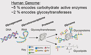
Monosaccharides
Eight sugars commonly found in glycoproteins.
Monosaccharides commonly found in eukaryotic glycoproteins include:[8]:526
The principal sugars found in human glycoproteins[9]
The two most common linkages in glycoproteins are N-linked and O-linked glycoproteins.[3] An N-linked glycoprotein has glycan bonds to the nitrogen containing an asparagine amino acid within the protein sequence.[4] An O-linked glycoprotein has the sugar is bonded to an oxygen atom of a serine or threonine amino acid in the protein.[4]
Glycoprotein size and composition can vary largely, with carbohydrate composition ranges from 1% to 70% of the total mass of the glycoprotein.[4] Within the cell, they appear in the blood, the extracellular matrix, or on the outer surface of the plasma membrane, and make up a large portion of the proteins secreted by eukaryotic cells.[4] They are very broad in their applications and can function as a variety of chemicals from antibodies to hormones.[4]
Glycomics
Glycomics is the study of the carbohydrate components of cells.[4] Though not exclusive to glycoproteins, it can reveal more information about different glycoproteins and their structure.[4] One of the purposes of this field of study is to determine which proteins are glycosylated and where in the amino acid sequence the glycosylation occurs.[4] Historically, mass spectrometry has been used to identify the structure of glycoproteins and characterize the carbohydrate chains attached.[4][10]
Examples
The unique interaction between the oligosaccharide chains have different applications. First, it aids in quality control by identifying misfolded proteins.[4] The oligosaccharide chains also change the solubility and polarity of the proteins that they are bonded to.[4] For example, if the oligosaccharide chains are negatively charged, with enough density around the protein, they can repulse proteolytic enzymes away from the bonded protein.[4] The diversity in interactions lends itself to different types of glycoproteins with different structures and functions.[5]
One example of glycoproteins found in the body is mucins, which are secreted in the mucus of the respiratory and digestive tracts. The sugars when attached to mucins give them considerable water-holding capacity and also make them resistant to proteolysis by digestive enzymes.
molecules such as antibodies (immunoglobulins), which interact directly with antigens.
molecules of the major histocompatibility complex (or MHC), which are expressed on the surface of cells and interact with T cells as part of the adaptive immune response.
sialyl Lewis X antigen on the surface of leukocytes.
H antigen of the ABO blood compatibility antigens. Other examples of glycoproteins include:
gonadotropins (luteinizing hormone and follicle-stimulating hormone)
components of the zona pellucida, which surrounds the oocyte, and is important for sperm-egg interaction.
structural glycoproteins, which occur in connective tissue. These help bind together the fibers, cells, and ground substance of connective tissue. They may also help components of the tissue bind to inorganic substances, such as calcium in bone.
Glycoprotein-41 (gp41) and glycoprotein-120 (gp120) are HIV viral coat proteins.
Variable surface glycoproteins allow the sleeping sickness Trypanosoma parasite to escape the immune response of the host.
The viral spike of the human immunodeficiency virus is heavily glycosylated.[12] Approximately half the mass of the spike is glycosylation and the glycans act to limit antibody recognition as the glycans are assembled by the host cell and so are largely 'self'. Over time, some patients can evolve antibodies to recognise the HIV glycans and almost all so-called 'broadly neutralising antibodies (bnAbs) recognise some glycans. This is possible mainly because the unusually high density of glycans hinders normal glycan maturation and they are therefore trapped in the premature, high-mannose, state.[13][14] This provides a window for immune recognition. In addition, as these glycans are much less variable than the underlying protein, they have emerged as promising targets for vaccine design.[15]
P-glycoproteins are critical for antitumor research due to its ability block the effects of antitumor drugs.[4][16] P-glycoprotein, or multidrug transporter (MDR1), is a type of ABC transporter that transports compounds out of cells.[4] This transportation of compounds out of cells includes drugs made to be delivered to the cell, causing a decrease in drug effectiveness.[4] Therefore, being able to inhibit this behavior would decrease P-glycoprotein interference in drug delivery, making this an important topic in drug discovery.[4] For example, P-Glycoprotein causes a decrease in anti-cancer drug accumulation within tumor cells, limiting the effectiveness of chemotherapies used to treat cancer.[16]
A glycoprotein is a compound containing carbohydrate (or glycan) covalently linked to protein. The carbohydrate may be in the form of a monosaccharide, disaccharide(s). oligosaccharide(s), polysaccharide(s), or their derivatives (e.g. sulfo- or phospho-substituted). One, a few, or many carbohydrate units may be present. Proteoglycans are a subclass of glycoproteins in which the carbohydrate units are polysaccharides that contain amino sugars. Such polysaccharides are also known as glycosaminoglycans.
Resultant shifts in electrophoretic migration help distinguish among proteins with N-glycan, O-glycan, or GPI linkages and also between high mannose and complex N-glycans.
Provides information on molecular mass, composition, sequence, and sometimes branching of a glycan chain. It can also be used for site-specific glycosylation profiling.[18]
In conjunction with size-exclusion chromatography, UV/Vis absorption and differential refractometry, provides information on molecular mass, protein-carbohydrate ratio, aggregation state, size, and sometimes branching of a glycan chain. In conjunction with composition-gradient analysis, analyzes self- and hetero-association to determine binding affinity and stoichiometry with proteins or carbohydrates in solution without labeling.
The glycosylation of proteins has an array of different applications from influencing cell to cell communication to changing the thermal stability and the folding of proteins.[4][19] Due to the unique abilities of glycoproteins, they can be used in many therapies.[19] By understanding glycoproteins and their synthesis, they can be made to treat cancer, Crohn's Disease, high cholesterol, and more.[3]
The process of glycosylation (binding a carbohydrate to a protein) is a post-translational modification, meaning it happens after the production of the protein.[3] Glycosylation is a process that roughly half of all human proteins undergo and heavily influences the properties and functions of the protein.[3] Within the cell, glycosylation occurs in the endoplasmic reticulum.[3]
Recombination
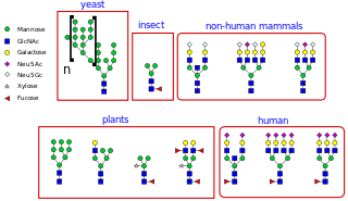
Depiction of differences in glycans amongst different animals.
There are several techniques for the assembly of glycoproteins. One technique utilizes recombination.[3] The first consideration for this method is the choice of host, as there are many different factors that can influence the success of glycoprotein recombination such as cost, the host environment, the efficacy of the process, and other considerations.[3] Some examples of host cells include E. coli, yeast, plant cells, insect cells, and mammalian cells.[3] Of these options, mammalian cells are the most common because their use does not face the same challenges that other host cells do such as different glycan structures, shorter half life, and potential unwanted immune responses in humans.[3] Of mammalian cells, the most common cell line used for recombinant glycoprotein production is the Chinese hamster ovary line.[3] However, as technologies develop, the most promising cell lines for recombinant glycoprotein production are human cell lines.[3]
Glycosylation
The formation of the link between the glycan and the protein is key element of the synthesis of glycoproteins.[5] The most common method of glycosylation of N-linked glycoproteins is through the reaction between a protected glycan and a protected Asparagine.[5] Similarly, an O-linked glycoprotein can be formed through the addition of a glycosyl donor with a protected Serine or Threonine.[5] These two methods are examples of natural linkage.[5] However, there are also methods of unnatural linkages.[5] Some methods include ligation and a reaction between a serine-derived sulfamidate and thiohexoses in water.[5] Once this linkage is complete, the amino acid sequence can be expanded upon using solid-phase peptide synthesis.[5]
A congenital disorder of glycosylation is one of several rare inborn errors of metabolism in which glycosylation of a variety of tissue proteins and/or lipids is deficient or defective. Congenital disorders of glycosylation are sometimes known as CDG syndromes. They often cause serious, sometimes fatal, malfunction of several different organ systems in affected infants. The most common sub-type is PMM2-CDG where the genetic defect leads to the loss of phosphomannomutase 2 (PMM2), the enzyme responsible for the conversion of mannose-6-phosphate into mannose-1-phosphate
Mannose is a sugar monomer of the aldohexose series of carbohydrates. It is a C-2 epimer of glucose. Mannose is important in human metabolism, especially in the glycosylation of certain proteins. Several congenital disorders of glycosylation are associated with mutations in enzymes involved in mannose metabolism.
The glycome is the entire complement of sugars, whether free or present in more complex molecules, of an organism. An alternative definition is the entirety of carbohydrates in a cell. The glycome may in fact be one of the most complex entities in nature. "Glycomics, analogous to genomics and proteomics, is the systematic study of all glycan structures of a given cell type or organism" and is a subset of glycobiology.
Glycosylation is the reaction in which a carbohydrate, i.e. a glycosyl donor, is attached to a hydroxyl or other functional group of another molecule in order to form a glycoconjugate. In biology, glycosylation usually refers to an enzyme-catalysed reaction, whereas glycation may refer to a non-enzymatic reaction.
An oligosaccharide is a saccharide polymer containing a small number of monosaccharides. Oligosaccharides can have many functions including cell recognition and cell adhesion.
Proteoglycans are proteins that are heavily glycosylated. The basic proteoglycan unit consists of a "core protein" with one or more covalently attached glycosaminoglycan (GAG) chain(s). The point of attachment is a serine (Ser) residue to which the glycosaminoglycan is joined through a tetrasaccharide bridge. The Ser residue is generally in the sequence -Ser-Gly-X-Gly-, although not every protein with this sequence has an attached glycosaminoglycan. The chains are long, linear carbohydrate polymers that are negatively charged under physiological conditions due to the occurrence of sulfate and uronic acid groups. Proteoglycans occur in connective tissue.
The terms glycans and polysaccharides are defined by IUPAC as synonyms meaning "compounds consisting of a large number of monosaccharides linked glycosidically". However, in practice the term glycan may also be used to refer to the carbohydrate portion of a glycoconjugate, such as a glycoprotein, glycolipid, or a proteoglycan, even if the carbohydrate is only an oligosaccharide. Glycans usually consist solely of O-glycosidic linkages of monosaccharides. For example, cellulose is a glycan composed of β-1,4-linked D-glucose, and chitin is a glycan composed of β-1,4-linked N-acetyl-D-glucosamine. Glycans can be homo- or heteropolymers of monosaccharide residues, and can be linear or branched.
Oligosaccharyltransferase or OST (EC 2.4.1.119) is a membrane protein complex that transfers a 14-sugar oligosaccharide from dolichol to nascent protein. It is a type of glycosyltransferase. The sugar Glc3Man9GlcNAc2 (where Glc=Glucose, Man=Mannose, and GlcNAc=N-acetylglucosamine) is attached to an asparagine (Asn) residue in the sequence Asn-X-Ser or Asn-X-Thr where X is any amino acid except proline. This sequence is called a glycosylation sequon. The reaction catalyzed by OST is the central step in the N-linked glycosylation pathway.
The mannose receptor is a C-type lectin primarily present on the surface of macrophages, immature dendritic cells and liver sinusoidal endothelial cells, but is also expressed on the surface of skin cells such as human dermal fibroblasts and keratinocytes. It is the first member of a family of endocytic receptors that includes Endo180 (CD280), M-type PLA2R, and DEC-205 (CD205).
The enzyme mannosyl-glycoprotein endo-β-N-acetylglucosaminidase (endoglycosidase H) (EC 3.2.1.96) has systematic name glycopeptide-D-mannosyl-N4-(N-acetyl-D-glucosaminyl)2-asparagine 1,4-N-acetyl-β-glucosaminohydrolase. It is a highly specific endoglycosidase which cleaves asparagine-linked mannose rich oligosaccharides, but not highly processed complex oligosaccharides from glycoproteins. It is used for research purposes to deglycosylate glycoproteins and to monitor intracellular protein trafficking through the secretory pathway.
UDP-N-acetylglucosamine—dolichyl-phosphate N-acetylglucosaminephosphotransferase is an enzyme that in humans is encoded by the DPAGT1 gene.
Glycopeptides are peptides that contain carbohydrate moieties (glycans) covalently attached to the side chains of the amino acid residues that constitute the peptide.
N-linked glycosylation, is the attachment of an oligosaccharide, a carbohydrate consisting of several sugar molecules, sometimes also referred to as glycan, to a nitrogen atom, in a process called N-glycosylation, studied in biochemistry. The resulting protein is called an N-linked glycan, or simply an N-glycan.
O-linked glycosylation is the attachment of a sugar molecule to the oxygen atom of serine (Ser) or threonine (Thr) residues in a protein. O-glycosylation is a post-translational modification that occurs after the protein has been synthesised. In eukaryotes, it occurs in the endoplasmic reticulum, Golgi apparatus and occasionally in the cytoplasm; in prokaryotes, it occurs in the cytoplasm. Several different sugars can be added to the serine or threonine, and they affect the protein in different ways by changing protein stability and regulating protein activity. O-glycans, which are the sugars added to the serine or threonine, have numerous functions throughout the body, including trafficking of cells in the immune system, allowing recognition of foreign material, controlling cell metabolism and providing cartilage and tendon flexibility. Because of the many functions they have, changes in O-glycosylation are important in many diseases including cancer, diabetes and Alzheimer's. O-glycosylation occurs in all domains of life, including eukaryotes, archaea and a number of pathogenic bacteria including Burkholderia cenocepacia, Neisseria gonorrhoeae and Acinetobacter baumannii.
Mannosyl-oligosaccharide glucosidase (MOGS) (EC 3.2.1.106, processing α-glucosidase I,Glc3Man9NAc2 oligosaccharide glucosidase, trimming glucosidase I, GCS1) is an enzyme with systematic name mannosyl-oligosaccharide glucohydrolase. MOGS is a transmembrane protein found in the membrane of the endoplasmic reticulum of eukaryotic cells. Biologically, it functions within the N-glycosylation pathway.
Peptide:N-glycosidase F, commonly referred to as PNGase F, is an amidase of the peptide-N4-(N-acetyl-beta-glucosaminyl)asparagine amidase class. PNGase F works by cleaving between the innermost GlcNAc and asparagine residues of high mannose, hybrid, and complex oligosaccharides from N-linked glycoproteins and glycopeptides. This results in a deaminated protein or peptide and a free glycan.
Translational glycobiology or applied glycobiology is the branch of glycobiology and glycochemistry that focuses on developing new pharmaceuticals through glycomics and glycoengineering. Although research in this field presents many difficulties, translational glycobiology presents applications with therapeutic glycoconjugates, with treating various bone diseases, and developing therapeutic cancer vaccines and other targeted therapies. Some mechanisms of action include using the glycan for drug targeting, engineering protein glycosylation for better efficacy, and glycans as drugs themselves.
N-glycosyltransferase is an enzyme in prokaryotes which transfers individual hexoses onto asparagine sidechains in substrate proteins, using a nucleotide-bound intermediary, within the cytoplasm. They are distinct from regular N-glycosylating enzymes, which are oligosaccharyltransferases that transfer pre-assembled oligosaccharides. Both enzyme families however target a shared amino acid sequence asparagine—-any amino acid except proline—serine or threonine (N–x–S/T), with some variations.
Glycan-Protein interactions represent a class of biomolecular interactions that occur between free or protein-bound glycans and their cognate binding partners. Intramolecular glycan-protein (protein-glycan) interactions occur between glycans and proteins that they are covalently attached to. Together with protein-protein interactions, they form a mechanistic basis for many essential cell processes, especially for cell-cell interactions and host-cell interactions. For instance, SARS-CoV-2, the causative agent of COVID-19, employs its extensively glycosylated spike (S) protein to bind to the ACE2 receptor, allowing it to enter host cells. The spike protein is a trimeric structure, with each subunit containing 22 N-glycosylation sites, making it an attractive target for vaccine search.
GlycoRNAs are small non-coding RNAs with sialylated glycans.
This page is based on this Wikipedia article Text is available under the CC BY-SA 4.0 license; additional terms may apply. Images, videos and audio are available under their respective licenses.