Affinity electrophoresis

Last updated
The quantitative principle of affinity electrophoresis illustrated with electrophoresis at pH 8.6 of concanavalin A into an agarose gel containing blood serum (3.6 microliter per square cm). The bar indicates 1 cm. Electrophoresis performed overnight at less than 10 V/cm. The analysis was performed early in the 1970s at the Protein Laboratory Affinityelectrophoreses1.jpg
The quantitative principle of affinity electrophoresis illustrated with electrophoresis at pH 8.6 of concanavalin A into an agarose gel containing blood serum (3.6 microliter per square cm). The bar indicates 1 cm. Electrophoresis performed overnight at less than 10 V/cm. The analysis was performed early in the 1970s at the Protein Laboratory

Affinity electrophoresis is a general name for many analytical methods used in biochemistry and biotechnology. Both qualitative and quantitative information may be obtained through affinity electrophoresis. [1] Cross electrophoresis, the first affinity electrophoresis method, was created by Nakamura et al. Enzyme-substrate complexes have been detected using cross electrophoresis. [2] [3] [4] [5] [6] The methods include the so-called electrophoretic mobility shift assay, charge shift electrophoresis and affinity capillary electrophoresis. [1] The methods are based on changes in the electrophoretic pattern of molecules (mainly macromolecules) through biospecific interaction or complex formation. The interaction or binding of a molecule, charged or uncharged, will normally change the electrophoretic properties of a molecule. [7] [1] Membrane proteins may be identified by a shift in mobility induced by a charged detergent. Nucleic acids or nucleic acid fragments may be characterized by their affinity to other molecules. The methods have been used for estimation of binding constants, as for instance in lectin affinity electrophoresis or characterization of molecules with specific features like glycan content or ligand binding. [1] For enzymes and other ligand-binding proteins, one-dimensional electrophoresis similar to counter electrophoresis or to "rocket immunoelectrophoresis", affinity electrophoresis may be used as an alternative quantification of the protein. [8] Some of the methods are similar to affinity chromatography by use of immobilized ligands.

Contents

Types and methods

Currently, there is ongoing research in developing new ways of utilizing the knowledge already associated with affinity electrophoresis to improve its functionality and speed, as well as attempts to improve already established methods and tailor them towards performing specific tasks.

Agarose gel electrophoresis

an example of an agarose gel after electrophoresis Figure displaying gel electrophoresis.png
an example of an agarose gel after electrophoresis

A type of electrophoretic mobility shift assay (AMSA), agarose gel electrophoresis is used to separate protein-bound amino acid complexes from free amino acids. Using a low voltage (~10 V/cm) to minimize the risk for heat damage, electricity is run across an agarose gel. When dissolved in a hot buffered solution (50 to 55 degrees Celsius), it produces a viscous solution, but when cooled, it solidifies as a gel. Serum proteins, hemoglobin, nucleic acids, polymerase chain reaction products, etc. are all separated using this method. Agarose's fixed sulfate groups can cause enhanced electroendosmosis, which lowers band resolution. Utilizing ultrapure agarose gel with little sulfate content can stop this.[ citation needed ]

Rapid agarose gel electrophoresis

This technique utilizes a high voltage (≥ 20 V/cm) with a 0.5× Tris-borate buffer run across an agarose gel. [9] This method differs from the traditional agarose gel electrophoresis by utilizing a higher voltage to facilitate a shorter run time as well as yield a higher band resolution. Other factors included in developing the technique of rapid agarose gel electrophoresis are gel thickness, and the percentage of agarose within the gel.

Boronate affinity electrophoresis

Boronate affinity electrophoresis utilizes boronic acid infused acrylamide gels to purify NAD-RNA. This purification allows for researchers to easily measure the kinetic activity of NAD-RNA decapping enzymes. [10]

Affinity capillary electrophoresis

Affinity capillary electrophoresis (ACE) refers to a number of techniques which rely on specific and nonspecific binding interactions to facilitate separation and detection through a formulary approach in accordance with the theory of electromigration. [11] [12] Using the intermolecular interactions between molecules occurring in free solution or mobilized onto a solid support, ACE allows for the separation and quantitation of analyte concentrations and binding and dissociation constants between molecules. [13] [14] As affinity probes in CAE, fluorophore-labeled compounds with affinities for the target molecules are employed. [15] With ACE, scientists hope to develop strong binding drug candidates, understand and measure enzymatic activity, and characterize the charges on proteins. [16] Affinity capillary electrophoresis can be divided into three distinct techniques: non-equilibrium electrophoresis of equilibrated sample mixtures, dynamic equilibrium ACE, and affinity-based ACE. [13]

Nonequilibrium electrophoresis of equilibrated sample mixtures is generally used in the separation and study of binding interactions of large proteins and involves combining both the analyte and its receptor molecule in a premixed sample.

These receptor molecules often take the form of affinity probes consisting of fluorophore-labeled molecules that will bind to target molecules that are mixed with the sample being tested. [13] This mixture, and its subsequent complexes, are then separated through capillary electrophoresis. [13] Because the original mixture of analyte and receptor molecule were bound together in an equilibrium, the slow dissociation of these two bound molecules during the electrophoretic experiment will result in their separation and a subsequent shift in equilibrium towards further dissociation. [17] The characteristic smear pattern produced by the slow release of the analyte from the complex during the experiment can be used to calculate the dissociation constant of the complex. [17]

Dynamic equilibrium ACE involves the combination of the analyte found in the sample and its receptor molecule found in the buffered solution in the capillary tube so that binding and separation only occur in the instrument. [13] It is assumed for dynamic equilibrium affinity capillary electrophoresis that ligand-receptor binding occurs rapidly when the analyte and buffer are mixed. Binding constants are generally derived from this technique based upon the peak migration shift of the receptor which is dependent upon the concentration of the analyte in the sample. [13]

Affinity-based capillary electrophoresis, also known as capillary electroaffinity chromatography (CEC), involves the binding of analyte in sample to an immobilized receptor molecule on the capillary wall, microbeads, or microchannels. [18] CEC offers the highest separation efficacy of all three ACE techniques as non-matrixed sample components are washed away and the ligand then be released and analyzed. [13]  

Affinity capillary electrophoresis takes the advantages of capillary electrophoresis and applies them to the study of protein interactions. [16] ACE is advantageous because it has a high separation efficiency, has a shorter analysis time, can be run at physiological pH, and involves low consumption of ligand/molecules. [19] [20] In addition, the composition of the protein of interest does not have to be known in order to run ACE studies. [16] The main disadvantage, though, is that it does not give much stoichiometric information about the reaction being studied. [20]

Affinity-trap polyacrylamide gel electrophoresis

Affinity-trap polyacrylamide gel electrophoresis (PAGE) has become one of the most popular methods of protein separation. This is not only due to its separation qualities, but also because it can be used in conjunction with a variety of other analytic methods, such as mass spectrometry, and western blotting. [14] In addition to helping isolate and purify proteins from biological samples, AT-PAGE is anticipated to be helpful in analyses of variations in the expression of particular proteins as well as in investigations of posttranslational modifications of proteins. [21] This method utilizes a two-step approach. First, a protein sample is run through a polyacrylamide gel using electrophoresis. Then, the sample is transferred to a different polyacrylamide gel (the affinity-trap gel) where affinity probes are immobilized. The proteins that do not have affinity for the affinity probes pass through the affinity-trap gel, and proteins with affinity for the probes will be "trapped" by the immobile affinity probes. These trapped proteins are then visualized and identified using mass spectrometry after in-gel digestion. [14]

Phosphate affinity electrophoresis

Phosphate affinity electrophoresis utilizes an affinity probe which consists of a molecule that binds specifically to divalent phosphate ions in neutral aqueous solution, known as a "Phos-Tag". This methods also utilizes a separation gel made of an acrylamide-pendent Phos-Tag monomer that is copolymerized. Phosphorylated proteins migrate slowly in the gel compared to non-phosphorylated proteins. This technique gives the researcher the ability to observe the differences in the phosphorylation states of any given protein. [14] This technique allows for the detection of distinct bands even in protein molecules that have the same amount of phosphorylated amino acid residues but are phosphorylated at different amino acid locations. [22] [23]

See also

Related Research Articles

<span class="mw-page-title-main">Agarose</span> Heteropolysaccharide found in red algae

Agarose is a heteropolysaccharide, generally extracted from certain red seaweed. It is a linear polymer made up of the repeating unit of agarobiose, which is a disaccharide made up of D-galactose and 3,6-anhydro-L-galactopyranose. Agarose is one of the two principal components of agar, and is purified from agar by removing agar's other component, agaropectin.

<span class="mw-page-title-main">Gel electrophoresis</span> Method for separation and analysis of biomolecules

Gel electrophoresis is a method for separation and analysis of biomacromolecules and their fragments, based on their size and charge. It is used in clinical chemistry to separate proteins by charge or size and in biochemistry and molecular biology to separate a mixed population of DNA and RNA fragments by length, to estimate the size of DNA and RNA fragments or to separate proteins by charge.

<span class="mw-page-title-main">Southern blot</span> DNA analysis technique

Southern blot is a method used for detection and quantification of a specific DNA sequence in DNA samples. This method is used in molecular biology. Briefly, purified DNA from a biological sample is digested with restriction enzymes, and the resulting DNA fragments are separated by using an electric current to move them through a sieve-like gel or matrix, which allows smaller fragments to move faster than larger fragments. The DNA fragments are transferred out of the gel or matrix onto a solid membrane, which is then exposed to a DNA probe labeled with a radioactive, fluorescent, or chemical tag. The tag allows any DNA fragments containing complementary sequences with the DNA probe sequence to be visualized within the Southern blot.

<span class="mw-page-title-main">Polyacrylamide gel electrophoresis</span> Analytical technique

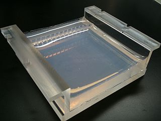
Polyacrylamide gel electrophoresis (PAGE) is a technique widely used in biochemistry, forensic chemistry, genetics, molecular biology and biotechnology to separate biological macromolecules, usually proteins or nucleic acids, according to their electrophoretic mobility. Electrophoretic mobility is a function of the length, conformation, and charge of the molecule. Polyacrylamide gel electrophoresis is a powerful tool used to analyze RNA samples. When polyacrylamide gel is denatured after electrophoresis, it provides information on the sample composition of the RNA species.

<span class="mw-page-title-main">Gel electrophoresis of nucleic acids</span>

Gel electrophoresis of nucleic acids is an analytical technique to separate DNA or RNA fragments by size and reactivity. Nucleic acid molecules are placed on a gel, where an electric field induces the nucleic acids to migrate toward the positively charged anode. The molecules separate as they travel through the gel based on the each molecule's size and shape. Longer molecules move more slowly because they the gel resists their movement more forcefully than it resists shorter molecules. After some time, the electricity is turned off and the positions of the different molecules are analyzed.

Protein purification is a series of processes intended to isolate one or a few proteins from a complex mixture, usually cells, tissues or whole organisms. Protein purification is vital for the specification of the function, structure and interactions of the protein of interest. The purification process may separate the protein and non-protein parts of the mixture, and finally separate the desired protein from all other proteins. Ideally, to study a protein of interest, it must be separated from other components of the cell so that contaminants will not interfere in the examination of the protein of interest's structure and function. Separation of one protein from all others is typically the most laborious aspect of protein purification. Separation steps usually exploit differences in protein size, physico-chemical properties, binding affinity and biological activity. The pure result may be termed protein isolate.

<span class="mw-page-title-main">Gel electrophoresis of proteins</span> Technique for separating proteins

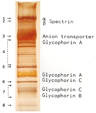
Protein electrophoresis is a method for analysing the proteins in a fluid or an extract. The electrophoresis may be performed with a small volume of sample in a number of alternative ways with or without a supporting medium, namely agarose or polyacrylamide. Variants of gel electrophoresis include SDS-PAGE, free-flow electrophoresis, electrofocusing, isotachophoresis, affinity electrophoresis, immunoelectrophoresis, counterelectrophoresis, and capillary electrophoresis. Each variant has many subtypes with individual advantages and limitations. Gel electrophoresis is often performed in combination with electroblotting or immunoblotting to give additional information about a specific protein.

<span class="mw-page-title-main">Blot (biology)</span>

A blot, in molecular biology and genetics, is a method of transferring proteins, DNA or RNA onto a carrier. In many instances, this is done after a gel electrophoresis, transferring the molecules from the gel onto the blotting membrane, and other times adding the samples directly onto the membrane. After the blotting, the transferred proteins, DNA or RNA are then visualized by colorant staining, autoradiographic visualization of radiolabelled molecules, or specific labelling of some proteins or nucleic acids. The latter is done with antibodies or hybridization probes that bind only to some molecules of the blot and have an enzyme joined to them. After proper washing, this enzymatic activity is visualized by incubation with proper reactive, rendering either a colored deposit on the blot or a chemiluminescent reaction which is registered by photographic film.

<span class="mw-page-title-main">Coomassie brilliant blue</span> Chemical compound

Coomassie brilliant blue is the name of two similar triphenylmethane dyes that were developed for use in the textile industry but are now commonly used for staining proteins in analytical biochemistry. Coomassie brilliant blue G-250 differs from Coomassie brilliant blue R-250 by the addition of two methyl groups. The name "Coomassie" is a registered trademark of Imperial Chemical Industries.

<span class="mw-page-title-main">Isoelectric focusing</span> Type of electrophoresis

Isoelectric focusing (IEF), also known as electrofocusing, is a technique for separating different molecules by differences in their isoelectric point (pI). It is a type of zone electrophoresis usually performed on proteins in a gel that takes advantage of the fact that overall charge on the molecule of interest is a function of the pH of its surroundings.

Capillary electrophoresis (CE) is a family of electrokinetic separation methods performed in submillimeter diameter capillaries and in micro- and nanofluidic channels. Very often, CE refers to capillary zone electrophoresis (CZE), but other electrophoretic techniques including capillary gel electrophoresis (CGE), capillary isoelectric focusing (CIEF), capillary isotachophoresis and micellar electrokinetic chromatography (MEKC) belong also to this class of methods. In CE methods, analytes migrate through electrolyte solutions under the influence of an electric field. Analytes can be separated according to ionic mobility and/or partitioning into an alternate phase via non-covalent interactions. Additionally, analytes may be concentrated or "focused" by means of gradients in conductivity and pH.

Affinity chromatography is a method of separating a biomolecule from a mixture, based on a highly specific macromolecular binding interaction between the biomolecule and another substance. The specific type of binding interaction depends on the biomolecule of interest; antigen and antibody, enzyme and substrate, receptor and ligand, or protein and nucleic acid binding interactions are frequently exploited for isolation of various biomolecules. Affinity chromatography is useful for its high selectivity and resolution of separation, compared to other chromatographic methods.

<span class="mw-page-title-main">Immunoelectrophoresis</span> Biochemical methods of separation and characterization of proteins

Immunoelectrophoresis is a general name for a number of biochemical methods for separation and characterization of proteins based on electrophoresis and reaction with antibodies. All variants of immunoelectrophoresis require immunoglobulins, also known as antibodies, reacting with the proteins to be separated or characterized. The methods were developed and used extensively during the second half of the 20th century. In somewhat chronological order: Immunoelectrophoretic analysis, crossed immunoelectrophoresis, rocket-immunoelectrophoresis, fused rocket immunoelectrophoresis ad modum Svendsen and Harboe, affinity immunoelectrophoresis ad modum Bøg-Hansen.

Electrophoresis is the motion of charged dispersed particles or dissolved charged molecules relative to a fluid under the influence of a spatially uniform electric field.

<span class="mw-page-title-main">Electrophoretic mobility shift assay</span>

An electrophoretic mobility shift assay (EMSA) or mobility shift electrophoresis, also referred as a gel shift assay, gel mobility shift assay, band shift assay, or gel retardation assay, is a common affinity electrophoresis technique used to study protein–DNA or protein–RNA interactions. This procedure can determine if a protein or mixture of proteins is capable of binding to a given DNA or RNA sequence, and can sometimes indicate if more than one protein molecule is involved in the binding complex. Gel shift assays are often performed in vitro concurrently with DNase footprinting, primer extension, and promoter-probe experiments when studying transcription initiation, DNA gang replication, DNA repair or RNA processing and maturation, as well as pre-mRNA splicing. Although precursors can be found in earlier literature, most current assays are based on methods described by Garner and Revzin and Fried and Crothers.

QPNC-PAGE, or QuantitativePreparativeNativeContinuousPolyacrylamideGel Electrophoresis, is a bioanalytical, one-dimensional, high-resolution and high-precision technique applied in biochemistry and bioinorganic chemistry to separate proteins quantitatively by isoelectric point and by continuous elution from a gel column. This standardized variant of native gel electrophoresis and subset of preparative polyacrylamide gel electrophoresis is used by biologists to isolate macromolecules in solution, for example, active or native metalloproteins in biological samples or properly and improperly folded metal cofactor-containing proteins or protein isoforms in complex protein mixtures.

Electrochromatography is a chemical separation technique in analytical chemistry, biochemistry and molecular biology used to resolve and separate mostly large biomolecules such as proteins. It is a combination of size exclusion chromatography and gel electrophoresis. These separation mechanisms operate essentially in superposition along the length of a gel filtration column to which an axial electric field gradient has been added. The molecules are separated by size due to the gel filtration mechanism and by electrophoretic mobility due to the gel electrophoresis mechanism. Additionally there are secondary chromatographic solute retention mechanisms.

<span class="mw-page-title-main">Electrophoretic color marker</span>

An electrophoretic color marker is a chemical used to monitor the progress of agarose gel electrophoresis and polyacrylamide gel electrophoresis (PAGE) since DNA, RNA, and most proteins are colourless. The color markers are made up of a mixture of dyes that migrate through the gel matrix alongside the sample of interest. They are typically designed to have different mobilities from the sample components and to generate colored bands that can be used to assess the migration and separation of sample components.

The history of electrophoresis for molecular separation and chemical analysis began with the work of Arne Tiselius in 1931, while new separation processes and chemical analysis techniques based on electrophoresis continue to be developed in the 21st century. Tiselius, with support from the Rockefeller Foundation, developed the "Tiselius apparatus" for moving boundary electrophoresis, which was described in 1937 in the well-known paper "A New Apparatus for Electrophoretic Analysis of Colloidal Mixtures". The method spread slowly until the advent of effective zone electrophoresis methods in the 1940s and 1950s, which used filter paper or gels as supporting media. By the 1960s, increasingly sophisticated gel electrophoresis methods made it possible to separate biological molecules based on minute physical and chemical differences, helping to drive the rise of molecular biology. Gel electrophoresis and related techniques became the basis for a wide range of biochemical methods, such as protein fingerprinting, Southern blot, other blotting procedures, DNA sequencing, and many more.

<span class="mw-page-title-main">SDS-PAGE</span> Biochemical technique

SDS-PAGE is a discontinuous electrophoretic system developed by Ulrich K. Laemmli which is commonly used as a method to separate proteins with molecular masses between 5 and 250 kDa. The combined use of sodium dodecyl sulfate and polyacrylamide gel eliminates the influence of structure and charge, and proteins are separated by differences in their size. At least up to 2012, the publication describing it was the most frequently cited paper by a single author, and the second most cited overall.

References

  1. 1 2 3 4 Kinoshita, Eiji; Kinoshita-Kikuta, Emiko; Koike, Tohru (2015-03-18). "The Cutting Edge of Affinity Electrophoresis Technology". Proteomes. 3 (1): 42–55. doi: 10.3390/proteomes3010042 . ISSN   2227-7382. PMC   5302491 . PMID   28248262.
  2. Nakamura, S.; Takeo, K.; Sasaki, I.; Murata, M. (August 1959). "Proof of the Formation of Enzyme-Substrate Complex by 'Crossing-Paper Electrophoresis'". Nature. 184 (4686): 638–639. Bibcode:1959Natur.184..638N. doi:10.1038/184638a0. ISSN   0028-0836. PMID   14425900. S2CID   4215250.
  3. Nakamura, S.; Takeo, K.; Sasaki, I. (January 1962). "Nachweis von Enzym-Substrat-Komplexen durch Kreuzungselektrophorese". Hoppe-Seyler's Zeitschrift für physiologische Chemie. 328 (Jahresband): 139–144. doi:10.1515/bchm2.1962.328.1.139. ISSN   0018-4888. PMID   14478156.
  4. Nakamura, Shojiro; Takeo, Kazusuke; Sasaki, Iwane (January 1963). "Nachweis von Enzym-Substrat-Komplexen bei inaktiven oder inaktivierten Enzymen". Hoppe-Seyler's Zeitschrift für physiologische Chemie. 334 (Jahresband): 95–102. doi:10.1515/bchm2.1963.334.1.95. ISSN   0018-4888. PMID   14136727.
  5. Takeo, Kazusuke; Nakamura, Shojiro (November 1972). "Dissociation constants of glucan phosphorylases of rabbit tissues studied by polyacrylamide gel disc electrophoresis". Archives of Biochemistry and Biophysics. 153 (1): 1–7. doi:10.1016/0003-9861(72)90413-4. PMID   4119570.
  6. Takeo, Kazusuke; Nitta, Kazuko; Nakamura, Shojiro (November 1974). "Demonstration of phosphorylase in urines by polyacrylamide gel disc electrophoresis". Clinica Chimica Acta. 57 (1): 45–54. doi:10.1016/0009-8981(74)90176-4. PMID   4430142.
  7. Aizpurua-Olaizola, Oier; Sastre Torano, Javier; Pukin, Aliaksei; Fu, Ou; Boons, Geert Jan; de Jong, Gerhardus J.; Pieters, Roland J. (January 2018). "Affinity capillary electrophoresis for the assessment of binding affinity of carbohydrate-based cholera toxin inhibitors". Electrophoresis. 39 (2): 344–347. doi:10.1002/elps.201700207. PMID   28905402. S2CID   33657660.
  8. Deeb, Sami El; Wätzig, Hermann; El-Hady, Deia Abd (July 2013). "Capillary electrophoresis to investigate biopharmaceuticals and pharmaceutically-relevant binding properties". TrAC Trends in Analytical Chemistry. 48: 112–131. doi:10.1016/j.trac.2013.04.005.
  9. Ream JA, Lewis LK, Lewis KA (October 2016). "Rapid agarose gel electrophoretic mobility shift assay for quantitating protein: RNA interactions". Analytical Biochemistry. 511: 36–41. doi:10.1016/j.ab.2016.07.027. PMC   5002362 . PMID   27495142.
  10. "Boronate affinity electrophoresis for the purification and analysis of cofactor-modified RNA". Institute of Pharmacy and Molecular Biotechnology, Heidelberg University, 69120 Heidelberg, Germany.
  11. Dubský P, Dvořák M, Ansorge M (2016). "Affinity capillary electrophoresis: the theory of electromigration". Analytical and Bioanalytical Chemistry. 408 (30): 8623–8641. doi:10.1007/s00216-016-9799-y. PMID   27558099. S2CID   31146155.
  12. Stutz, Hanno (January 2023). "Advances and applications of capillary electromigration methods in the analysis of therapeutic and diagnostic recombinant proteins – A Review". Journal of Pharmaceutical and Biomedical Analysis. 222: 115089. doi: 10.1016/j.jpba.2022.115089 . PMID   36279846. S2CID   252675814.
  13. 1 2 3 4 5 6 7 Heegaard, Niels H. H; Nilsson, Staffan; Guzman, Norberto A (1998-09-11). "Affinity capillary electrophoresis: important application areas and some recent developments". Journal of Chromatography B: Biomedical Sciences and Applications. 715 (1): 29–54. doi:10.1016/S0378-4347(98)00258-8. ISSN   0378-4347. PMID   9792496.
  14. 1 2 3 4 Kinoshita, Eiji; Kinoshita-Kikuta, Emiko; Koike, Tohru (2015-03-18). "The Cutting Edge of Affinity Electrophoresis Technology". Proteomes. 3 (1): 42–55. doi: 10.3390/proteomes3010042 . PMC   5302491 . PMID   28248262.
  15. Shimura, Kiyohito.; Karger, Barry L. (1994-01-01). "Affinity probe capillary electrophoresis: analysis of recombinant human growth hormone with a fluorescent labeled antibody fragment". Analytical Chemistry. 66 (1): 9–15. doi:10.1021/ac00073a004. ISSN   0003-2700. PMID   8116876.
  16. 1 2 3 Chu, Yen-Ho; Avila, Luis Z.; Gao, Jinming; Whitesides, George M. (November 1995). "Affinity Capillary Electrophoresis". Accounts of Chemical Research. 28 (11): 461–468. doi:10.1021/ar00059a004. ISSN   0001-4842.
  17. 1 2 Krylov, Sergey N. (2006). "Nonequilibrium Capillary Electrophoresis of Equilibrium Mixtures (NECEEM): A Novel Method for Biomolecular Screening". Journal of Biomolecular Screening. 11 (2): 115–122. doi: 10.1177/1087057105284339 . ISSN   1087-0571. PMID   16418314.
  18. Dinges, Meredith M.; Solakyildirim, Kemal; Larive, Cynthia K. (May 2014). "Affinity capillary electrophoresis for the determination of binding affinities for low molecular weight heparins and antithrombin-III: CE and CEC". Electrophoresis. 35 (10): 1469–1477. doi:10.1002/elps.201300549. PMID   24616065. S2CID   20484201.
  19. Yu, Fangzhi; Zhao, Qiang; Zhang, Dapeng; Yuan, Zheng; Wang, Hailin (2019-01-02). "Affinity Interactions by Capillary Electrophoresis: Binding, Separation, and Detection". Analytical Chemistry. 91 (1): 372–387. doi:10.1021/acs.analchem.8b04741. ISSN   0003-2700. PMID   30392351. S2CID   53217680.
  20. 1 2 "Affinity Capillary Electrophoresis | MyBioSource Learning Center". Mybiosource Learning Center. Retrieved 2019-12-13.
  21. Awada, Chihiro; Sato, Takashi; Takao, Toshifumi (2010-01-15). "Affinity-Trap Polyacrylamide Gel Electrophoresis: A Novel Method of Capturing Specific Proteins by Electro-Transfer". Analytical Chemistry. 82 (2): 755–761. doi:10.1021/ac902290q. ISSN   0003-2700. PMID   20038085.
  22. Kinoshita, Eiji; Kinoshita-Kikuta, Emiko; Matsubara, Mamoru; Aoki, Yuri; Ohie, Shiori; Mouri, Yuka; Koike, Tohru (February 2009). "Two-dimensional phosphate-affinity gel electrophoresis for the analysis of phosphoprotein isotypes". Electrophoresis. 30 (3): 550–559. doi: 10.1002/elps.200800386 . PMID   19156764. S2CID   39358740.
  23. Kinoshita, Eiji; Kinoshita-Kikuta, Emiko; Matsubara, Mamoru; Yamada, Seiji; Nakamura, Hiro; Shiro, Yoshitsugu; Aoki, Yuri; Okita, Kouki; Koike, Tohru (August 2008). "Separation of phosphoprotein isotypes having the same number of phosphate groups using phosphate-affinity SDS-PAGE". Proteomics. 8 (15): 2994–3003. doi:10.1002/pmic.200800243. PMID   18615432. S2CID   32263510.