Activin and inhibin

Last updated
inhibin, alpha
Identifiers
Symbol INHA
NCBI gene 3623
HGNC 6065
OMIM 147380
RefSeq NM_002191
UniProt P05111
Other data
Locus Chr. 2 q33-qter
Search for
Structures Swiss-model
Domains InterPro
inhibin, beta A
Peptide hormones - 2ARV.png
The Activin dimer, from 2ARV.pdb
Identifiers
Symbol INHBA
Alt. symbolsactivin A
NCBI gene 3624
HGNC 6066
OMIM 147290
RefSeq NM_002192
UniProt P08476
Other data
Locus Chr. 7 p15-p13
Search for
Structures Swiss-model
Domains InterPro
inhibin, beta B
Identifiers
Symbol INHBB
Alt. symbolsactivin B
NCBI gene 3625
HGNC 6067
OMIM 147390
RefSeq NM_002193
UniProt P09529
Other data
Locus Chr. 2 cen-q13
Search for
Structures Swiss-model
Domains InterPro
inhibin, beta C
Identifiers
Symbol INHBC
Alt. symbolsactivin C
NCBI gene 3626
HGNC 6068
OMIM 601233
RefSeq NM_005538
UniProt P55103
Other data
Locus Chr. 12 q13
Search for
Structures Swiss-model
Domains InterPro
inhibin, beta E
Identifiers
SymbolINHBE
Alt. symbolsactivin E
NCBI gene 83729
HGNC 24029
OMIM 612031
RefSeq NM_031479
UniProt P58166
Other data
Locus Chr. 12 q13.2
Search for
Structures Swiss-model
Domains InterPro

Activin and inhibin are two closely related protein complexes that have almost directly opposite biological effects. Identified in 1986, [1] [2] activin enhances FSH biosynthesis and secretion, and participates in the regulation of the menstrual cycle. Many other functions have been found to be exerted by activin, including roles in cell proliferation, differentiation, apoptosis, [3] metabolism, homeostasis, immune response, wound repair, [4] and endocrine function. Conversely, inhibin downregulates FSH synthesis and inhibits FSH secretion. [5] The existence of inhibin was hypothesized as early as 1916; however, it was not demonstrated to exist until Neena Schwartz and Cornelia Channing's work in the mid-1970s, after which both proteins were molecularly characterized ten years later. [6]

Contents

Activin is a dimer composed of two identical or very similar beta subunits. Inhibin is also a dimer wherein the first component is a beta subunit similar or identical to the beta subunit in activin. However, in contrast to activin, the second component of the inhibin dimer is a more distantly-related alpha subunit. [7] [8] Activin, inhibin and a number of other structurally related proteins such as anti-Müllerian hormone, bone morphogenetic protein, and growth differentiation factor belong to the TGF-β protein superfamily. [9]

Structure

The activin and inhibin protein complexes are both dimeric in structure, and, in each complex, the two monomers are linked to one another by a single disulfide bond. [10] In addition, both complexes are derived from the same family of related genes and proteins but differ in their subunit composition. [7] Below is a list of the most common inhibin and activin complexes and their subunit composition:

The alpha and beta subunits share approximately 25% sequence similarity, whereas the similarity between beta subunits is approximately 65%. [9]

In mammals, four beta subunits have been described, called activin βA, activin βB, activin βC and activin βE. Activin βA and βB are identical to the two beta subunits of inhibin. A fifth subunit, activin βD, has been described in Xenopus laevis . Two activin βA subunits give rise to activin A, one βA, and one βB subunit gives rise to activin AB, and so on. Various, but not all theoretically possible, heterodimers have been described. [11] [12] The subunits are linked by a single covalent disulfide bond.

The βC subunit is able to form activin heterodimers with βA or βB subunits but is unable to dimerize with inhibin α. [13]

Function

Activin

Activin is produced in the gonads, pituitary gland, placenta, and other organs:

Inhibin

In both females and males, inhibin inhibits FSH production. Inhibin does not inhibit the secretion of GnRH from the hypothalamus. [16] [17] However, the overall mechanism differs between the sexes:

In females

Inhibin is produced in the gonads, pituitary gland, placenta, corpus luteum and other organs.

FSH stimulates the secretion of inhibin from the granulosa cells of the ovarian follicles in the ovaries. In turn, inhibin suppresses FSH.

Inhibin secretion is diminished by GnRH, and enhanced by insulin-like growth factor-1 (IGF-1).

In males

It is secreted from the Sertoli cells, [18] located in the seminiferous tubules inside the testes. Androgens stimulate inhibin production; this protein may also help to locally regulate spermatogenesis. [19]

Mechanism of action

Activin

As with other members of the superfamily, activins interact with two types of cell surface transmembrane receptors (Types I and II) which have intrinsic serine/threonine kinase activities in their cytoplasmic domains:

Activin binds to the Type II receptor and initiates a cascade reaction that leads to the recruitment, phosphorylation, and activation of Type I activin receptor. This then interacts with and then phosphorylates SMAD2 and SMAD3, two of the cytoplasmic SMAD proteins.

Smad3 then translocates to the nucleus and interacts with SMAD4 through multimerization, resulting in their modulation as transcription factor complexes responsible for the expression of a large variety of genes.

Inhibin

In contrast to activin, much less is known about the mechanism of action of inhibin, but may involve competing with activin for binding to activin receptors and/or binding to inhibin-specific receptors. [8]

Clinical significance

Activin

Activin A is more plentiful in the adipose tissue of obese, compared to lean persons. [20] Activin A promotes the proliferation of adipocyte progenitor cells, while inhibiting their differentiation into adipocytes. [20] Activin A also increases inflammatory cytokines in macrophages. [20]

A mutation in the gene for the activin receptor ACVR1 results in fibrodysplasia ossificans progressiva, a fatal disease that causes muscle and soft tissue to gradually be replaced by bone tissue. [21] This condition is characterized by the formation of an extra skeleton that produces immobilization and eventually death by suffocation. [21] The mutation in ACVR1 causes activin A, which normally acts as an antagonist of the receptor and blocks osteogenesis (bone growth), to behave as an agonist of the receptor and to induce hyperactive bone growth. [21] On 2 September 2015, Regeneron Pharmaceuticals announced that they had developed an antibody for activin A that effectively cures the disease in an animal model of the condition. [22]

Mutations in the ACVR1 gene have also been linked to cancer, especially diffuse intrinsic pontine glioma(DIPG). [23] [24] [25]

Elevated Activin B levels with normal Activin A levels provided a possible biomarker for myalgic encephalomyelitis/chronic fatigue syndrome. [26]

Activin A is overexpressed in many cancers. It was shown to promote tumorigenesis by hampering the adaptive anti-tumor immune response in melanoma. [27]

Inhibin

Quantification of inhibin A is part of the prenatal quad screen that can be administered during pregnancy at a gestational age of 16–18 weeks. An elevated inhibin A (along with an increased beta-hCG, decreased AFP, and a decreased estriol) is suggestive of the presence of a fetus with Down syndrome. [28] As a screening test, abnormal quad screen test results need to be followed up with more definitive tests.

It also has been used as a marker for ovarian cancer. [29] [30]

Inhibin B may be used as a marker of spermatogenesis function and male infertility. The mean serum inhibin B level is significantly higher among fertile men (approximately 140 pg/mL) than in infertile men (approximately 80 pg/mL). [31] In men with azoospermia, a positive test for inhibin B slightly raises the chances for successfully achieving pregnancy through testicular sperm extraction (TESE), although the association is not very substantial, having a sensitivity of 0.65 (95% confidence interval [CI]: 0.56–0.74) and a specificity of 0.83 (CI: 0.64–0.93) for prediction the presence of sperm in the testes in non-obstructive azoospermia. [32]

Related Research Articles

<span class="mw-page-title-main">Luteinizing hormone</span> Gonadotropin secreted by the adenohypophysis

Luteinizing hormone is a hormone produced by gonadotropic cells in the anterior pituitary gland. The production of LH is regulated by gonadotropin-releasing hormone (GnRH) from the hypothalamus. In females, an acute rise of LH known as an LH surge, triggers ovulation and development of the corpus luteum. In males, where LH had also been called interstitial cell–stimulating hormone (ICSH), it stimulates Leydig cell production of testosterone. It acts synergistically with follicle-stimulating hormone (FSH).

<span class="mw-page-title-main">Follicle-stimulating hormone</span> Gonadotropin that regulates the development of reproductive processes

Follicle-stimulating hormone (FSH) is a gonadotropin, a glycoprotein polypeptide hormone. FSH is synthesized and secreted by the gonadotropic cells of the anterior pituitary gland and regulates the development, growth, pubertal maturation, and reproductive processes of the body. FSH and luteinizing hormone (LH) work together in the reproductive system.

<span class="mw-page-title-main">Sertoli cell</span> Cells found in human testes which help produce sperm

Sertoli cells are a type of sustentacular "nurse" cell found in human testes which contribute to the process of spermatogenesis as a structural component of the seminiferous tubules. They are activated by follicle-stimulating hormone (FSH) secreted by the adenohypophysis and express FSH receptor on their membranes.

<span class="mw-page-title-main">Estrogen receptor</span> Proteins activated by the hormone estrogen

Estrogen receptors (ERs) are a group of proteins found inside cells. They are receptors that are activated by the hormone estrogen (17β-estradiol). Two classes of ER exist: nuclear estrogen receptors, which are members of the nuclear receptor family of intracellular receptors, and membrane estrogen receptors (mERs), which are mostly G protein-coupled receptors. This article refers to the former (ER).

<span class="mw-page-title-main">Hypothalamic–pituitary–gonadal axis</span> Concept of regarding the hypothalamus, pituitary gland and gonadal glands as a single entity

The hypothalamic–pituitary–gonadal axis refers to the hypothalamus, pituitary gland, and gonadal glands as if these individual endocrine glands were a single entity. Because these glands often act in concert, physiologists and endocrinologists find it convenient and descriptive to speak of them as a single system.

<span class="mw-page-title-main">Growth differentiation factor-9</span> Protein-coding gene in the species Homo sapiens

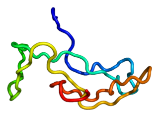
Growth/differentiation factor 9 is a protein that in humans is encoded by the GDF9 gene.

<span class="mw-page-title-main">Mothers against decapentaplegic homolog 7</span> Protein-coding gene in the species Homo sapiens

Mothers against decapentaplegic homolog 7 or SMAD7 is a protein that in humans is encoded by the SMAD7 gene.

The transforming growth factor beta (TGFB) signaling pathway is involved in many cellular processes in both the adult organism and the developing embryo including cell growth, cell differentiation, cell migration, apoptosis, cellular homeostasis and other cellular functions. The TGFB signaling pathways are conserved. In spite of the wide range of cellular processes that the TGFβ signaling pathway regulates, the process is relatively simple. TGFβ superfamily ligands bind to a type II receptor, which recruits and phosphorylates a type I receptor. The type I receptor then phosphorylates receptor-regulated SMADs (R-SMADs) which can now bind the coSMAD SMAD4. R-SMAD/coSMAD complexes accumulate in the nucleus where they act as transcription factors and participate in the regulation of target gene expression.

<span class="mw-page-title-main">Follicle-stimulating hormone receptor</span> Protein-coding gene in the species Homo sapiens

The follicle-stimulating hormone receptor or FSH receptor (FSHR) is a transmembrane receptor that interacts with the follicle-stimulating hormone (FSH) and represents a G protein-coupled receptor (GPCR). Its activation is necessary for the hormonal functioning of FSH. FSHRs are found in the ovary, testis, and uterus.

<span class="mw-page-title-main">Follistatin</span> Mammalian protein found in Homo sapiens

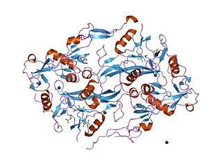
Follistatin, also known as activin-bindings protein, is a protein that in humans is encoded by the FST gene. Follistatin is an autocrine glycoprotein that is expressed in nearly all tissues of higher animals.

<span class="mw-page-title-main">ACVR1</span> Protein-coding gene

Activin A receptor, type I (ACVR1) is a protein which in humans is encoded by the ACVR1 gene; also known as ALK-2. ACVR1 has been linked to the 2q23-24 region of the genome. This protein is important in the bone morphogenic protein (BMP) pathway which is responsible for the development and repair of the skeletal system. While knock-out models with this gene are in progress, the ACVR1 gene has been connected to fibrodysplasia ossificans progressiva, an extremely rare progressive genetic disease characterized by heterotopic ossification of muscles, tendons and ligaments. It is a bone morphogenetic protein receptor, type 1.

<span class="mw-page-title-main">ACVR2A</span> Protein-coding gene in the species Homo sapiens

Activin receptor type-2A is a protein that in humans is encoded by the ACVR2A gene. ACVR2A is an activin type 2 receptor.

<span class="mw-page-title-main">ACVR2B</span> Protein-coding gene in the species Homo sapiens

Activin receptor type-2B is a protein that in humans is encoded by the ACVR2B gene. ACVR2B is an activin type 2 receptor.

<span class="mw-page-title-main">INHBA</span> Protein-coding gene in the species Homo sapiens

Inhibin, beta A, also known as INHBA, is a protein which in humans is encoded by the INHBA gene. INHBA is a subunit of both activin and inhibin, two closely related glycoproteins with opposing biological effects.

<span class="mw-page-title-main">INHA</span> Protein and coding gene in humans

Inhibin, alpha, also known as INHA, is a protein which in humans is encoded by the INHA gene.

<span class="mw-page-title-main">FSHB</span> Protein-coding gene in the species Homo sapiens

Follitropin subunit beta also known as follicle-stimulating hormone beta subunit (FSH-B) is a protein that in humans is encoded by the FSHB gene. Alternative splicing results in two transcript variants encoding the same protein.

<span class="mw-page-title-main">INHBC</span> Protein-coding gene in the species Homo sapiens

Inhibin beta C chain is a protein that in humans is encoded by the INHBC gene.

<span class="mw-page-title-main">INHBB</span> Protein-coding gene in the species Homo sapiens

Inhibin, beta B, also known as INHBB, is a protein which in humans is encoded by the INHBB gene. INHBB is a subunit of both activin and inhibin, two closely related glycoproteins with opposing biological effects.

Hypogonadotropic hypogonadism (HH), is due to problems with either the hypothalamus or pituitary gland affecting the hypothalamic-pituitary-gonadal axis. Hypothalamic disorders result from a deficiency in the release of gonadotropic releasing hormone (GnRH), while pituitary gland disorders are due to a deficiency in the release of gonadotropins from the anterior pituitary. GnRH is the central regulator in reproductive function and sexual development via the HPG axis. GnRH is released by GnRH neurons, which are hypothalamic neuroendocrine cells, into the hypophyseal portal system acting on gonadotrophs in the anterior pituitary. The release of gonadotropins, LH and FSH, act on the gonads for the development and maintenance of proper adult reproductive physiology. LH acts on Leydig cells in the male testes and theca cells in the female. FSH acts on Sertoli cells in the male and follicular cells in the female. Combined this causes the secretion of gonadal sex steroids and the initiation of folliculogenesis and spermatogenesis. The production of sex steroids forms a negative feedback loop acting on both the anterior pituitary and hypothalamus causing a pulsatile secretion of GnRH. GnRH neurons lack sex steroid receptors and mediators such as kisspeptin stimulate GnRH neurons for pulsatile secretion of GnRH.

Follicle-stimulating hormone (FSH) insensitivity, or ovarian insensitivity to FSH in females, also referable to as ovarian follicle hypoplasia or granulosa cell hypoplasia in females, is a rare autosomal recessive genetic and endocrine syndrome affecting both females and males, with the former presenting with much greater severity of symptomatology. It is characterized by a resistance or complete insensitivity to the effects of follicle-stimulating hormone (FSH), a gonadotropin which is normally responsible for the stimulation of estrogen production by the ovaries in females and maintenance of fertility in both sexes. The condition manifests itself as hypergonadotropic hypogonadism, reduced or absent puberty, amenorrhea, and infertility in females, whereas males present merely with varying degrees of infertility and associated symptoms.

References

  1. Vale W, Rivier J, Vaughan J, McClintock R, Corrigan A, Woo W, Karr D, Spiess J (1986). "Purification and characterization of an FSH releasing protein from porcine ovarian follicular fluid". Nature. 321 (6072): 776–9. Bibcode:1986Natur.321..776V. doi:10.1038/321776a0. PMID   3012369. S2CID   4365045.
  2. Ling N, Ying SY, Ueno N, Shimasaki S, Esch F, Hotta M, Guillemin R (1986). "Pituitary FSH is released by a heterodimer of the beta-subunits from the two forms of inhibin". Nature. 321 (6072): 779–82. Bibcode:1986Natur.321..779L. doi:10.1038/321779a0. PMID   3086749. S2CID   38100413.
  3. Chen YG, Wang Q, Lin SL, Chang CD, Chuang J, Chung J, Ying SY (May 2006). "Activin signaling and its role in regulation of cell proliferation, apoptosis, and carcinogenesis". Experimental Biology and Medicine. 231 (5): 534–44. doi:10.1177/153537020623100507. PMID   16636301. S2CID   39050907.
  4. Sulyok S, Wankell M, Alzheimer C, Werner S (October 2004). "Activin: an important regulator of wound repair, fibrosis, and neuroprotection". Molecular and Cellular Endocrinology. 225 (1–2): 127–32. doi:10.1016/j.mce.2004.07.011. PMID   15451577. S2CID   6943949.
  5. van Zonneveld P, Scheffer GJ, Broekmans FJ, Blankenstein MA, de Jong FH, Looman CW, Habbema JD, te Velde ER (March 2003). "Do cycle disturbances explain the age-related decline of female fertility? Cycle characteristics of women aged over 40 years compared with a reference population of young women". Human Reproduction. 18 (3): 495–501. doi: 10.1093/humrep/deg138 . PMID   12615813.
  6. Makanji Y, Zhu J, Mishra R, Holmquist C, Wong WP, Schwartz NB, Mayo KE, Woodruff TK (October 2014). "Inhibin at 90: from discovery to clinical application, a historical review". Endocrine Reviews. 35 (5): 747–94. doi:10.1210/er.2014-1003. PMC   4167436 . PMID   25051334.
  7. 1 2 Burger HG, Igarashi M (April 1988). "Inhibin: definition and nomenclature, including related substances". The Journal of Clinical Endocrinology and Metabolism. 66 (4): 885–6. PMID   3346366.
  8. 1 2 Robertson DM, Burger HG, Fuller PJ (March 2004). "Inhibin/activin and ovarian cancer". Endocrine-Related Cancer. 11 (1): 35–49. doi: 10.1677/erc.0.0110035 . PMID   15027884. S2CID   12202820.
  9. 1 2 Kingsley DM (January 1994). "The TGF-beta superfamily: new members, new receptors, and new genetic tests of function in different organisms". Genes & Development. 8 (2): 133–46. doi: 10.1101/gad.8.2.133 . PMID   8299934.
  10. Ying SY (December 1987). "Inhibins and activins: chemical properties and biological activity". Proceedings of the Society for Experimental Biology and Medicine. 186 (3): 253–64. doi:10.3181/00379727-186-42611a. PMID   3122219. S2CID   36872324.
  11. Xu P, Hall AK (November 2006). "The role of activin in neuropeptide induction and pain sensation". Developmental Biology. 299 (2): 303–9. doi:10.1016/j.ydbio.2006.08.026. PMID   16973148.
  12. Deli A, Kreidl E, Santifaller S, Trotter B, Seir K, Berger W, Schulte-Hermann R, Rodgarkia-Dara C, Grusch M (March 2008). "Activins and activin antagonists in hepatocellular carcinoma". World Journal of Gastroenterology. 14 (11): 1699–709. doi: 10.3748/wjg.14.1699 . PMC   2695910 . PMID   18350601.
  13. Mellor SL, Cranfield M, Ries R, Pedersen J, Cancilla B, de Kretser D, Groome NP, Mason AJ, Risbridger GP (December 2000). "Localization of activin beta(A)-, beta(B)-, and beta(C)-subunits in humanprostate and evidence for formation of new activin heterodimers of beta(C)-subunit". The Journal of Clinical Endocrinology and Metabolism. 85 (12): 4851–8. doi: 10.1210/jcem.85.12.7052 . PMID   11134153.
  14. Bamberger, Casimir; Schärer, Agnes; Antsiferova, Maria; Tychsen, Birte; Pankow, Sandra; Müller, Mischa; Rülicke, Thomas; Paus, Ralf; Werner, Sabine (9 March 2021). "Activin Controls Skin Morphogenesis and Wound Repair Predominantly via Stromal Cells and in a Concentration-Dependent Manner via Keratinocytes". The American Journal of Pathology. 167 (3): 733–747. doi:10.1016/S0002-9440(10)62047-0. PMC   1698729 . PMID   16127153.
  15. Pauklin S, Vallier L (2015). "Activin/Nodal signalling in stem cells". Development. 142 (4): 607–19. doi: 10.1242/dev.091769 . PMID   25670788.
  16. Luisi S, Florio P, Reis FM, Petraglia F (2005). "Inhibins in female and male reproductive physiology: role in gametogenesis, conception, implantation and early pregnancy". Human Reproduction Update. 11 (2): 123–35. doi:10.1093/humupd/dmh057. PMID   15618291.
  17. Le T, Bhushan V, Hofmann J (2012). First Aid for the USMLE Step 1 . McGraw Hill. p.  534. ISBN   978-0-07-177636-3.
  18. Skinner MK, McLachlan RI, Bremner WJ (October 1989). "Stimulation of Sertoli cell inhibin secretion by the testicular paracrine factor PModS". Molecular and Cellular Endocrinology. 66 (2): 239–49. doi:10.1016/0303-7207(89)90036-1. hdl: 1773/4395 . PMID   2515083. S2CID   21885326.
  19. Meachem SJ, Nieschlag E, Simoni M (November 2001). "Inhibin B in male reproduction: pathophysiology and clinical relevance". European Journal of Endocrinology. 145 (5): 561–71. doi: 10.1530/eje.0.1450561 . PMID   11720872.
  20. 1 2 3 Zaragosi LE, Wdziekonski B, Villageois P, Keophiphath M, Maumus M, Tchkonia T, Bourlier V, Mohsen-Kanson T, Ladoux A, Elabd C, Scheideler M, Trajanoski Z, Takashima Y, Amri EZ, Lacasa D, Sengenes C, Ailhaud G, Clément K, Bouloumie A, Kirkland JL, Dani C (2010). "Activin a plays a critical role in proliferation and differentiation of human adipose progenitors". Diabetes . 59 (10): 2513–2521. doi:10.2337/db10-0013. PMC   3279533 . PMID   20530742.
  21. 1 2 3 Shore EM, Xu M, Feldman GJ, Fenstermacher DA, Cho TJ, Choi IH, Connor JM, Delai P, Glaser DL, LeMerrer M, Morhart R, Rogers JG, Smith R, Triffitt JT, Urtizberea JA, Zasloff M, Brown MA, Kaplan FS (May 2006). "A recurrent mutation in the BMP type I receptor ACVR1 causes inherited and sporadic fibrodysplasia ossificans progressiva". Nature Genetics. 38 (5): 525–527. doi:10.1038/ng1783. PMID   16642017. S2CID   41579747.
  22. Julie Steenhuysen (2 September 2015). "Regeneron scientists discover key to excess bone growth in rare disease". Reuters.
  23. Taylor KR, Mackay A, Truffaux N, Butterfield YS, Morozova O, Philippe C, Castel D, Grasso CS, Vinci M, Carvalho D, Carcaboso AM, de Torres C, Cruz O, Mora J, Entz-Werle N, Ingram WJ, Monje M, Hargrave D, Bullock AN, Puget S, Yip S, Jones C, Grill J (May 2014). "Recurrent activating ACVR1 mutations in diffuse intrinsic pontine glioma". Nature Genetics. 46 (5): 457–61. doi:10.1038/ng.2925. PMC   4018681 . PMID   24705252.
  24. "Cure Brain Cancer - News - Multiple Breakthroughs in Childhood Brain Cancer DIPG". Cure Brain Cancer Foundation.
  25. Buczkowicz P, Hoeman C, Rakopoulos P, Pajovic S, Letourneau L, Dzamba M, et al. (May 2014). "Genomic analysis of diffuse intrinsic pontine gliomas identifies three molecular subgroups and recurrent activating ACVR1 mutations". Nature Genetics. 46 (5): 451–6. doi:10.1038/ng.2936. PMC   3997489 . PMID   24705254.
  26. Lidbury BA, Kita B, Lewis DP, Hayward S, Ludlow H, Hedger MP, de Kretser DM (March 2017). "Activin B is a novel biomarker for chronic fatigue syndrome/myalgic encephalomyelitis (CFS/ME) diagnosis: a cross sectional study". Journal of Translational Medicine. 15 (1): 60. doi: 10.1186/s12967-017-1161-4 . PMC   5353946 . PMID   28302133.
  27. Donovan P, Dubey OA, Kallioinen S, Rogers KW, Muehlethaler K, Müller P, et al. (December 2017). "Paracrine Activin-A Signaling Promotes Melanoma Growth and Metastasis through Immune Evasion". The Journal of Investigative Dermatology. 137 (12): 2578–2587. doi: 10.1016/j.jid.2017.07.845 . PMID   28844941.
  28. Aitken DA, Wallace EM, Crossley JA, Swanston IA, van Pareren Y, van Maarle M, Groome NP, Macri JN, Connor JM (May 1996). "Dimeric inhibin A as a marker for Down's syndrome in early pregnancy". The New England Journal of Medicine. 334 (19): 1231–6. doi: 10.1056/NEJM199605093341904 . PMID   8606718.
  29. Robertson DM, Pruysers E, Jobling T (April 2007). "Inhibin as a diagnostic marker for ovarian cancer". Cancer Letters. 249 (1): 14–7. doi:10.1016/j.canlet.2006.12.017. PMID   17320281.
  30. Robertson DM, Pruysers E, Burger HG, Jobling T, McNeilage J, Healy D (October 2004). "Inhibins and ovarian cancer". Molecular and Cellular Endocrinology. 225 (1–2): 65–71. doi:10.1016/j.mce.2004.02.014. PMID   15451569. S2CID   33801243.
  31. Myers GM, Lambert-Messerlian GM, Sigman M (December 2009). "Inhibin B reference data for fertile and infertile men in Northeast America". Fertility and Sterility. 92 (6): 1920–3. doi: 10.1016/j.fertnstert.2008.09.033 . PMID   19006797.
  32. Toulis KA, Iliadou PK, Venetis CA, Tsametis C, Tarlatzis BC, Papadimas I, Goulis DG (2010). "Inhibin B and anti-Mullerian hormone as markers of persistent spermatogenesis in men with non-obstructive azoospermia: a meta-analysis of diagnostic accuracy studies". Human Reproduction Update. 16 (6): 713–24. doi: 10.1093/humupd/dmq024 . PMID   20601364.