Corpus luteum

Last updated
Corpus luteum
Gray1163.png
Section of the ovary. 1. Outer covering. 1’. Attached border. 2. Central stroma. 3. Peripheral stroma. 4. Bloodvessels. 5. Vesicular follicles in their earliest stage. 6, 7, 8. More advanced follicles. 9. An almost mature follicle. 9’. Follicle from which the ovum has escaped. 10. Corpus luteum.
Details
System Reproductive system
Location Ovary
Identifiers
Latin corpus luteum
MeSH D003338
TA98 A09.1.01.015
TA2 3484
FMA 18619
Anatomical terms of microanatomy

The corpus luteum (Latin for "yellow body"; pl.: corpora lutea) is a temporary endocrine structure in female ovaries involved in the production of relatively high levels of progesterone, and moderate levels of estradiol, and inhibin A. [1] [2] It is the remains of the ovarian follicle that has released a mature ovum during a previous ovulation. [3]

Contents

The corpus luteum is colored as a result of concentrating carotenoids (including lutein) from the diet and secretes a moderate amount of estrogen that inhibits further release of gonadotropin-releasing hormone (GnRH) and thus secretion of luteinizing hormone (LH) and follicle-stimulating hormone (FSH). A new corpus luteum develops with each menstrual cycle.

Development and structure

The corpus luteum develops from an ovarian follicle during the luteal phase of the menstrual cycle or oestrous cycle, following the release of a secondary oocyte from the follicle during ovulation. The follicle first forms a corpus hemorrhagicum before it becomes a corpus luteum, but the term refers to the visible collection of blood, left after rupture of the follicle, that secretes progesterone. While the oocyte (later the zygote if fertilization occurs) traverses the fallopian tube into the uterus, the corpus luteum remains in the ovary.[ citation needed ]

The corpus luteum is typically very large relative to the size of the ovary; in humans, the size of the structure ranges from under 2 cm to 5 cm in diameter. [4]

Its cells develop from the follicular cells surrounding the ovarian follicle. [5] The follicular theca cells luteinize into small luteal cells (thecal-lutein cells) and follicular granulosa cells luteinize into large luteal cells (granulosal-lutein cells) forming the corpus luteum. Progesterone is synthesized from cholesterol by both the large and small luteal cells upon luteal maturation. Cholesterol-LDL complexes bind to receptors on the plasma membrane of luteal cells and are internalized. Cholesterol is released and stored within the cell as cholesterol ester. LDL is recycled for further cholesterol transport. Large luteal cells produce more progesterone due to uninhibited/basal levels of protein kinase A (PKA) activity within the cell. Small luteal cells have LH receptors that regulate PKA activity within the cell. PKA actively phosphorylates steroidogenic acute regulatory protein (StAR) and translocator protein to transport cholesterol from the outer mitochondrial membrane to the inner mitochondrial membrane. [6]

The development of the corpus luteum is accompanied by an increase in the level of the steroidogenic enzyme P450scc that converts cholesterol to pregnenolone in the mitochondria. [7] Pregnenolone is then converted to progesterone that is secreted out of the cell and into the blood stream. During the bovine estrous cycle, plasma levels of progesterone increase in parallel to the levels of P450scc and its electron donor adrenodoxin, indicating that progesterone secretion is a result of enhanced expression of P450scc in the corpus luteum. [7]

The mitochondrial P450 system electron transport chain including adrenodoxin reductase and adrenodoxin has been shown to leak electrons leading to the formation of superoxide radical. [8] [9] Apparently to cope with the radicals produced by this system and by enhanced mitochondrial metabolism, the levels of antioxidant enzymes catalase and superoxide dismutase also increase in parallel with the enhanced steroidogenesis in the corpus luteum. [7]

Follicular structureLuteal structureSecretion
Theca cells Theca lutein cells androgens, [10] progesterone [10]
Granulosa cells Granulosa lutein cellsprogesterone, [5] estrogen(majority), [5] and inhibin A [5] [10]
Steroidogenesis, with progesterone in yellow field at upper center. The androgens are shown in blue field, and aromatase at lower center - the enzyme present in granulosa lutein cells that convert androgens into estrogens (shown in pink triangle). Steroidogenesis.svg
Steroidogenesis, with progesterone in yellow field at upper center. The androgens are shown in blue field, and aromatase at lower center – the enzyme present in granulosa lutein cells that convert androgens into estrogens (shown in pink triangle).

Like the previous theca cells, the theca lutein cells lack the aromatase enzyme that is necessary to produce estrogen, so they can only perform steroidogenesis until formation of androgens. The granulosa lutein cells do have aromatase, and use it to produce estrogens, using the androgens previously synthesized by the theca lutein cells, as the granulosa lutein cells in themselves do not have the 17α-hydroxylase or 17,20 lyase to produce androgens. [5] Once the corpus luteum regresses the remnant is known as corpus albicans. [12]

Function

The corpus luteum is essential for establishing and maintaining pregnancy in females. The corpus luteum secretes progesterone, which is a steroid hormone responsible for the decidualization of the endometrium (its development) and maintenance, respectively. It also produces relaxin, a hormone responsible for softening of the pubic symphysis which helps in parturition. [13]

Unsuccessful fertilisation

If the egg is not fertilised, the corpus luteum stops secreting progesterone and decays (after approximately 10 days in humans). It then degenerates into a corpus albicans, which is a mass of fibrous scar tissue. [14]

With cessation of progesterone release, the uterine lining (functional, inner layer of the endometrium) is expelled through the vagina (in mammals that go through a menstrual cycle). Across an estrous cycle, the functional layer regenerates to provide nourishing tissue for potential fertilisation and implantation. [15] [16]

Successful fertilisation

Vaginal ultrasound showing a corpus luteum in a pregnant woman, with a fluid-filled cavity in its center Ultrasonography of corpus luteum.jpg
Vaginal ultrasound showing a corpus luteum in a pregnant woman, with a fluid-filled cavity in its center

If the egg is fertilised and implantation occurs, the syncytiotrophoblast (derived from trophoblast) cells of the blastocyst secrete the hormone human chorionic gonadotropin (hCG, or a similar hormone in other species) by day 9 post-fertilisation.[ citation needed ]

Human chorionic gonadotropin signals the corpus luteum to continue progesterone secretion, thereby maintaining the thick lining (endometrium) of the uterus and providing an area rich in blood vessels in which the zygote(s) can develop. From this point on, the corpus luteum is called the corpus luteum graviditatis. [17]

The introduction of prostaglandins at this point causes the degeneration of the corpus luteum and the abortion of the fetus. However, in placental animals such as humans, the placenta eventually takes over progesterone production and the corpus luteum degrades into a corpus albicans without embryo/fetus loss.[ citation needed ]

Luteal support refers to the administration of medication (generally progestins) for the purpose of increasing the success of implantation and early embryogenesis, thereby complementing the function of the corpus luteum.

Content of carotenoids

The yellow color and name of the corpus luteum, like that of the macula lutea of the retina, is due to its concentration of certain carotenoids, especially lutein. In 1968, a report indicated that beta-carotene was synthesized in laboratory conditions in slices of corpus luteum from cows. However, attempts have been made to replicate these findings, but have not succeeded. The idea is not presently accepted by the scientific community. [18] Rather, the corpus luteum concentrates carotenoids from the diet of the mammal.

In animals

Similar structures and functions of the corpus luteum exist in some reptiles. [19] Dairy cattle also follow a similar cycle. [20]

Additional images

Pathology

See also

Related Research Articles

<span class="mw-page-title-main">Ovary</span> Female reproductive organ that produces egg cells

The ovary is a gonad in the female reproductive system that produces ova. When an ovum is released, this travels through the fallopian tube into the uterus. There is an ovary found on the left and the right side of the body. The ovaries also secrete hormones that play a role in the menstrual cycle and fertility. The ovary progresses through many stages beginning in the prenatal period through menopause. It is also an endocrine gland because of the various hormones that it secretes.

<span class="mw-page-title-main">Menstrual cycle</span> Natural changes in the human female reproductive system

The menstrual cycle is a series of natural changes in hormone production and the structures of the uterus and ovaries of the female reproductive system that makes pregnancy possible. The ovarian cycle controls the production and release of eggs and the cyclic release of estrogen and progesterone. The uterine cycle governs the preparation and maintenance of the lining of the uterus (womb) to receive an embryo. These cycles are concurrent and coordinated, normally last between 21 and 35 days, with a median length of 28 days, and continue for about 30–45 years.

<span class="mw-page-title-main">Ovulation</span> Release of egg cells from the ovaries

Ovulation is the release of eggs from the ovaries. In women, this event occurs when the ovarian follicles rupture and release the secondary oocyte ovarian cells. After ovulation, during the luteal phase, the egg will be available to be fertilized by sperm. In addition, the uterine lining (endometrium) is thickened to be able to receive a fertilized egg. If no conception occurs, the uterine lining as well as the egg will be shed during menstruation.

<span class="mw-page-title-main">Luteinizing hormone</span> Gonadotropin secreted by the adenohypophysis

Luteinizing hormone is a hormone produced by gonadotropic cells in the anterior pituitary gland. The production of LH is regulated by gonadotropin-releasing hormone (GnRH) from the hypothalamus. In females, an acute rise of LH known as an LH surge, triggers ovulation and development of the corpus luteum. In males, where LH had also been called interstitial cell–stimulating hormone (ICSH), it stimulates Leydig cell production of testosterone. It acts synergistically with follicle-stimulating hormone (FSH).

<span class="mw-page-title-main">Follicle-stimulating hormone</span> Gonadotropin that regulates the development of reproductive processes

Follicle-stimulating hormone (FSH) is a gonadotropin, a glycoprotein polypeptide hormone. FSH is synthesized and secreted by the gonadotropic cells of the anterior pituitary gland and regulates the development, growth, pubertal maturation, and reproductive processes of the body. FSH and luteinizing hormone (LH) work together in the reproductive system.

Israel Hanukoglu is a Turkish-born Israeli scientist. He is a full professor of biochemistry and molecular biology at Ariel University and former science and technology adviser to the prime minister of Israel (1996–1999). He is founder of Israel Science and Technology Directory.

<span class="mw-page-title-main">Ovarian follicle</span> Structure containing a single egg cell

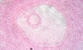
An ovarian follicle is a roughly spheroid cellular aggregation set found in the ovaries. It secretes hormones that influence stages of the menstrual cycle. At the time of puberty, women have approximately 200,000 to 300,000 follicles, each with the potential to release an egg cell (ovum) at ovulation for fertilization. These eggs are developed once every menstrual cycle with around 450–500 being ovulated during a woman's reproductive lifetime.

<span class="mw-page-title-main">Corpus albicans</span> Structure derived from the corpus luteum

The corpus albicans is the regressed form of the corpus luteum. As the corpus luteum is being broken down by macrophages, fibroblasts lay down type I collagen, forming the corpus albicans. This process is called "luteolysis". The remains of the corpus albicans may persist as a scar on the surface of the ovary.

<span class="mw-page-title-main">Granulosa cell</span>

A granulosa cell or follicular cell is a somatic cell of the sex cord that is closely associated with the developing female gamete in the ovary of mammals.

The estrous cycle is a set of recurring physiological changes induced by reproductive hormones in females of mammalian subclass Theria. Estrous cycles start after sexual maturity in females and are interrupted by anestrous phases, otherwise known as "rest" phases, or by pregnancies. Typically, estrous cycles repeat until death. These cycles are widely variable in duration and frequency depending on the species. Some animals may display bloody vaginal discharge, often mistaken for menstruation. Many mammals used in commercial agriculture, such as cattle and sheep, may have their estrous cycles artificially controlled with hormonal medications for optimum productivity. The male equivalent, seen primarily in ruminants, is called rut.

<span class="mw-page-title-main">Folliculogenesis</span> Process of maturation of primordial follicles

In biology, folliculogenesis is the maturation of the ovarian follicle, a densely packed shell of somatic cells that contains an immature oocyte. Folliculogenesis describes the progression of a number of small primordial follicles into large preovulatory follicles that occurs in part during the menstrual cycle.

Luteolysis is the structural and functional degradation of the corpus luteum, which occurs at the end of the luteal phase of both the estrous and menstrual cycles in the absence of pregnancy.

<span class="mw-page-title-main">Luteal phase</span> The latter part of the menstrual cycle associated with ovulation and an increase in progesterone

The menstrual cycle is on average 28 days in length. It begins with menses during the follicular phase, followed by ovulation and ending with the luteal phase. Unlike the follicular phase which can vary in length among individuals, the luteal phase is typically fixed at approximately 14 days and is characterized by changes to hormone levels, such as an increase in progesterone and estrogen levels, decrease in gonadotropins such as follicle-stimulating hormone (FSH) and luteinizing hormone (LH), changes to the endometrial lining to promote implantation of the fertilized egg, and development of the corpus luteum. In the absence of fertilization by sperm, the corpus luteum atrophies leading to a decrease in progesterone and estrogen, an increase in FSH and LH, and shedding of the endometrial lining (menses) to begin the menstrual cycle again.

<span class="mw-page-title-main">Follicular phase</span> Phase of the estrous or menstrual cycle

The follicular phase, also known as the preovulatory phase or proliferative phase, is the phase of the estrous cycle during which follicles in the ovary mature from primary follicle to a fully mature graafian follicle. It ends with ovulation. The main hormones controlling this stage are secretion of gonadotropin-releasing hormones, which are follicle-stimulating hormones and luteinising hormones. They are released by pulsatile secretion. The duration of the follicular phase can differ depending on the length of the menstrual cycle, while the luteal phase is usually stable, does not really change and lasts 14 days.

<span class="mw-page-title-main">Follicular atresia</span>

Follicular atresia refers to the process in which a follicle fails to develop, thus preventing it from ovulating and releasing an egg. It is a normal, naturally occurring progression that occurs as mammalian ovaries age. Approximately 1% of mammalian follicles in ovaries undergo ovulation and the remaining 99% of follicles go through follicular atresia as they cycle through the growth phases. In summary, follicular atresia is a process that leads to the follicular loss and loss of oocytes, and any disturbance or loss of functionality of this process can lead to many other conditions.

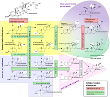
Theca interna cells express receptors for luteinizing hormone (LH) to produce androstenedione, which via a few steps, gives the granulosa the precursor for estrogen manufacturing.

<span class="mw-page-title-main">Cholesterol side-chain cleavage enzyme</span> Mammalian protein found in Homo sapiens

Cholesterol side-chain cleavage enzyme is commonly referred to as P450scc, where "scc" is an acronym for side-chain cleavage. P450scc is a mitochondrial enzyme that catalyzes conversion of cholesterol to pregnenolone. This is the first reaction in the process of steroidogenesis in all mammalian tissues that specialize in the production of various steroid hormones.

The theca folliculi comprise a layer of the ovarian follicles. They appear as the follicles become secondary follicles.

Theca lutein cyst is a type of bilateral functional ovarian cyst filled with clear, straw-colored fluid. These cysts result from exaggerated physiological stimulation due to elevated levels of beta-human chorionic gonadotropin (beta-hCG) or hypersensitivity to beta-hCG. On ultrasound and MRI, theca lutein cysts appear in multiples on ovaries that are enlarged.

Gonadotropin surge-attenuating factor (GnSAF) is a nonsteroidal ovarian hormone produced by the granulosa cells of small antral ovarian follicles in females. GnSAF is involved in regulating the secretion of luteinizing hormone (LH) from the anterior pituitary and the ovarian cycle. During the early to mid-follicular phase of the ovarian cycle, GnSAF acts on the anterior pituitary to attenuate LH release, limiting the secretion of LH to only basal levels. At the transition between follicular and luteal phase, GnSAF bioactivity declines sufficiently to permit LH secretion above basal levels, resulting in the mid-cycle LH surge that initiates ovulation. In normally ovulating women, the LH surge only occurs when the oocyte is mature and ready for extrusion. GnSAF bioactivity is responsible for the synchronised, biphasic nature of LH secretion.

References

  1. "Histology Laboratory Manual". www.columbia.edu. Archived from the original on 6 May 2017. Retrieved 3 May 2018.
  2. Inquiry Into Biology (Textbook). McGraw-Hill Ryerson. 2007. p. 497. ISBN   978-0-07-096052-7.
  3. Karch 2017, p. 657.
  4. Vegetti W, Alagna F (2006). "FSH and follucogenesis: from physiology to ovarian stimulation". Reproductive biomedicine Online. Archived from the original on 2011-07-15. Retrieved 2009-05-26.
  5. 1 2 3 4 5 Boron 2005, p. 1300.
  6. Niswender GD (March 2002). "Molecular control of luteal secretion of progesterone". Reproduction. 123 (3): 333–9. doi: 10.1530/rep.0.1230333 . PMID   11882010.
  7. 1 2 3 Rapoport R, Sklan D, Wolfenson D, Shaham-Albalancy A, Hanukoglu I (March 1998). "Antioxidant capacity is correlated with steroidogenic status of the corpus luteum during the bovine estrous cycle". Biochim. Biophys. Acta. 1380 (1): 133–40. doi:10.1016/S0304-4165(97)00136-0. PMID   9545562.
  8. Hanukoglu I, Rapoport R, Weiner L, Sklan D (September 1993). "Electron leakage from the mitochondrial NADPH-adrenodoxin reductase-adrenodoxin-P450scc (cholesterol side chain cleavage) system". Arch. Biochem. Biophys. 305 (2): 489–98. doi:10.1006/abbi.1993.1452. PMID   8396893.
  9. Rapoport R, Sklan D, Hanukoglu I (March 1995). "Electron leakage from the adrenal cortex mitochondrial P450scc and P450c11 systems: NADPH and steroid dependence". Arch. Biochem. Biophys. 317 (2): 412–6. doi:10.1006/abbi.1995.1182. PMID   7893157.
  10. 1 2 3 The IUPS Physiome Project --> Female Reproductive System – Cells Archived 2009-12-10 at the Wayback Machine Retrieved on Nov 9, 2009
  11. Häggström, Mikael; Richfield, David (2014). "Diagram of the pathways of human steroidogenesis". WikiJournal of Medicine. 1 (1). doi: 10.15347/wjm/2014.005 . ISSN   2002-4436.
  12. Marieb, Elaine (2013). Anatomy & physiology. Benjamin-Cummings. p. 915. ISBN   9780321887603.
  13. Wang, Yan; Li, Yong-Qiang; Tian, Mei-Rong; Wang, Nan; Zheng, Zun-Cheng (2021-01-06). "Role of relaxin in diastasis of the pubic symphysis peripartum". World Journal of Clinical Cases. 9 (1): 91–101. doi: 10.12998/wjcc.v9.i1.91 . ISSN   2307-8960. PMC   7809669 . PMID   33511175.
  14. Kirkendoll, Shelbie D.; Bacha, Dhouha (2023), "Histology, Corpus Albicans", StatPearls, Treasure Island (FL): StatPearls Publishing, PMID   31424854 , retrieved 2023-11-16
  15. Critchley, Hilary O. D.; Babayev, Elnur; Bulun, Serdar E.; Clark, Sandy; Garcia-Grau, Iolanda; Gregersen, Peter K.; Kilcoyne, Aoife; Kim, Ji-Yong Julie; Lavender, Missy; Marsh, Erica E.; Matteson, Kristen A.; Maybin, Jacqueline A.; Metz, Christine N.; Moreno, Inmaculada; Silk, Kami (November 2020). "Menstruation: science and society". American Journal of Obstetrics and Gynecology. 223 (5): 624–664. doi:10.1016/j.ajog.2020.06.004. ISSN   1097-6868. PMC   7661839 . PMID   32707266.
  16. Campo, Hannes; Murphy, Alina; Yildiz, Sule; Woodruff, Teresa; Cervelló, Irene; Kim, J. Julie (July 2020). "Microphysiological Modeling of the Human Endometrium". Tissue Engineering. Part A. 26 (13–14): 759–768. doi:10.1089/ten.tea.2020.0022. ISSN   1937-335X. PMC   7398432 . PMID   32348708.
  17. Oliver, Rebecca; Pillarisetty, Leela Sharath (2023), "Anatomy, Abdomen and Pelvis, Ovary Corpus Luteum", StatPearls, Treasure Island (FL): StatPearls Publishing, PMID   30969526 , retrieved 2023-11-16
  18. Brian Davis. Carotenoid metabolism as a preparation for function. Pure and Applied Chemistry, Vol. 63, No. 1, pp. 131–140, 1991. available online. Archived 2011-07-26 at the Wayback Machine Accessed April 30, 2010.
  19. Trauth, Stanley E. (1978). "Ovarian Cycle of Crotaphytus collaris (Reptilia, Lacertilia, Iguanidae) from Arkansas with Emphasis on Corpora Albicantia, Follicular Atresia, and Reproductive Potential". Journal of Herpetology. 12 (4): 461–470. doi:10.2307/1563350. ISSN   0022-1511. JSTOR   1563350.
  20. Dairy cattle fertility & sterility. Fort Atkinson, Wis: W.D. Hoard & Sons. 1996. ISBN   978-0932147271.{{cite book}}: |work= ignored (help)

Bibliography