Ovarian follicle

Last updated
Ovarian follicle
Graafian Follicle, Human Ovary (3595817584).jpg
Histology section of a mature ovarian follicle. The oocyte is the large, round, pink-staining cell at top center of the image.
Details
Precursor Cortical cords
Identifiers
Latin folliculus ovaricus
MeSH D006080
TA98 A09.1.01.013
TA2 3482
FMA 18640
Anatomical terminology

An ovarian follicle is a roughly spheroid cellular aggregation set found in the ovaries. It secretes hormones that influence stages of the menstrual cycle. At the time of puberty, women have approximately 200,000 to 300,000 follicles, [1] [2] each with the potential to release an egg cell (ovum) at ovulation for fertilization. [3] These eggs are developed once every menstrual cycle with around 450–500 being ovulated during a woman's reproductive lifetime. [4]

Contents

Structure

Section of vesicular ovarian follicle of cat. X 50. Gray1164.png
Section of vesicular ovarian follicle of cat. X 50.

Ovarian follicles are the basic units of female reproductive biology. Each of them contains a single oocyte (immature ovum or egg cell). These structures are periodically initiated to grow and develop, culminating in ovulation of usually a single competent oocyte in humans. [5] They also consist of granulosa cells and theca of follicle.

Oocyte

Once a month, one of the ovaries releases a mature egg (ovum), known as an oocyte. The nucleus of such an oocyte is called a germinal vesicle [6] (see picture).

Cumulus oophorus

Cumulus oophorus is a cluster of cells (called cumulus cells) that surround the oocyte both in the ovarian follicle and after ovulation.

Membrana granulosa

It contains numerous granulosa cells.

Granulosa cell

Granulosa cells or follicular cells are cells that surround the oocyte within the follicle; their numbers increase directly in response to heightened levels of circulating gonadotropins or decrease in response to testosterone. They also produce peptides involved in ovarian hormone synthesis regulation. Follicle-stimulating hormone (FSH) induces granulosa cells to express luteinizing hormone (LH) receptors on their surfaces; when circulating LH binds to these receptors, proliferation stops. [7]

Theca of follicle

The granulosa cells, in turn, are enclosed in a thin layer of extracellular matrix – the follicular basement membrane or basal lamina (fibro-vascular coat in picture). Outside the basal lamina, the layers theca interna and theca externa are found.

Development

Primordial follicles are indiscernible to the naked eye. However, these eventually develop into primary, secondary and tertiary vesicular follicles. Tertiary vesicular follicles (also called "mature vesicular follicles" or "ripe vesicular follicles") are sometimes called Graafian follicles (after Regnier de Graaf).

In humans, oocytes are established in the ovary before birth and may lie dormant awaiting initiation for up to 50 years. [8]

After rupturing, the follicle is turned into a corpus luteum.

Development of oocytes in ovarian follicles

In a larger perspective, the whole folliculogenesis from primordial to preovulatory follicle is located in the stage of meiosis I of ootidogenesis in oogenesis.

Embryonic development in males and females follows a common pathway before gametogenesis. Once gametogonia enter the gonadal ridge, however, they attempt to associate with these somatic cells. Development proceeds and the gametogonia turn into oogonia, which become fully surrounded by a layer of cells (pre-granulosa cells).

The oogonia multiply by dividing mitotically; this proliferation ends when the oogonia enter meiosis. The amount of time that oogonia multiply by mitosis is not species specific. In the human fetus, cells undergoing mitosis are seen until the second and third trimester of pregnancy. [9] [10] After beginning the meiotic process, the oogonia (now called primary oocytes) can no longer replicate. Therefore, the total number of gametes is established at this time. Once the primary oocytes stop dividing the cells enter a prolonged 'resting phase'. This 'resting phase' or dictyate stage can last anywhere up to fifty years in the human.

For several primary oocytes that complete meiosis I each month, only one or a few functional oocyte, the dominant follicles, completes maturation and undergoes ovulation. The other follicles that begin to mature will regress and become atretic follicles, eventually deteriorating.

The primary oocyte turns into a secondary oocyte in mature ovarian follicles. Unlike the sperm, the egg is arrested in the secondary stage of meiosis until fertilization.

Upon fertilization by sperm, the secondary oocyte continues the second part of meiosis and becomes a zygote.

Clinical significance

Any ovarian follicle that is larger than about three centimeters is termed an ovarian cyst.

Ovarian function may be measured by gynecologic ultrasonography of follicular volume. Presently, ovarian follicle volumes can be measured rapidly and automatically from three-dimensionally reconstructed ultrasound images. [11]

Rupture of the follicle can result in abdominal pain (mittelschmerz) and is to be considered in the differential diagnosis in people of childbearing age. [12]

Cryopreservation and culture tissue after cryopreservation. Cryopreservation of ovarian tissue is of interest to people who want to preserve their reproductive function beyond the natural limit, or whose reproductive potential is threatened by cancer therapy, [13] for example in hematologic malignancies or breast cancer. [14]

For in vitro culture of follicles, there are various techniques to optimize the growth of follicles, including the use of defined media, growth factors and three-dimensional extracellular matrix support. [15] Molecular methods and immunoassay can evaluate stage of maturation and guide adequate differentiation. [15] Animal studies have generally shown correct imprinted DNA methylation establishment in oocytes resulting from follicle culture. [16]

Additional images

See also

Related Research Articles

<span class="mw-page-title-main">Ovary</span> Female reproductive organ that produces egg cells

The ovary is a gonad in the female reproductive system that produces ova. When an ovum is released, this travels through the fallopian tube into the uterus. There is an ovary found on the left and the right side of the body. The ovaries also secrete hormones that play a role in the menstrual cycle and fertility. The ovary progresses through many stages beginning in the prenatal period through menopause. It is also an endocrine gland because of the various hormones that it secretes.

<span class="mw-page-title-main">Menstrual cycle</span> Natural changes in the human female reproductive system

The menstrual cycle is a series of natural changes in hormone production and the structures of the uterus and ovaries of the female reproductive system that makes pregnancy possible. The ovarian cycle controls the production and release of eggs and the cyclic release of estrogen and progesterone. The uterine cycle governs the preparation and maintenance of the lining of the uterus (womb) to receive an embryo. These cycles are concurrent and coordinated, normally last between 21 and 35 days, with a median length of 28 days, and continue for about 30–45 years.

<span class="mw-page-title-main">Ovulation</span> Release of egg cells from the ovaries

Ovulation is the release of eggs from the ovaries. In women, this event occurs when the ovarian follicles rupture and release the secondary oocyte ovarian cells. After ovulation, during the luteal phase, the egg will be available to be fertilized by sperm. In addition, the uterine lining (endometrium) is thickened to be able to receive a fertilized egg. If no conception occurs, the uterine lining as well as the egg will be shed during menstruation.

<span class="mw-page-title-main">Germ cell</span> Gamete-producing cell

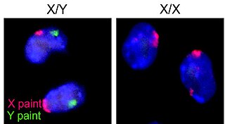
A germ cell is any cell that gives rise to the gametes of an organism that reproduces sexually. In many animals, the germ cells originate in the primitive streak and migrate via the gut of an embryo to the developing gonads. There, they undergo meiosis, followed by cellular differentiation into mature gametes, either eggs or sperm. Unlike animals, plants do not have germ cells designated in early development. Instead, germ cells can arise from somatic cells in the adult, such as the floral meristem of flowering plants.

<span class="mw-page-title-main">Corpus luteum</span> Temporary endocrine structure in ovaries

The corpus luteum is a temporary endocrine structure in female ovaries involved in the production of relatively high levels of progesterone, and moderate levels of estradiol, and inhibin A. It is the remains of the ovarian follicle that has released a mature ovum during a previous ovulation.

<span class="mw-page-title-main">Oogenesis</span> Egg cell production process

Oogenesis, ovogenesis, or oögenesis is the differentiation of the ovum into a cell competent to further develop when fertilized. It is developed from the primary oocyte by maturation. Oogenesis is initiated in the embryonic stage.

<span class="mw-page-title-main">Granulosa cell</span>

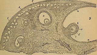
A granulosa cell or follicular cell is a somatic cell of the sex cord that is closely associated with the developing female gamete in the ovary of mammals.

<span class="mw-page-title-main">Folliculogenesis</span> Process of maturation of primordial follicles

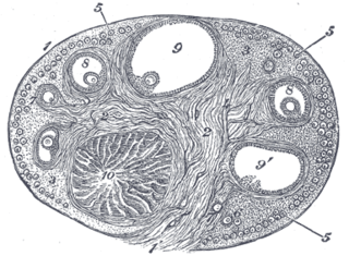
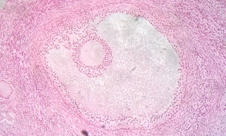
In biology, folliculogenesis is the maturation of the ovarian follicle, a densely packed shell of somatic cells that contains an immature oocyte. Folliculogenesis describes the progression of a number of small primordial follicles into large preovulatory follicles that occurs in part during the menstrual cycle.

<span class="mw-page-title-main">Growth differentiation factor-9</span> Protein-coding gene in the species Homo sapiens

Growth/differentiation factor 9 is a protein that in humans is encoded by the GDF9 gene.

<span class="mw-page-title-main">Follicular atresia</span>

Follicular atresia refers to the process in which a follicle fails to develop, thus preventing it from ovulating and releasing an egg. It is a normal, naturally occurring progression that occurs as mammalian ovaries age. Approximately 1% of mammalian follicles in ovaries undergo ovulation and the remaining 99% of follicles go through follicular atresia as they cycle through the growth phases. In summary, follicular atresia is a process that leads to the follicular loss and loss of oocytes, and any disturbance or loss of functionality of this process can lead to many other conditions.

The theca folliculi comprise a layer of the ovarian follicles. They appear as the follicles become secondary follicles.

<span class="mw-page-title-main">Antral follicle</span>

An antral or secondary follicle, also known as Graafian follicle and tertiary follicle, is an ovarian follicle during a certain latter stage of folliculogenesis.

<span class="mw-page-title-main">In vitro maturation</span> Artificial maturation of harvested immature egg cells

In vitro maturation (IVM) is the technique of letting the contents of ovarian follicles and the oocytes inside mature in vitro. It can be offered to women with infertility problems, combined with In Vitro Fertilization (IVF), offering women pregnancy without ovarian stimulation.

Transvaginal oocyte retrieval (TVOR), also referred to as oocyte retrieval (OCR), is a technique used in in vitro fertilization (IVF) in order to remove oocytes from an ovary, enabling fertilization outside the body. Transvaginal oocyte retrieval is more properly referred to as transvaginal ovum retrieval when the oocytes have matured into ova, as is normally the case in IVF. It can be also performed for egg donation, oocyte cryopreservation and other assisted reproduction technology such as ICSI.

Ovarian follicle activation can be defined as primordial follicles in the ovary moving from a quiescent (inactive) to a growing phase. The primordial follicle in the ovary is what makes up the “pool” of follicles that will be induced to enter growth and developmental changes that change them into pre-ovulatory follicles, ready to be released during ovulation. The process of development from a primordial follicle to a pre-ovulatory follicle is called folliculogenesis.

Gonadotropin surge-attenuating factor (GnSAF) is a nonsteroidal ovarian hormone produced by the granulosa cells of small antral ovarian follicles in females. GnSAF is involved in regulating the secretion of luteinizing hormone (LH) from the anterior pituitary and the ovarian cycle. During the early to mid-follicular phase of the ovarian cycle, GnSAF acts on the anterior pituitary to attenuate LH release, limiting the secretion of LH to only basal levels. At the transition between follicular and luteal phase, GnSAF bioactivity declines sufficiently to permit LH secretion above basal levels, resulting in the mid-cycle LH surge that initiates ovulation. In normally ovulating women, the LH surge only occurs when the oocyte is mature and ready for extrusion. GnSAF bioactivity is responsible for the synchronised, biphasic nature of LH secretion.

Ovarian follicle dominance is the process where one or more follicles are selected per cycle to ovulate.

<span class="mw-page-title-main">Artificial ovary</span>

An artificial ovary is a potential fertility preservation treatment that aims to mimic the function of the natural ovary.

Ovarian culture is an in-vitro process that allows for the investigation of the development, toxicology and pathology of the ovary. This technique can also be used to study possible applications of fertility treatments e.g. isolating oocytes from primordial ovarian follicles that could be used for fertilisation.

<span class="mw-page-title-main">Ovarian stem cell</span>

Ovarian stem cells are oocytes formed in ovarian follicle before birth in female mammals. They do not form post-natally, and are depleted throughout reproductive life. In humans it is estimated that 500,000–1,000,000 primordial follicles are present at birth, decreasing rapidly with age until roughly age 51 when ovulation stops, resulting in menopause. The origin of these oocytes remains under discussion. The publication of a study in 2004 proposing germ cell renewal in adult mice sparked a debate on the possibility of stem cells in the postnatal ovary. An increasing number of studies suggest that stem cells exist within the mammalian ovary and can be manipulated in vitro to produce oocytes, but whether such ovarian stem cells have the potential to differentiate into oocytes remains uncertain.

References

  1. McGee, Elizabeth A.; Hsueh, Aaron J. W. (2000). "Initial and Cyclic Recruitment of Ovarian Follicles". Endocrine Reviews. 21 (2): 200–214. doi: 10.1210/edrv.21.2.0394 . PMID   10782364.
  2. Krogh D (2010). Biology: A Guide to the Natural World. Benjamin-Cummings Publishing Company. p. 638. ISBN   978-0-321-61655-5.
  3. "What Is an Ovarian Follicle?". wiseGEEK.org. wiseGEEK. Archived from the original on 24 May 2015. Retrieved 24 May 2015.
  4. "Your Guide to the Female Reproductive System".
  5. Luijkx T. "Ovarian follicle". radiopaedia.org. radiopaedia.org. Archived from the original on 26 May 2015. Retrieved 24 May 2015.
  6. "Germinal vesicle - Biology-Online Dictionary". www.biology-online.org. Archived from the original on 2007-10-08.
  7. Katz: Comprehensive Gynecology, 5th ed.
  8. McGee EA, Hsueh AJ (April 2000). "Initial and cyclic recruitment of ovarian follicles". Endocrine Reviews. 21 (2): 200–14. doi: 10.1210/edrv.21.2.0394 . PMID   10782364.
  9. Baker, T. G. (1982). Oogenesis and ovulation. In "Book 1: Germ cells and fertilization" (C. R. Austin and R. V. Short, Eds.), pp. 17-45. Cambridge University Press, Cambridge.
  10. Byskov AG, Hoyer PE (1988). "Embryology of mammalian gonads and ducts.". In Knobil E, Neill J (eds.). The physiology of reproduction. New York: Raven Press, Ltd. pp. 265–302.
  11. Salama S, Arbo E, Lamazou F, Levailllant JM, Frydman R, Fanchin R (April 2010). "Reproducibility and reliability of automated volumetric measurement of single preovulatory follicles using SonoAVC". Fertility and Sterility. 93 (6): 2069–73. doi: 10.1016/j.fertnstert.2008.12.115 . PMID   19342038.
  12. "Acute Abdominal Pain - Gastrointestinal Disorders - Merck Manuals Professional Edition". merck.com. Archived from the original on 2010-05-24.
  13. Isachenko V, Lapidus I, Isachenko E, Krivokharchenko A, Kreienberg R, Woriedh M, et al. (August 2009). "Human ovarian tissue vitrification versus conventional freezing: morphological, endocrinological, and molecular biological evaluation". Reproduction. 138 (2): 319–27. doi: 10.1530/REP-09-0039 . PMID   19439559.
  14. Oktay K, Oktem O (February 2010). "Ovarian cryopreservation and transplantation for fertility preservation for medical indications: report of an ongoing experience". Fertility and Sterility. 93 (3): 762–8. doi: 10.1016/j.fertnstert.2008.10.006 . PMID   19013568.
  15. 1 2 Smitz J, Dolmans MM, Donnez J, Fortune JE, Hovatta O, Jewgenow K, et al. (February 2010). "Current achievements and future research directions in ovarian tissue culture, in vitro follicle development and transplantation: implications for fertility preservation". Human Reproduction Update. 16 (4): 395–414. doi:10.1093/humupd/dmp056. PMC   2880913 . PMID   20124287.
  16. Anckaert E, De Rycke M, Smitz J (2012). "Culture of oocytes and risk of imprinting defects". Human Reproduction Update. 19 (1): 52–66. doi: 10.1093/humupd/dms042 . PMID   23054129.