Immunoassay

Last updated
Immunoassay
Immunoassay.svg
Illustration of the basic components of an immunoassay, which includes an analyte (green), an antibody (black), and a detectable label (yellow)
MeSH D007118

An immunoassay (IA) is a biochemical test that measures the presence or concentration of a macromolecule or a small molecule in a solution through the use of an antibody (usually) or an antigen (sometimes). The molecule detected by the immunoassay is often referred to as an "analyte" and is in many cases a protein, although it may be other kinds of molecules, of different sizes and types, as long as the proper antibodies that have the required properties for the assay are developed. Analytes in biological liquids such as serum or urine are frequently measured using immunoassays for medical and research purposes. [1]

Contents

Immunoassays come in many different formats and variations. Immunoassays may be run in multiple steps with reagents being added and washed away or separated at different points in the assay. Multi-step assays are often called separation immunoassays or heterogeneous immunoassays. Some immunoassays can be carried out simply by mixing the reagents and samples and making a physical measurement. Such assays are called homogeneous immunoassays, or less frequently non-separation immunoassays.

The use of a calibrator is often employed in immunoassays. Calibrators are solutions that are known to contain the analyte in question, and the concentration of that analyte is generally known. Comparison of an assay's response to a real sample against the assay's response produced by the calibrators makes it possible to interpret the signal strength in terms of the presence or concentration of analyte in the sample.

Principle

Immunoassays rely on the ability of an antibody to recognize and bind a specific macromolecule in what might be a complex mixture of macromolecules. In immunology the particular macromolecule bound by an antibody is referred to as an antigen and the area on an antigen to which the antibody binds is called an epitope.

In some cases, an immunoassay may use an antigen to detect for the presence of antibodies, which recognize that antigen, in a solution. In other words, in some immunoassays, the analyte may be an antibody rather than an antigen.

In addition to the binding of an antibody to its antigen, the other key feature of all immunoassays is a means to produce a measurable signal in response to the binding. Most, though not all, immunoassays involve chemically linking antibodies or antigens with some kind of detectable label. A large number of labels exist in modern immunoassays, and they allow for detection through different means. Many labels are detectable because they either emit radiation, produce a color change in a solution, fluoresce under light, or can be induced to emit light.

History

Rosalyn Sussman Yalow and Solomon Berson are credited with the development of the first immunoassays in the 1950s. Yalow accepted the Nobel Prize for her work in immunoassays in 1977, becoming the second American woman to have won the award. [2]

Immunoassays became considerably simpler to perform and more popular when techniques for chemically linked enzymes to antibodies were demonstrated in the late 1960s. [3]

In 1983, Professor Anthony Campbell [4] at Cardiff University replaced radioactive iodine used in immunoassay with an acridinium ester that makes its own light: chemiluminescence. This type of immunoassay is now used in around 100 million clinical tests every year worldwide, enabling clinicians to measure a wide range of proteins, pathogens and other molecules in blood samples. [5]

By 2012, the commercial immunoassay industry earned US$ 17,000,000,000 and was thought to have prospects of slow annual growth in the 2 to 3 percent range. [6]

Labels

Immunoassays employ a variety of different labels to allow for detection of antibodies and antigens. Labels are typically chemically linked or conjugated to the desired antibody or antigen.

A sandwich ELISA run on a microtitre plate ELISA.jpg
A sandwich ELISA run on a microtitre plate

Enzymes

Possibly one of the most popular labels to use in immunoassays is enzymes. Immunoassays which employ enzymes are referred to as enzyme immunoassays (EIAs), of which enzyme-linked immunosorbent assays (ELISAs) and enzyme multiplied immunoassay technique (EMIT) are the most common types.

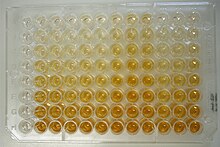
ELISA plate showing various cortisol levels ELISA Plate image.jpg
ELISA plate showing various cortisol levels

Enzymes used in ELISAs include horseradish peroxidase (HRP), alkaline phosphatase (AP) or glucose oxidase. These enzymes allow for detection often because they produce an observable color change in the presence of certain reagents. In some cases these enzymes are exposed to reagents which cause them to produce light or chemiluminescence. There are several types of ELISA: direct, indirect, sandwich, competitive. [7]

Radioactive isotopes

Radioactive isotopes can be incorporated into immunoassay reagents to produce a radioimmunoassay (RIA). Radioactivity emitted by bound antibody-antigen complexes can be easily detected using conventional methods.

RIAs were some of the earliest immunoassays developed, but have fallen out of favor largely due to the difficulty and potential dangers presented by working with radioactivity. [8] [9]

DNA reporters

A newer approach to immunoassays involves combining real-time quantitative polymerase chain reaction (RT qPCR) and traditional immunoassay techniques. Called real-time immunoquantitative PCR (iqPCR) the label used in these assays is a DNA probe. [10] [11]

Fluorogenic reporters

Fluorogenic reporters like phycoerythrin are used in a number of modern immunoassays. [12] Protein microarrays are a type of immunoassay that often employ fluorogenic reporters. [13]

Electrochemiluminescent tags

Some labels work via electrochemiluminescence (ECL), in which the label emits detectable light in response to electric current. [14] [15]

Label-free immunoassays

While some kind of label is generally employed in immunoassays, there are certain kinds of assays which do not rely on labels, but instead employ detection methods that do not require the modification or labeling the components of the assay. Surface plasmon resonance is an example of technique that can detect binding between an unlabeled antibody and antigens. [16] Another demonstrated labeless immunoassay involves measuring the change in resistance on an electrode as antigens bind to it. [17]

Classifications and formats

In a competitive, homogeneous immunoassay unlabeled analyte displaces bound labelled analyte, which is then detected or measured. Competitive homogeneous immunoassay.svg
In a competitive, homogeneous immunoassay unlabeled analyte displaces bound labelled analyte, which is then detected or measured.

Immunoassays can be run in a number of different formats. Generally, an immunoassay will fall into one of several categories depending on how it is run. [18]

Competitive, homogeneous immunoassays

In a competitive, homogeneous immunoassay, unlabelled analyte in a sample competes with labeled analyte to bind an antibody. The amount of labelled, unbound analyte is then measured. In theory, the more analyte in the sample, the more labelled analyte gets displaced and then measured; hence, the amount of labelled, unbound analyte is proportional to the amount of analyte in the sample.

Homogeneous competitive assays: FPIA, EMIT, LOCI, KIMS and CEDIA. See section text for details. Homogeneous competitive assays (FPIA, EMIT, LOCI, KIMS and CEDIA).png
Homogeneous competitive assays: FPIA, EMIT, LOCI, KIMS and CEDIA. See section text for details.
Two-site, noncompetitive immunoassays usually consist of an analyte "sandwiched" between two antibodies. ELISAs are often run in this format. Sandwich Immunoassay, ELISA.jpg
Two-site, noncompetitive immunoassays usually consist of an analyte "sandwiched" between two antibodies. ELISAs are often run in this format.

Competitive, heterogeneous immunoassays

As in a competitive, homogeneous immunoassay, unlabelled analyte in a sample competes with labelled analyte to bind an antibody. In the heterogeneous assays, the labelled, unbound analyte is separated or washed away, and the remaining labelled, bound analyte is measured.

One-site, noncompetitive immunoassays

The unknown analyte in the sample binds with labelled antibodies. The unbound, labelled antibodies are washed away, and the bound, labelled antibodies are measured. The intensity of the signal is directly proportional to the amount of unknown analyte.

Two-site, noncompetitive immunoassays

The analyte in the unknown sample is bound to the antibody site, then the labelled antibody is bound to the analyte. The amount of labelled antibody on the site is then measured. It will be directly proportional to the concentration of the analyte because the labelled antibody will not bind if the analyte is not present in the unknown sample. This type of immunoassay is also known as a sandwich assay as the analyte is "sandwiched" between two antibodies.

Examples

Clinical tests

A wide range of medical tests are immunoassays, called immunodiagnostics in this context. Many home pregnancy tests are immunoassays, which detect the pregnancy marker human chorionic gonadotropin. [20] More specifically, they are qualitative tests that detect whether hCG is present, using a lateral flow setup. [21] The COVID-19 rapid antigen test is also a qualitative, lateral-flow test. [22]

Other clinical immunoassays are quantitative; they measure amounts. Immunoassays can measure levels of CK-MB to assess heart disease, insulin to assess hypoglycemia, prostate-specific antigen to detect prostate cancer, and some are also used for the detection and/or quantitative measurement of some pharmaceutical compounds (see Enzyme multiplied immunoassay technique for more details). [23]

Drug testing also starts with a quick qualtitative immunoassay. [24]

Sports anti-doping analysis

Immunoassays are used in sports anti-doping laboratories to test athletes' blood samples for prohibited recombinant human growth hormone (rhGH, rGH, hGH, GH). [25]

Research

Photoacoustic Immunoassay

The photoacoustic immunoassay measures low-frequency acoustic signals generated by metal nanoparticle tags. Illuminated by a modulated light at a plasmon resonance wavelength, the nanoparticles generate strong acoustic signal, which can be measured using a microphone. [26] The photoacoustic immunoassay can be applied to lateral flow tests, which use colloidal nanoparticles. [27]

See also

Related Research Articles

<span class="mw-page-title-main">ELISA</span> Method to detect an antigen using an antibody and enzyme

The enzyme-linked immunosorbent assay (ELISA) is a commonly used analytical biochemistry assay, first described by Eva Engvall and Peter Perlmann in 1971. The assay uses a solid-phase type of enzyme immunoassay (EIA) to detect the presence of a ligand in a liquid sample using antibodies directed against the protein to be measured. ELISA has been used as a diagnostic tool in medicine, plant pathology, and biotechnology, as well as a quality control check in various industries.

A biosensor is an analytical device, used for the detection of a chemical substance, that combines a biological component with a physicochemical detector. The sensitive biological element, e.g. tissue, microorganisms, organelles, cell receptors, enzymes, antibodies, nucleic acids, etc., is a biologically derived material or biomimetic component that interacts with, binds with, or recognizes the analyte under study. The biologically sensitive elements can also be created by biological engineering. The transducer or the detector element, which transforms one signal into another one, works in a physicochemical way: optical, piezoelectric, electrochemical, electrochemiluminescence etc., resulting from the interaction of the analyte with the biological element, to easily measure and quantify. The biosensor reader device connects with the associated electronics or signal processors that are primarily responsible for the display of the results in a user-friendly way. This sometimes accounts for the most expensive part of the sensor device, however it is possible to generate a user friendly display that includes transducer and sensitive element. The readers are usually custom-designed and manufactured to suit the different working principles of biosensors.

An assay is an investigative (analytic) procedure in laboratory medicine, mining, pharmacology, environmental biology and molecular biology for qualitatively assessing or quantitatively measuring the presence, amount, or functional activity of a target entity. The measured entity is often called the analyte, the measurand, or the target of the assay. The analyte can be a drug, biochemical substance, chemical element or compound, or cell in an organism or organic sample. An assay usually aims to measure an analyte's intensive property and express it in the relevant measurement unit.

<span class="mw-page-title-main">Immunostaining</span> Biochemical technique

In biochemistry, immunostaining is any use of an antibody-based method to detect a specific protein in a sample. The term "immunostaining" was originally used to refer to the immunohistochemical staining of tissue sections, as first described by Albert Coons in 1941. However, immunostaining now encompasses a broad range of techniques used in histology, cell biology, and molecular biology that use antibody-based staining methods.

<span class="mw-page-title-main">Biochip</span> Substrates performing biochemical reactions

In molecular biology, biochips are engineered substrates that can host large numbers of simultaneous biochemical reactions. One of the goals of biochip technology is to efficiently screen large numbers of biological analytes, with potential applications ranging from disease diagnosis to detection of bioterrorism agents. For example, digital microfluidic biochips are under investigation for applications in biomedical fields. In a digital microfluidic biochip, a group of (adjacent) cells in the microfluidic array can be configured to work as storage, functional operations, as well as for transporting fluid droplets dynamically.

<span class="mw-page-title-main">Radioimmunoassay</span> Immunoassay that uses radiolabeled molecules

A radioimmunoassay (RIA) is an immunoassay that uses radiolabeled molecules in a stepwise formation of immune complexes. A RIA is a very sensitive in vitro assay technique used to measure concentrations of substances, usually measuring antigen concentrations by use of antibodies.

Heterophile antibodies are antibodies induced by external antigens.

<span class="mw-page-title-main">Digoxigenin</span> Chemical compound

Digoxigenin (DIG) is a steroid found exclusively in the flowers and leaves of the plants Digitalis purpurea, Digitalis orientalis and Digitalis lanata (foxgloves), where it is attached to sugars, to form the glycosides.

<span class="mw-page-title-main">Antibody microarray</span>

An antibody microarray is a specific form of protein microarray. In this technology, a collection of captured antibodies are spotted and fixed on a solid surface such as glass, plastic, membrane, or silicon chip, and the interaction between the antibody and its target antigen is detected. Antibody microarrays are often used for detecting protein expression from various biofluids including serum, plasma and cell or tissue lysates. Antibody arrays may be used for both basic research and medical and diagnostic applications.

<span class="mw-page-title-main">Lateral flow test</span> Immunochromatographic testing devices

A lateral flow test (LFT), is an assay also known as a lateral flow device (LFD), lateral flow immunochromatographic assay, or rapid test. It is a simple device intended to detect the presence of a target substance in a liquid sample without the need for specialized and costly equipment. LFTs are widely used in medical diagnostics in the home, at the point of care, and in the laboratory. For instance, the home pregnancy test is an LFT that detects a specific hormone. These tests are simple and economical and generally show results in around five to thirty minutes. Many lab-based applications increase the sensitivity of simple LFTs by employing additional dedicated equipment. Because the target substance is often a biological antigen, many lateral flow tests are rapid antigen tests.

Magnetic immunoassay (MIA) is a type of diagnostic immunoassay using magnetic beads as labels in lieu of conventional enzymes (ELISA), radioisotopes (RIA) or fluorescent moieties to detect a specified analyte. MIA involves the specific binding of an antibody to its antigen, where a magnetic label is conjugated to one element of the pair. The presence of magnetic beads is then detected by a magnetic reader (magnetometer) which measures the magnetic field change induced by the beads. The signal measured by the magnetometer is proportional to the analyte concentration in the initial sample.

A reverse phase protein lysate microarray (RPMA) is a protein microarray designed as a dot-blot platform that allows measurement of protein expression levels in a large number of biological samples simultaneously in a quantitative manner when high-quality antibodies are available.

<span class="mw-page-title-main">Surround optical-fiber immunoassay</span>

Surround optical-fiber immunoassay (SOFIA) is an ultrasensitive, in vitro diagnostic platform incorporating a surround optical-fiber assembly that captures fluorescence emissions from an entire sample. The technology's defining characteristics are its extremely high limit of detection, sensitivity, and dynamic range. SOFIA's sensitivity is measured at the attogram level (10−18 g), making it about one billion times more sensitive than conventional diagnostic techniques. Based on its enhanced dynamic range, SOFIA is able to discriminate levels of analyte in a sample over 10 orders of magnitude, facilitating accurate titering.

Flow-through tests or immunoconcentration assays are a type of diagnostic assay that allows users to test for the presence of a biomarker, usually a specific antibody, in a sample such as blood. They are a type of point of care test, a test designed to be used by a healthcare provider at patient contact. Point of care tests often allow for rapid detection of a specific biomarker without specialized lab equipment and training; this aids in diagnosis and allows therapeutic action to be initiated more quickly. Flow-through tests began development in the early 1980s and were the first type of immunostrip to be developed, although lateral flow tests have subsequently become the dominant immunostrip point of care device.

Mass spectrometric immunoassay (MSIA) is a rapid method is used to detect and/ or quantify antigens and or antibody analytes. This method uses an analyte affinity isolation to extract targeted molecules and internal standards from biological fluid in preparation for matrix assisted laser desorption ionization-time of flight mass spectrometry (MALDI-TOF-MS). This method allows for "top down" and "bottom up" analysis. This sensitive method allows for a new and improved process for detecting multiple antigens and antibodies in a single assay. This assay is also capable of distinguishing mass shifted forms of the same molecule via a panantibody, as well as distinguish point mutations in proteins. Each specific form is detected uniquely based on their characteristic molecular mass. MSIA has dual specificity because of the antibody-antigen reaction coupled with the power of a mass spectrometer.

A ligand binding assay (LBA) is an assay, or an analytic procedure, which relies on the binding of ligand molecules to receptors, antibodies or other macromolecules. A detection method is used to determine the presence and extent of the ligand-receptor complexes formed, and this is usually determined electrochemically or through a fluorescence detection method. This type of analytic test can be used to test for the presence of target molecules in a sample that are known to bind to the receptor.

A hybridization assay comprises any form of quantifiable hybridization i.e. the quantitative annealing of two complementary strands of nucleic acids, known as nucleic acid hybridization.

<span class="mw-page-title-main">CD/DVD based immunoassay</span>

A compact disk/digital versatile disk (CD/DVD) based immunoassay is a method for determining the concentration of a compound in research and diagnostic laboratories by performing the test on an adapted CD/DVD surface using an adapted optical disc drive; these methods have been discussed and prototyped in research labs since 1991.

<span class="mw-page-title-main">Fluorescence polarization immunoassay</span> Class of invitro biochemical test

Fluorescence polarization immunoassay (FPIA) is a class of in vitro biochemical test used for rapid detection of antibody or antigen in sample. FPIA is a competitive homogenous assay, that consists of a simple prepare and read method, without the requirement of separation or washing steps.

The enzyme-linked immunosorbent spot (ELISpot) is a type of assay that focuses on quantitatively measuring the frequency of cytokine secretion for a single cell. The ELISpot Assay is also a form of immunostaining since it is classified as a technique that uses antibodies to detect a protein analyte, with the word analyte referring to any biological or chemical substance being identified or measured.

References

  1. Yetisen A. K. (2013). "Paper-based microfluidic point-of-care diagnostic devices". Lab on a Chip. 13 (12): 2210–2251. doi:10.1039/C3LC50169H. PMID   23652632.
  2. Rall JE. Solomon A. Berson. In "Biographical Memoirs". National Academy of Sciences 1990;59:54-71. ISBN   0-309-04198-8. Fulltext.
  3. Lequin R (2005). "Enzyme immunoassay (EIA)/enzyme-linked immunosorbent assay (ELISA)". Clin. Chem. 51 (12): 2415–8. doi: 10.1373/clinchem.2005.051532 . PMID   16179424.
  4. Prof Anthony Campbell - MA PhD, Cardiff University , retrieved 29 December 2012
  5. "NPS Focus", Rainbow makers, Royal Society of Chemistry (RSC), 2003, retrieved 29 December 2012
  6. Carlson, Bruce (15 February 2014). "Seizing Immunoassay Opportunities". Gen. Eng. Biotechnol. News . Vol. 34, no. 4. pp. 12–13.
  7. Aydin, Suleyman (2015). "A short history, principles, and types of ELISA, and our laboratory experience with peptide/protein analyses using ELISA". Peptides. 72: 4–15. doi:10.1016/j.peptides.2015.04.012. PMID   25908411. S2CID   36486495.
  8. Susan J. Landers (3 April 2006). "ELISA test marks 35 years of answering medical questions". American Medical News . Retrieved 9 December 2012.
  9. "Rosalyn Sussman Yalow". America.gov. April 27, 2008. Retrieved June 26, 2010.
  10. Rajkovic; El-Moualij (2006). "Immunoquantitative real-time PCR for detection and quantification of Staphylococcus aureus enterotoxin B in foods". Applied and Environmental Microbiology . 72 (10): 6593–9. Bibcode:2006ApEnM..72.6593R. doi:10.1128/AEM.03068-05. PMC   1610299 . PMID   17021210.
  11. Gofflot; El (2004). "Immuno-quantitative polymerase chain reaction for detection and quantitation of prion protein". Journal of Immunoassay and Immunochemistry. 25 (3): 241–58. doi:10.1081/ias-200028044. PMID   15461386. S2CID   37548553.
  12. "Luminex xMAP Technology". Millipore Corporation . Retrieved 13 December 2012.
  13. Chatterjee; Sitaraman (2008). "Protein microarray on-demand: a novel protein microarray system". PLOS ONE . 3 (9): e3265. Bibcode:2008PLoSO...3.3265C. doi: 10.1371/journal.pone.0003265 . PMC   2533396 . PMID   18813342.
  14. Zanut, A.; Fiorani, A.; Canola, S.; Saito, T.; Ziebart, N.; Rapino, S.; Rebeccani, S.; Barbon, A.; Irie, T.; Josel, H.; Negri, F.; Marcaccio, M.; Windfuhr, M.; Imai, K.; Valenti, G.; Paolucci, F. (2020). "Insights into the mechanism of coreactant electrochemiluminescence facilitating enhanced bioanalytical performance". Nat. Commun. 11 (1): 2668. Bibcode:2020NatCo..11.2668Z. doi:10.1038/s41467-020-16476-2. PMC   7260178 . PMID   32472057.
  15. Forster RJ, Bertoncello P, Keyes TE (2009). "Electrogenerated Chemiluminescence". Annual Review of Analytical Chemistry. 2: 359–85. Bibcode:2009ARAC....2..359F. doi:10.1146/annurev-anchem-060908-155305. PMID   20636067.
  16. J. B. González-Díaz; et al. (2008). "Plasmonic Au/Co/Au nanosandwiches with Enhanced Magneto-Optical Activity". Small. 4 (2): 202–5. doi:10.1002/smll.200700594. hdl: 10261/17402 . PMID   18196506. S2CID   206490102.
  17. Georgios Tsekenis (2008). "Label-free immunosensor assay for myelin basic protein based upon an ac impedance protocol". Analytical Chemistry . 80 (6): 2058–62. doi:10.1021/ac702070e. PMID   18260654.
  18. Goldys, Ewa (2009-08-24). Fluorescence Applications in Biotechnology and Life Sciences. Wiley-Blackwell. p. 311. ISBN   978-0470083703 . Retrieved 11 December 2012. License: "CC BY 4.0"
  19. 1 2 3 4 5 6 Sanavio B, Krol S (2015). "On the Slow Diffusion of Point-of-Care Systems in Therapeutic Drug Monitoring". Front Bioeng Biotechnol. 3: 20. doi: 10.3389/fbioe.2015.00020 . PMC   4341557 . PMID   25767794.
  20. "ELISA for Home Pregnancy Test". Archived from the original on 21 September 2017. Retrieved 11 December 2012.
  21. Butler, SA (2001). "Detection of early pregnancy forms of human chorionic gonadotropin by home pregnancy test devices". Clinical Chemistry . 47 (12): 2131–6. doi: 10.1093/clinchem/47.12.2131 . PMID   11719477.
  22. Guglielmi G (September 2020). "Fast coronavirus tests: what they can and can't do". Nature. 585 (7826): 496–498. Bibcode:2020Natur.585..496G. doi:10.1038/d41586-020-02661-2. PMID   32939084. S2CID   221768935.
  23. Twyman, SA (2001). "Immunoassays, applications: Clinica" (PDF). Encyclopedia of Analytical Science. 4: 317–324. Archived from the original (PDF) on 24 March 2012. Retrieved 11 December 2012.
  24. "Principles of Drug Testing Technology," Drug Tests Direct, Retrieved online Dec 07, 2012.
  25. Tsivou M; Kioukia-Fougia N; Lyris E; Aggelis Y; Fragkaki A; Kiousi X; Simitsek Ph; Dimopoulou H; Leontiou I-P; Stamou M; Spyridaki M-H; Georgakopoulos C (2006). "An overview of the doping control analysis during the Olympic Games of 2004 in Athens, Greece". Analytica Chimica Acta. 555: 1–13. doi:10.1016/j.aca.2005.08.068.
  26. Zhao Y, Cao M, McClelland JF, Lu M (2016). "A photoacoustic immunoassay for biomarker detection". Biosensors and Bioelectronics. 85: 261–66. doi:10.1016/j.bios.2016.05.028. PMID   27183276.
  27. Zhao Y, Huang Y, Zhao X, McClelland JF, Lu M (2016). "Nanoparticle-based photoacoustic analysis for highly sensitive lateral flow assays". Nanoscale. 8 (46): 19204–19210. doi:10.1039/C6NR05312B. PMID   27834971.

"The Immunoassay Handbook", 3rd Edition, David Wild, Ed., Elsevier,2008