Hemagglutinin

Last updated

Illustration showing influenza virus attaching to cell membrane via the surface protein hemagglutinin CSIRO ScienceImage 354 Influenza Protein Attaching to Cell Membrane.jpg
Illustration showing influenza virus attaching to cell membrane via the surface protein hemagglutinin

In molecular biology, hemagglutinins (alternatively spelled haemagglutinin, from the Greek haima, 'blood' + Latin gluten, 'glue') are receptor-binding membrane fusion glycoproteins produced by viruses in the Paramyxoviridae and Orthomyxoviridae families. [1] [2] Hemagglutinins are responsible for binding to receptors on red blood cells to initiate viral attachment and infection. [3] The agglutination of red cells occurs when antibodies on one cell bind to those on others, causing amorphous aggregates of clumped cells.

Contents

Hemagglutinins recognize cell-surface glycoconjugates containing sialic acid on the surface of host red blood cells with a low affinity, and use them to enter the endosome of host cells. [4] In the endosome, hemagglutinins are activated at a pH of 5–6.5 to undergo conformational changes that enable viral attachment through a fusion peptide. [5]

Virologist George K. Hirst discovered agglutination and hemagglutinins in 1941. [6] Alfred Gottschalk proved in 1957 that hemagglutinins bind a virus to a host cell by attaching to sialic acids on carbohydrate side chains of cell-membrane glycoproteins and glycolipids. [7]

Types

Structure

Hemagglutinins are small proteins that project from the virus membrane surface as 135 Angstrom (Å) long spikes with a diameter of 30–50 Å. [13] Each spike is made up of three identical monomer subunits, making the protein a homotrimer. These monomers are formed of two glycopeptides, HA1 and HA2, and linked by two disulphide polypeptides, including membrane-distal HA1 and the smaller membrane-proximal HA2. X-ray crystallography and spectroscopy were used to identify that the majority of the protein structures is made of α-helical proteins. [14] In addition to the homotrimeric core structure, hemagglutinins have four subdomains: the membrane-distal receptor binding R subdomain, the vestigial domain E, that functions as a receptor-destroying esterase, the fusion domain F, and the membrane anchor subdomain M. The membrane anchor subdomain forms elastic protein chains linking the hemagglutinin to the ectodomain. [15]

Uses in serology

A schematic diagram of the experimental setup to detect hemagglutination for blood typing. Experimental setup to detect hemagglutination.png
A schematic diagram of the experimental setup to detect hemagglutination for blood typing.

See also

Related Research Articles

<span class="mw-page-title-main">Lectin</span> Carbohydrate-binding protein

Lectins are carbohydrate-binding proteins that are highly specific for sugar groups that are part of other molecules, so cause agglutination of particular cells or precipitation of glycoconjugates and polysaccharides. Lectins have a role in recognition at the cellular and molecular level and play numerous roles in biological recognition phenomena involving cells, carbohydrates, and proteins. Lectins also mediate attachment and binding of bacteria, viruses, and fungi to their intended targets.

<i>Influenza A virus</i> Species of virus

Influenza A virus (IAV) is a pathogen that causes the flu in birds and some mammals, including humans. It is an RNA virus whose subtypes have been isolated from wild birds. Occasionally, it is transmitted from wild to domestic birds, and this may cause severe disease, outbreaks, or human influenza pandemics.

<span class="mw-page-title-main">Hemagglutinin (influenza)</span> Hemagglutinin of influenza virus

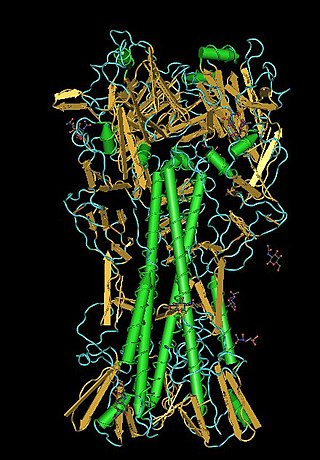
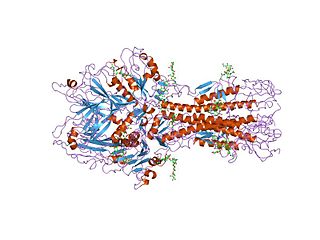
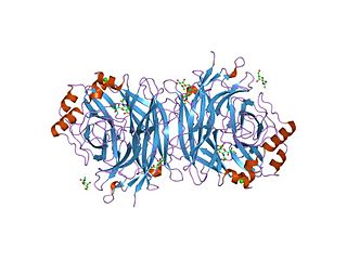
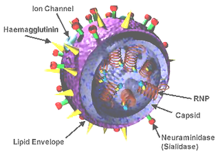
Influenza hemagglutinin (HA) or haemagglutinin[p] is a homotrimeric glycoprotein found on the surface of influenza viruses and is integral to its infectivity.

<i>Orthomyxoviridae</i> Family of RNA viruses including the influenza viruses

Orthomyxoviridae is a family of negative-sense RNA viruses. It includes seven genera: Alphainfluenzavirus, Betainfluenzavirus, Gammainfluenzavirus, Deltainfluenzavirus, Isavirus, Thogotovirus, and Quaranjavirus. The first four genera contain viruses that cause influenza in birds and mammals, including humans. Isaviruses infect salmon; the thogotoviruses are arboviruses, infecting vertebrates and invertebrates. The Quaranjaviruses are also arboviruses, infecting vertebrates (birds) and invertebrates (arthropods).

Antigenic drift is a kind of genetic variation in viruses, arising from the accumulation of mutations in the virus genes that code for virus-surface proteins that host antibodies recognize. This results in a new strain of virus particles that is not effectively inhibited by the antibodies that prevented infection by previous strains. This makes it easier for the changed virus to spread throughout a partially immune population. Antigenic drift occurs in both influenza A and influenza B viruses.

<span class="mw-page-title-main">Hemagglutination assay</span> Measure of virus or bacteria concentration

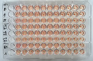
The hemagglutination assay or haemagglutination assay (HA) and the hemagglutination inhibition assay were developed in 1941–42 by American virologist George Hirst as methods for quantifying the relative concentration of viruses, bacteria, or antibodies.

<span class="mw-page-title-main">Hemagglutinin esterase</span> Glycoprotein present in some enveloped viruses

Hemagglutinin esterase (HEs) is a glycoprotein that certain enveloped viruses possess and use as an invading mechanism. HEs helps in the attachment and destruction of certain sialic acid receptors that are found on the host cell surface. Viruses that possess HEs include influenza C virus, toroviruses, and coronaviruses of the subgenus Embecovirus. HEs is a dimer transmembrane protein consisting of two monomers, each monomer is made of three domains. The three domains are: membrane fusion, esterase, and receptor binding domains.

Hemagglutination, or haemagglutination, is a specific form of agglutination that involves red blood cells (RBCs). It has two common uses in the laboratory: blood typing and the quantification of virus dilutions in a haemagglutination assay.

<i>Influenza C virus</i> Genus of viruses in the family Orthomyxoviridae

Influenza C virus is the only species in the genus Gammainfluenzavirus, in the virus family Orthomyxoviridae, which like other influenza viruses, causes influenza.

<span class="mw-page-title-main">H5N1 genetic structure</span>

H5N1 genetic structure is the molecular structure of the H5N1 virus's RNA.

<span class="mw-page-title-main">Virosome</span> Drug or vaccine delivery mechanism

A virosome is a drug or vaccine delivery mechanism consisting of unilamellar phospholipid membrane vesicle incorporating virus derived proteins to allow the virosomes to fuse with target cells. Viruses are infectious agents that can replicate in their host organism, however virosomes do not replicate. The properties that virosomes share with viruses are based on their structure; virosomes are essentially safely modified viral envelopes that contain the phospholipid membrane and surface glycoproteins. As a drug or vaccine delivery mechanism they are biologically compatible with many host organisms and are also biodegradable. The use of reconstituted virally derived proteins in the formation of the virosome allows for the utilization of what would otherwise be the immunogenic properties of a live-attenuated virus, but is instead a safely killed virus. A safely killed virus can serve as a promising vector because it won't cause infection and the viral structure allows the virosome to recognize specific components of its target cells.

Antigenic variation or antigenic alteration refers to the mechanism by which an infectious agent such as a protozoan, bacterium or virus alters the proteins or carbohydrates on its surface and thus avoids a host immune response, making it one of the mechanisms of antigenic escape. It is related to phase variation. Antigenic variation not only enables the pathogen to avoid the immune response in its current host, but also allows re-infection of previously infected hosts. Immunity to re-infection is based on recognition of the antigens carried by the pathogen, which are "remembered" by the acquired immune response. If the pathogen's dominant antigen can be altered, the pathogen can then evade the host's acquired immune system. Antigenic variation can occur by altering a variety of surface molecules including proteins and carbohydrates. Antigenic variation can result from gene conversion, site-specific DNA inversions, hypermutation, or recombination of sequence cassettes. The result is that even a clonal population of pathogens expresses a heterogeneous phenotype. Many of the proteins known to show antigenic or phase variation are related to virulence.

<span class="mw-page-title-main">Spike protein</span> Glycoprotein spike on a viral capsid or viral envelope

In virology, a spike protein or peplomer protein is a protein that forms a large structure known as a spike or peplomer projecting from the surface of an enveloped virus. The proteins are usually glycoproteins that form dimers or trimers.

<span class="mw-page-title-main">Viral neuraminidase</span> InterPro Family

Viral neuraminidase is a type of neuraminidase found on the surface of influenza viruses that enables the virus to be released from the host cell. Neuraminidases are enzymes that cleave sialic acid groups from glycoproteins. Viral neuraminidase was discovered by Alfred Gottschalk at the Walter and Eliza Hall Institute in 1957. Neuraminidase inhibitors are antiviral agents that inhibit influenza viral neuraminidase activity and are of major importance in the control of influenza.

<span class="mw-page-title-main">Hemagglutinin-neuraminidase</span>

Hemagglutinin-neuraminidase refers to a single viral protein that has both hemagglutinin and (endo) neuraminidase EC 3.2.1.18 activity. This is in contrast to the proteins found in influenza, where both functions exist but in two separate proteins. Its neuraminidase domain has the CAZy designation glycoside hydrolase family 83 (GH83).

FI6 is an antibody that targets a protein found on the surface of all influenza A viruses called hemagglutinin. FI6 is the only known antibody found to bind all 16 subtypes of the influenza A virus hemagglutinin and is hoped to be useful for a universal influenza virus therapy.

<span class="mw-page-title-main">George Hirst (virologist)</span> American virologist and science administrator

George Keble Hirst, M.D. was an American virologist and science administrator who was among the first to study the molecular biology and genetics of animal viruses, especially influenza virus. He directed the Public Health Research Institute in New York City (1956–1981), and was also the founding editor-in-chief of Virology, the first English-language journal to focus on viruses. He is particularly known for inventing the hemagglutination assay, a simple method for quantifying viruses, and adapting it into the hemagglutination inhibition assay, which measures virus-specific antibodies in serum. He was the first to discover that viruses can contain enzymes, and the first to propose that virus genomes can consist of discontinuous segments. The New York Times described him as "a pioneer in molecular virology."

<span class="mw-page-title-main">Universal flu vaccine</span> Vaccine that prevents infection from all strains of the flu

A universal flu vaccine is a flu vaccine that is effective against all influenza strains regardless of the virus sub type, antigenic drift or antigenic shift. Hence it should not require modification from year to year. As of 2021 no universal flu vaccine had been approved for general use, several were in development, and one was in clinical trial.

<i>Influenza D virus</i> Species of virus

Influenza D virus is a species in the virus genus Deltainfluenzavirus, in the family Orthomyxoviridae, that causes influenza.

Bat mumps orthorubulavirus, formerly Bat mumps rubulavirus (BMV), is a member of genus Orthorubulavirus, family Paramyxoviridae, and order Mononegavirales. Paramyxoviridae viruses were first isolated from bats using heminested PCR with degenerate primers. This process was then followed by Sanger sequencing. A specific location of this virus is not known because it was isolated from bats worldwide. Although multiple paramyxoviridae viruses have been isolated worldwide, BMV specifically has not been isolated thus far. However, BMV was detected in African fruit bats, but no infectious form has been isolated to date. It is known that BMV is transmitted through saliva in the respiratory system of bats. While the virus was considered its own species for a few years, phylogenetic analysis has since shown that it is a member of Mumps orthorubulavirus.

References

  1. Couch, Robert B. (1996), Baron, Samuel (ed.), "Orthomyxoviruses", Medical Microbiology (4th ed.), Galveston (TX): University of Texas Medical Branch at Galveston, ISBN   978-0-9631172-1-2, PMID   21413353 , retrieved 30 January 2024
  2. "Paramyxoviridae - an overview | ScienceDirect Topics". www.sciencedirect.com. Retrieved 30 January 2024.
  3. Nobusawa, E. (October 1997). "[Structure and function of the hemagglutinin of influenza viruses]". Nihon Rinsho. Japanese Journal of Clinical Medicine. 55 (10): 2562–2569. ISSN   0047-1852. PMID   9360372.
  4. Bangaru, Sandhya; Lang, Shanshan; Schotsaert, Michael; Vanderven, Hillary A.; Zhu, Xueyong; Kose, Nurgun; Bombardi, Robin; Finn, Jessica A.; Kent, Stephen J.; Gilchuk, Pavlo; Gilchuk, Iuliia (2019). "A Site of Vulnerability on the Influenza Virus Hemagglutinin Head Domain Trimer Interface". Cell. 177 (5): 1136–1152.e18. doi:10.1016/j.cell.2019.04.011. PMC   6629437 . PMID   31100268.
  5. Medeiros, R.; Escriou, N.; Naffakh, N.; Manuguerra, J. C.; van der Werf, S. (10 October 2001). "Hemagglutinin residues of recent human A(H3N2) influenza viruses that contribute to the inability to agglutinate chicken erythrocytes". Virology. 289 (1): 74–85. doi: 10.1006/viro.2001.1121 . ISSN   0042-6822. PMID   11601919.
  6. Kolata, Gina (26 January 1994). "George Keble Hirst, 84, Is Dead; A Pioneer in Molecular Virology". The New York Times. ISSN   0362-4331 . Retrieved 14 May 2024.
  7. Henry, Ronnie; Murphy, Frederick A. (October 2018). "Etymologia: Hemagglutinin and Neuraminidase". Emerging Infectious Diseases. 24 (10): 1849. doi:10.3201/eid2410.ET2410. PMC   6154157 .
  8. CDC (2 November 2021). "Types of Influenza Viruses". Centers for Disease Control and Prevention. Retrieved 18 October 2022.
  9. Hashiguchi, Takao; Maenaka, Katsumi; Yanagi, Yusuke (16 December 2011). "Measles Virus Hemagglutinin: Structural Insights into Cell Entry and Measles Vaccine". Frontiers in Microbiology. 2: 247. doi: 10.3389/fmicb.2011.00247 . ISSN   1664-302X. PMC   3267179 . PMID   22319511.
  10. Pan CH, Jimenez GS, Nair N (21 August 2014) [August, 2008]. "Use of Vaxfectin Adjuvant with DNA Vaccine Encoding the Measles Virus Hemagglutinin and Fusion Proteins Protects Juvenile and Infant Rhesus Macaques against Measles Virus". Clinical and Vaccine Immunology. 15 (8): 1214–1221. doi:10.1128/CVI.00120-08. PMC   2519314 . PMID   18524884.{{cite journal}}: CS1 maint: multiple names: authors list (link)
  11. Tappert, Mary M.; Porterfield, J. Zachary; Mehta-D'Souza, Padmaja; Gulati, Shelly; Air, Gillian M. (August 2013). "Quantitative Comparison of Human Parainfluenza Virus Hemagglutinin-Neuraminidase Receptor Binding and Receptor Cleavage". Journal of Virology. 87 (16): 8962–8970. doi:10.1128/JVI.00739-13. ISSN   0022-538X. PMC   3754076 . PMID   23740997.
  12. Kubota, Marie; Hashiguchi, Takao (2020). "Large-Scale Expression and Purification of Mumps Virus Hemagglutinin-Neuraminidase for Structural Analyses and Glycan-Binding Assays". Lectin Purification and Analysis. Methods in Molecular Biology. Vol. 2132. pp. 641–652. doi:10.1007/978-1-0716-0430-4_55. ISBN   978-1-0716-0429-8. ISSN   1940-6029. PMID   32306363. S2CID   216030421.
  13. Gamblin, Steven J.; Vachieri, Sébastien G.; Xiong, Xiaoli; Zhang, Jie; Martin, Stephen R.; Skehel, John J. (1 October 2021). "Hemagglutinin Structure and Activities". Cold Spring Harbor Perspectives in Medicine. 11 (10): a038638. doi:10.1101/cshperspect.a038638. ISSN   2157-1422. PMC   8485738 . PMID   32513673.
  14. Gamblin, Steven J.; Vachieri, Sébastien G.; Xiong, Xiaoli; Zhang, Jie; Martin, Stephen R.; Skehel, John J. (1 October 2021). "Hemagglutinin Structure and Activities". Cold Spring Harbor Perspectives in Medicine. 11 (10): a038638. doi:10.1101/cshperspect.a038638. ISSN   2157-1422. PMC   8485738 . PMID   32513673.
  15. Donald J. Benton, Andrea Nans, Lesley J. Calder, Jack Turner, Ursula Neu, Yi Pu Lin, Esther Ketelaars, Nicole L. Kallewaard, Davide Corti, Antonio Lanzavecchia, Steven J. Gamblin, Peter B. Rosenthal, John J. Skehel (2 October 2018) [Sep 17, 2018]. "Hemagglutinin membrane anchor". Proceedings of the National Academy of Sciences of the United States of America. 115 (40): 10112–10117. doi: 10.1073/pnas.1810927115 . PMC   6176637 . PMID   30224494.{{cite journal}}: CS1 maint: multiple names: authors list (link)
  16. Payne, Susan (2017). "Methods to Study Viruses". Viruses. pp. 37–52. doi:10.1016/B978-0-12-803109-4.00004-0. ISBN   978-0-12-803109-4. S2CID   89981392.
  17. Ashiba, Hiroki; Fujimaki, Makoto; Awazu, Koichi; Fu, Mengying; Ohki, Yoshimichi; Tanaka, Torahiko; Makishima, Makoto (March 2015). "Hemagglutination detection for blood typing based on waveguide-mode sensors". Sensing and Bio-Sensing Research. 3: 59–64. doi: 10.1016/j.sbsr.2014.12.003 .
  18. Theis, Samuel R.; Hashmi, Muhammad F. (2022), "Coombs Test", StatPearls, Treasure Island (FL): StatPearls Publishing, PMID   31613487 , retrieved 16 December 2022
  19. Focosi, Daniele; Franchini, Massimo; Maggi, Fabrizio (8 March 2022). "Modified Hemagglutination Tests for COVID-19 Serology in Resource-Poor Settings: Ready for Prime-Time?". Vaccines. 10 (3): 406. doi: 10.3390/vaccines10030406 . ISSN   2076-393X. PMC   8953758 . PMID   35335038.