Parathyroid gland

Last updated

Parathyroid glands
Parathyroid es.svg
Diagram showing structures in the human neck. The four green shaded areas represent the most common position of the parathyroid glands, which are generally four in number and situated behind the lateral lobes of the thyroid gland (shaded orange).
Illu thyroid parathyroid.jpg
Thyroid and parathyroids as viewed from the front of the neck
Details
Precursor Neural crest mesenchyme and third and fourth Pharyngeal pouch endoderm
System Endocrine
Artery Superior thyroid artery, inferior thyroid artery,
Vein Superior thyroid vein, middle thyroid vein, inferior thyroid vein,
Nerve Middle cervical ganglion, inferior cervical ganglion
Lymph Pretracheal, prelaryngeal, jugulodigastric lymph nodes
Identifiers
Latin glandula parathyreoidea inferior, glandula parathyreoidea superior
MeSH D010280
TA98 A11.4.00.001
TA2 3870
FMA 13890
Anatomical terminology

Parathyroid glands are small endocrine glands in the neck of humans and other tetrapods. Humans usually have four parathyroid glands, located on the back of the thyroid gland in variable locations. The parathyroid gland produces and secretes parathyroid hormone in response to low blood calcium, which plays a key role in regulating the amount of calcium in the blood and within the bones.

Contents

Parathyroid glands share a similar blood supply, venous drainage, and lymphatic drainage to the thyroid glands. Parathyroid glands are derived from the epithelial lining of the third and fourth pharyngeal pouches, with the superior glands arising from the fourth pouch and the inferior glands arising from the higher third pouch. The relative position of the inferior and superior glands, which are named according to their final location, changes because of the migration of embryological tissues.

Hyperparathyroidism and hypoparathyroidism, characterized by alterations in the blood calcium levels and bone metabolism, are states of either surplus or deficient parathyroid function.

Structure

The parathyroid glands are two pairs of glands usually positioned behind the left and right lobes of the thyroid. Each gland is a yellowish-brown flat ovoid that resembles a lentil seed, usually about 6 mm long and 3 to 4 mm wide, and 1 to 2 mm anteroposteriorly. [1] There are typically four parathyroid glands. The two parathyroid glands on each side which are positioned higher are called the superior parathyroid glands, while the lower two are called the inferior parathyroid glands. Healthy parathyroid glands generally weigh about 30 mg in men and 35 mg in women. [2] These glands are not visible or able to be felt during examination of the neck. [3]

Each parathyroid vein drains into the superior, middle and inferior thyroid veins. The superior and middle thyroid veins drain into the internal jugular vein, and the inferior thyroid vein drains into the brachiocephalic vein. [4]

Lymphatic drainage

Lymphatic vessels from the parathyroid glands drain into deep cervical lymph nodes and paratracheal lymph nodes. [4]

Variation

The parathyroid glands are variable in number: three or more small glands, [5] and can usually be located on the posterior surface of the thyroid gland. [5] Occasionally, some individuals may have six, eight, or even more parathyroid glands. [3] Rarely, the parathyroid glands may be within the thyroid gland itself, the chest, or even the thymus. [5]

Microanatomy

The parathyroid glands are named for their proximity to the thyroid—and serve a completely different role than the thyroid gland. The parathyroid glands are quite easily recognizable from the thyroid as they have densely packed cells, in contrast with the follicular structure of the thyroid. [6] Two unique types of cells are present in the parathyroid gland:

Development

In the early development of the human embryo, a series of five pharyngeal arches and four pharyngeal pouches form that give rise to the face, neck, and surrounding structures. The pouches are numbered such that the first pouch is the closest to the top of the embryo's head and the fourth is the furthest from it. The parathyroid glands originate from the interaction of the endoderm of the third and fourth pouch and neural crest mesenchyme. [5] The position of the glands reverses during embryological development. The pair of glands which is ultimately inferior develops from the third pouch with the thymus, whereas the pair of glands which is ultimately superior develops from the fourth pouch. During embryological development, the thymus migrates downwards, dragging the inferior glands with it. The superior pair are not dragged downwards by the fourth pouch to the same degree. The glands are named after their final, not embryological, positions. [9] Since the thymus's ultimate destination is in the mediastinum of the chest, it is occasionally possible to have ectopic parathyroids derived from the third pouch within the chest cavity if they fail to detach in the neck.

Parathyroid development is regulated by a number of genes, including those coding for several transcription factors. [10] [lower-alpha 1]

Function

The major function of the parathyroid glands is to maintain the body's calcium and phosphate levels within a very narrow range, so that the nervous and muscular systems can function properly. The parathyroid glands do this by secreting parathyroid hormone (PTH). [11]

Parathyroid hormone (also known as parathormone) is a small protein that takes part in the control of calcium and phosphate homeostasis, as well as bone physiology. Parathyroid hormone has effects antagonistic to those of calcitonin. [12]

Disorders

Parathyroid disease is conventionally divided into states where the parathyroid is overactive (hyperparathyroidism), and states where the parathyroid is under- or hypoactive (hypoparathyroidism). Both states are characterised by their symptoms, which relate to the excess or deficiency of parathyroid hormone in the blood. [13]

Hyperparathyroidism

Primary

3D medical animation image showing hyperparathyroidism (right) 3D Medical Animation still shot Hyperparathyroidism.jpg
3D medical animation image showing hyperparathyroidism (right)

Hyperparathyroidism is the state in which there is excess parathyroid hormone circulating in the blood. This may cause bone pain and tenderness, due to increased bone resorption. With increased circulating calcium, there may be other symptoms associated with hypercalcemia, most commonly dehydration. Hyperparathyroidism is most commonly caused by a benign proliferation of chief cells in one parathyroid gland, and rarely in MEN syndrome. This is known as primary hyperparathyroidism, [13] which is generally managed by surgical removal of the abnormal parathyroid gland. [14]

Secondary

Renal disease may lead to hyperparathyroidism. When too much calcium is lost from the blood via urination, there is a compensation by the parathyroid, and parathyroid hormone is released. The glands enlarge (hypertrophy) to synthesize more parathyroid hormone. This is known as secondary hyperparathyroidism.

Tertiary

If secondary hyperparathyroidism persists over months, the parathyroid tissue may become unresponsive to the blood calcium levels, and begin to autonomously release parathyroid hormone. This is known as tertiary hyperparathyroidism. [15]

Hypoparathyroidism

The state of decreased parathyroid activity is known as hypoparathyroidism. This is most commonly associated with damage to the glands or their blood supply during thyroid surgery  it may be associated with rarer genetic syndromes such as DiGeorge syndrome, which is inherited as an autosomal dominant syndrome. Hypoparathyroidism will occur after surgical removal of the parathyroid glands. [16]

Occasionally, an individual's tissues are resistant to the effects of parathyroid hormone. This is known as pseudohypoparathyroidism. In this case the parathyroid glands are fully functional, and the hormone itself is not able to function, resulting in a decrease in blood calcium levels. Pseudohypoparathyroidism is often associated with the genetic condition Albright's hereditary osteodystrophy. Pseudo-pseudohypoparathyroidism, one of the longest words in the English language, is used to describe an individual with Albright's hereditary osteodystrophy with normal parathyroid hormone and serum calcium levels. [16]

Hypoparathyroidism may present with symptoms associated with decreased calcium, and is generally treated with Vitamin D analogues. [16]

History

The parathyroid glands were first discovered in the Indian rhinoceros by Richard Owen in 1852. [17] In his description of the neck anatomy, Owen referred to the glands as "a small compact yellow glandular body attached to the thyroid at the point where the veins emerged". The glands were first discovered in humans by Ivar Viktor Sandström (1852–1889), a Swedish medical student, in 1880 at Uppsala University. [18] Unaware of Owen's description, he described the glands in his monograph "On a New Gland in Man and Fellow Animals" as the "glandulae parathyroidae", noting its existence in dogs, cats, rabbits, oxen, horses and humans. [19] [20] For several years, Sandström's description received little attention. [21]

Eugene Gley, Giulio Vassale, and others documented the putative function of the glands in 1891, noting the connection between their removal and the development of muscular tetany. William G. MacCallum in 1908, investigating tumours of the parathyroid, proposed their role in calcium metabolism. [20] He noted that "Tetany occurs spontaneously in many forms and may be produced by the destruction of the parathyroid glands". [22]

The first successful removal of the parathyroid may have been carried out in 1928 by medical doctor Isaac Y Olch, whose intern had noticed elevated calcium levels in an elderly patient with muscle weakness. Prior to this surgery, patients with removed parathyroid glands typically died from muscular tetany. [20]

Parathyroid hormone was isolated in 1923 by Adolph M. Hanson and 1925 by James B. Collip. Studies of parathyroid hormone levels by Roger Guillemin, Andrew Schally and Rosalyn Sussman Yalow led to the development of immunoassays capable of measuring body substances and a Nobel Prize in 1977. [18] [20]

Other animals

Parathyroid glands are found in all adult tetrapods; they vary in their number and position. Mammals typically have four parathyroid glands, while other types of animals typically have six. The removal of parathyroid glands in animals produces a condition resembling acute poisoning with irregular muscle contractions. [23]

Fish do not possess parathyroid glands; several species have been found to express parathyroid hormone. Developmental genes and calcium-sensing receptors in fish gills are similar to those within the parathyroid glands of birds and mammals. It has been suggested that the tetrapod glands may have been evolutionarily derived from these fish gills. [10] [24]

Additional images

See also

Notes

  1. Namely the co-activator EYA1, the homeobox transcription factor SIX1, and the transcription factor GCM2 [10]

Related Research Articles

<span class="mw-page-title-main">Endocrine system</span> Hormone-producing glands of a body

The endocrine system is a messenger system in an organism comprising feedback loops of hormones that are released by internal glands directly into the circulatory system and that target and regulate distant organs. In vertebrates, the hypothalamus is the neural control center for all endocrine systems.

<span class="mw-page-title-main">Thyroid</span> Endocrine gland in the neck; secretes hormones that influence metabolism

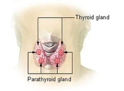
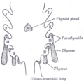
The thyroid, or thyroid gland, is an endocrine gland in vertebrates. In humans, it is in the neck and consists of two connected lobes. The lower two thirds of the lobes are connected by a thin band of tissue called the isthmus (pl.: isthmi). The thyroid gland is a butterfly-shaped gland located in the neck below the Adam's apple. Microscopically, the functional unit of the thyroid gland is the spherical thyroid follicle, lined with follicular cells (thyrocytes), and occasional parafollicular cells that surround a lumen containing colloid. The thyroid gland secretes three hormones: the two thyroid hormones – triiodothyronine (T3) and thyroxine (T4) – and a peptide hormone, calcitonin. The thyroid hormones influence the metabolic rate and protein synthesis and growth and development in children. Calcitonin plays a role in calcium homeostasis. Secretion of the two thyroid hormones is regulated by thyroid-stimulating hormone (TSH), which is secreted from the anterior pituitary gland. TSH is regulated by thyrotropin-releasing hormone (TRH), which is produced by the hypothalamus.

<span class="mw-page-title-main">Parathyroid hormone</span> Mammalian protein found in Homo sapiens

Parathyroid hormone (PTH), also called parathormone or parathyrin, is a peptide hormone secreted by the parathyroid glands that regulates the serum calcium concentration through its effects on bone, kidney, and intestine.

<span class="mw-page-title-main">Calcium metabolism</span> Movement and regulation of calcium ions in and out of the body

Calcium metabolism is the movement and regulation of calcium ions (Ca2+) in (via the gut) and out (via the gut and kidneys) of the body, and between body compartments: the blood plasma, the extracellular and intracellular fluids, and bone. Bone acts as a calcium storage center for deposits and withdrawals as needed by the blood via continual bone remodeling.

<span class="mw-page-title-main">Hypocalcemia</span> Low calcium levels in ones blood serum

Hypocalcemia is a medical condition characterized by low calcium levels in the blood serum. The normal range of blood calcium is typically between 2.1–2.6 mmol/L, while levels less than 2.1 mmol/L are defined as hypocalcemic. Mildly low levels that develop slowly often have no symptoms. Otherwise symptoms may include numbness, muscle spasms, seizures, confusion, or in extreme cases cardiac arrest.

Disorders of calcium metabolism occur when the body has too little or too much calcium. The serum level of calcium is closely regulated within a fairly limited range in the human body. In a healthy physiology, extracellular calcium levels are maintained within a tight range through the actions of parathyroid hormone, vitamin D and the calcium sensing receptor. Disorders in calcium metabolism can lead to hypocalcemia, decreased plasma levels of calcium or hypercalcemia, elevated plasma calcium levels.

<span class="mw-page-title-main">Parathyroid chief cell</span>

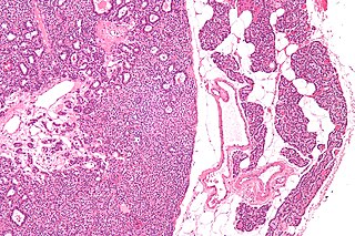
Parathyroid chief cells are one of the two cell types of the parathyroid glands, along with oxyphil cells. The chief cells are much more prevalent in the parathyroid gland than the oxyphil cells. It is perceived that oxyphil cells may be derived from chief cells at puberty, as they are not present at birth like chief cells.

<span class="mw-page-title-main">Oxyphil cell (parathyroid)</span> Cell type

Parathyroid oxyphil cells are one out of the two types of cells found in the parathyroid gland, the other being parathyroid chief cell. Oxyphil cells are only found in a select few number of species and humans are one of them.

Hypoparathyroidism is decreased function of the parathyroid glands with underproduction of parathyroid hormone (PTH). This can lead to low levels of calcium in the blood, often causing cramping and twitching of muscles or tetany, and several other symptoms. It is a very rare disease. The condition can be inherited, but it is also encountered after thyroid or parathyroid gland surgery, and it can be caused by immune system-related damage as well as a number of rarer causes. The diagnosis is made with blood tests, and other investigations such as genetic testing depending on the results. The primary treatment of hypoparathyroidism is calcium and vitamin D supplementation. Calcium replacement or vitamin D can ameliorate the symptoms but can increase the risk of kidney stones and chronic kidney disease. Additionally, medications such as recombinant human parathyroid hormone or teriparatide may be given by injection to replace the missing hormone.

<span class="mw-page-title-main">Hyperparathyroidism</span> Increase in parathyroid hormone levels in the blood

Hyperparathyroidism is an increase in parathyroid hormone (PTH) levels in the blood. This occurs from a disorder either within the parathyroid glands or as response to external stimuli. Symptoms of hyperparathyroidism are caused by inappropriately normal or elevated blood calcium excreted from the bones and flowing into the blood stream in response to increased production of parathyroid hormone. In healthy people, when blood calcium levels are high, parathyroid hormone levels should be low. With long-standing hyperparathyroidism, the most common symptom is kidney stones. Other symptoms may include bone pain, weakness, depression, confusion, and increased urination. Both primary and secondary may result in osteoporosis.

<span class="mw-page-title-main">Hyperphosphatemia</span> Medical condition

Hyperphosphatemia is an electrolyte disorder in which there is an elevated level of phosphate in the blood. Most people have no symptoms while others develop calcium deposits in the soft tissue. Often there is also low calcium levels which can result in muscle spasms.

<span class="mw-page-title-main">Parathyroidectomy</span> Surgical removal of one or more of the parathyroid glands

Parathyroidectomy is the surgical removal of one or more of the (usually) four parathyroid glands. This procedure is used to remove an adenoma or hyperplasia of these glands when they are producing excessive parathyroid hormone (PTH): hyperparathyroidism. The glands are usually four in number and located adjacent to the posterior surface of the thyroid gland, but their exact location is variable. When an elevated PTH level is found, a sestamibi scan or an ultrasound may be performed in order to confirm the presence and location of abnormal parathyroid tissue.

<span class="mw-page-title-main">Endocrine gland</span> Glands of the endocrine system that secrete hormones to blood

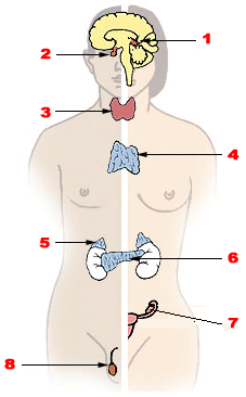
Endocrine glands are ductless glands of the endocrine system that secrete their products, hormones, directly into the blood. The major glands of the endocrine system include the pineal gland, pituitary gland, pancreas, ovaries, testicles, thyroid gland, parathyroid gland, hypothalamus and adrenal glands. The hypothalamus and pituitary glands are neuroendocrine organs.

<span class="mw-page-title-main">Osteitis fibrosa cystica</span> Medical condition

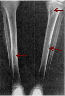
Osteitis fibrosa cystica is a skeletal disorder resulting in a loss of bone mass, a weakening of the bones as their calcified supporting structures are replaced with fibrous tissue, and the formation of cyst-like brown tumors in and around the bone. Osteitis fibrosis cystica (OFC), also known as osteitis fibrosa, osteodystrophia fibrosa, and von Recklinghausen's disease of bone, is caused by hyperparathyroidism, which is a surplus of parathyroid hormone from over-active parathyroid glands. This surplus stimulates the activity of osteoclasts, cells that break down bone, in a process known as osteoclastic bone resorption. The hyperparathyroidism can be triggered by a parathyroid adenoma, hereditary factors, parathyroid carcinoma, or renal osteodystrophy. Osteoclastic bone resorption releases minerals, including calcium, from the bone into the bloodstream, causing both elevated blood calcium levels, and the structural changes which weaken the bone. The symptoms of the disease are the consequences of both the general softening of the bones and the excess calcium in the blood, and include bone fractures, kidney stones, nausea, moth-eaten appearance in the bones, appetite loss, and weight loss.

<span class="mw-page-title-main">Albright's hereditary osteodystrophy</span> Form of osteodystrophy and a rare human disease

Albright's hereditary osteodystrophy is a form of osteodystrophy, and is classified as the phenotype of pseudohypoparathyroidism type 1A; this is a condition in which the body does not respond to parathyroid hormone.

Pseudohypoparathyroidism is a rare autosomal dominant genetic condition associated primarily with resistance to the parathyroid hormone. Those with the condition have a low serum calcium and high phosphate, but the parathyroid hormone level (PTH) is inappropriately high. Its pathogenesis has been linked to dysfunctional G proteins. Pseudohypoparathyroidism is a very rare disorder, with estimated prevalence between 0.3 and 1.1 cases per 100000 population depending on geographic location.

<span class="mw-page-title-main">Tertiary hyperparathyroidism</span> Medical condition

Tertiary hyperparathyroidism is a condition involving the overproduction of the hormone, parathyroid hormone, produced by the parathyroid glands. The parathyroid glands are involved in monitoring and regulating blood calcium levels and respond by either producing or ceasing to produce parathyroid hormone.

<span class="mw-page-title-main">Parathyroid adenoma</span> Medical condition

A parathyroid adenoma is a benign tumor of the parathyroid gland. It generally causes hyperparathyroidism; there are very few reports of parathyroid adenomas that were not associated with hyperparathyroidism.

<span class="mw-page-title-main">Parathyroid disease</span> Medical condition

Many conditions are associated with disorders of the function of the parathyroid gland. Some disorders may be purely anatomical resulting in an enlarged gland which will raise concern. Such benign disorders, such as parathyroid cyst, are not discussed here. Parathyroid diseases can be divided into those causing hyperparathyroidism, and those causing hypoparathyroidism.

<span class="mw-page-title-main">Sestamibi parathyroid scan</span> Procedure in nuclear medicine

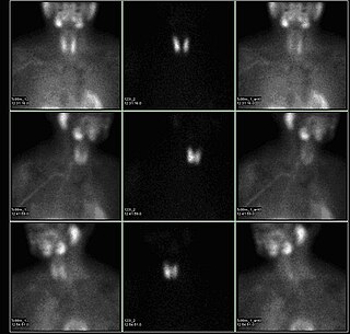
A sestamibi parathyroid scan is a procedure in nuclear medicine which is performed to localize parathyroid adenoma, which causes Hyperparathyroidism. Adequate localization of parathyroid adenoma allows the surgeon to use a minimally invasive surgical approach.

References

  1. Gray, Henry (1980). Williams, Peter L; Warwick, Roger (eds.). Gray's Anatomy (36th ed.). Churchill Livingstone. p. 1453. ISBN   0-443-01505-8.
  2. Johnson, S J (1 April 2005). "Best Practice No 183: Examination of parathyroid gland specimens". Journal of Clinical Pathology. 58 (4): 338–342. doi:10.1136/jcp.2002.002550. PMC   1770637 . PMID   15790694.
  3. 1 2 Illustrated Anatomy of the Head and Neck, Fehrenbach and Herring, Elsevier, 2012, p. 159
  4. 1 2 Drake, Richard L.; Vogl, Wayne; Tibbitts, Adam W.M. Mitchell; illustrations by Richard; Richardson, Paul (2005). Gray's anatomy for students. Philadelphia: Elsevier/Churchill Livingstone. p. 918. ISBN   978-0-8089-2306-0.
  5. 1 2 3 4 Williams, S. Jacob; dissections by David J. Hinchcliffe; photography by Mick A. Turton; illustrated by Amanda (2007). Human anatomy: a clinically-orientated approach (New ed.). Edinburgh: Churchill Livingstone. ISBN   978-0-443-10373-5.{{cite book}}: CS1 maint: multiple names: authors list (link)[ page needed ]
  6. Lappas D, Noussios G, Anagnostis P, Adamidou F, Chatzigeorgiou A, Skandalakis P (September 2012). "Location, number and morphology of parathyroid glands: results from a large anatomical series". Anat Sci Int. 87 (3): 160–4. doi:10.1007/s12565-012-0142-1. PMID   22689148. S2CID   207431255.
  7. 1 2 3 Young, Barbara; Heath, John W.; Stevens, Alan; Burkitt, H. George (2006). Wheater's functional histology: a text and colour atlas (5th ed.). Edinburgh: Churchill Livingstone/Elsevier. p.  337. ISBN   978-0-443-06850-8.
  8. Ritter, Cynthia S.; Haughey, Bruce H.; Miller, Brent; Brown, Alex J. (August 2012). "Differential Gene Expression by Oxyphil and Chief Cells of Human Parathyroid Glands". The Journal of Clinical Endocrinology & Metabolism. 97 (8): E1499–E1505. doi:10.1210/jc.2011-3366. PMC   3591682 . PMID   22585091.
  9. Larsen, William J. (2001). Human embryology (3rd ed.). Philadelphia, Pa.: Churchill Livingstone. pp. 377–8. ISBN   0-443-06583-7.
  10. 1 2 3 Zajac, Jeffrey D; Danks, Janine A (July 2008). "The development of the parathyroid gland: from fish to human". Current Opinion in Nephrology and Hypertension. 17 (4): 353–356. doi:10.1097/MNH.0b013e328304651c. PMID   18660669. S2CID   7971353.
  11. Young, Barbara; Heath, John W.; Stevens, Alan; Burkitt, H. George (2006). Wheater's functional histology: a text and colour atlas (5th ed.). Edinburgh: Churchill Livingstone/Elsevier. p.  336. ISBN   978-0-443-06850-8.
  12. 1 2 3 Hall, Arthur C. Guyton, John E. (2005). Textbook of medical physiology (11th ed.). Philadelphia: W.B. Saunders. pp. 985–8. ISBN   978-0-7216-0240-0.{{cite book}}: CS1 maint: multiple names: authors list (link)
  13. 1 2 Colledge, Nicki R.; Walker, Brian R.; Ralston, Stuart H., eds. (2010). Davidson's principles and practice of medicine (21st ed.). Edinburgh: Churchill Livingstone/Elsevier. pp.  766–7. ISBN   978-0-7020-3084-0.
  14. Colledge, Nicki R.; Walker, Brian R.; Ralston, Stuart H., eds. (2010). Davidson's principles and practice of medicine (21st ed.). Edinburgh: Churchill Livingstone/Elsevier. p.  767. ISBN   978-0-7020-3084-0.
  15. Staren, Prinz; Richard, A., eds. (2000). Endocrine surgery . Georgetown TX: Landes Bioscience. pp.  98–114. ISBN   978-1-57059-574-5.
  16. 1 2 3 Colledge, Nicki R.; Walker, Brian R.; Ralston, Stuart H., eds. (2010). Davidson's principles and practice of medicine (21st ed.). Edinburgh: Churchill Livingstone/Elsevier. p.  768. ISBN   978-0-7020-3084-0.
  17. Cave, A.J.E. (1953). "Richard Owen and the discovery of the parathyroid glands" (PDF). In E. Ashworth Underwood (ed.). Science, Medicine and History. Essays on the Evolution of Scientific Thought and Medical Practice. Vol. 2. Oxford University Press. pp. 217–222. Retrieved 2009-07-20.
  18. 1 2 Eknoyan G (November 1995). "A history of the parathyroid glands". American Journal of Kidney Diseases . 26 (5): 801–7. doi:10.1016/0272-6386(95)90447-6. PMID   7485136.
  19. "On a New Gland in Man and Several Mammals (Glandulæ Parathyreoideæ)". Journal of the American Medical Association. 111 (2): 197. 9 July 1938. doi:10.1001/jama.1938.02790280087037.
  20. 1 2 3 4 DuBose, Joseph; Ragsdale, Timothy; Morvant, Jason (January 2005). ""Bodies so tiny": The history of parathyroid surgery". Current Surgery. 62 (1): 91–95. doi:10.1016/j.cursur.2004.07.012. PMID   15708157.
  21. Carney, JA (Sep 1996). "The glandulae parathyroideae of Ivar Sandström. Contributions from two continents". The American Journal of Surgical Pathology. 20 (9): 1123–44. doi:10.1097/00000478-199609000-00010. PMID   8764749.
  22. Maccallum, W. G; Voegtlin, C. (Jan 9, 1909). "On the Relation of Tetant to the Parathyroid Glands and to Calcium Metabolism". The Journal of Experimental Medicine. 11 (1): 118–51. doi:10.1084/jem.11.1.118. PMC   2124703 . PMID   19867238.
  23. Councilman, WT (1913). "Chapter 1". Disease and Its Causes. United States: New York Henry Holt and Company London Williams and Norgate The University Press, Cambridge, U.S. ASIN   B0065T4O6Q. OCLC   654587300.
  24. Okabe, Masataka; Graham, Anthony (2004). "The origin of the parathyroid gland". Proceedings of the National Academy of Sciences. 101 (51): 17716–9. Bibcode:2004PNAS..10117716O. doi: 10.1073/pnas.0406116101 . JSTOR   3374034. PMC   539734 . PMID   15591343.

Further reading