Pharyngeal arch

Last updated
Pharyngeal arch
Gray41.png
Schematic of developing human fetus with first, second and third arches labelled
Details
Carnegie stage 10
Identifiers
Latin arcus pharyngei
MeSH D001934
TE arch_by_E5.4.2.0.0.0.2 E5.4.2.0.0.0.2
Anatomical terminology
Gray979.png
Floor of the pharynx of human embryo at about 26 days old
PharyngealArchHuman.jpg
Scheme of the pharyngeal arches
Kiemenbogen.jpg
Scheme of the pharyngeal arches

The pharyngeal arches, also known as visceral arches, are structures seen in the embryonic development of vertebrates that are recognisable precursors for many structures. In fish, the arches are known as the branchial arches, or gill arches.

Contents

In the human embryo, the arches are first seen during the fourth week of development. They appear as a series of outpouchings of mesoderm on both sides of the developing pharynx. The vasculature of the pharyngeal arches is known as the aortic arches.

In fish, the branchial arches support the gills.

Structure

In vertebrates, the pharyngeal arches are derived from all three germ layers (the primary layers of cells that form during embryogenesis). [1] Neural crest cells enter these arches where they contribute to features of the skull and facial skeleton such as bone and cartilage. [1] However, the existence of pharyngeal structures before neural crest cells evolved is indicated by the existence of neural crest-independent mechanisms of pharyngeal arch development. [2] The first, most anterior pharyngeal arch gives rise to the oral jaw. The second arch becomes the hyoid and jaw support. [1] In fish, the other posterior arches contribute to the branchial skeleton, which support the gills; in tetrapods the anterior arches develop into components of the ear, tonsils, and thymus. [3] The genetic and developmental basis of pharyngeal arch development is well characterized. It has been shown that Hox genes and other developmental genes such as DLX are important for patterning the anterior/posterior and dorsal/ventral axes of the branchial arches. [4] Some fish species have a second set of jaws in their throat, known as pharyngeal jaws, which develop using the same genetic pathways involved in oral jaw formation. [5]

During human, and all vertebrate development, a series of pharyngeal arch pairs form in the developing embryo. These project forward from the back of the embryo toward the front of the face and neck. Each arch develops its own artery, nerve that controls a distinct muscle group, and skeletal tissue. The arches are numbered from 1 to 6, with 1 being the arch closest to the head of the embryo, and arch 5 existing only transiently. [6]

These grow and join in the ventral midline. The first arch, as the first to form, separates the mouth pit or stomodeum from the pericardium. By differential growth the neck elongates and new arches form, so the pharynx has six arches ultimately.

Each pharyngeal arch has a cartilaginous stick, a muscle component that differentiates from the cartilaginous tissue, an artery, and a cranial nerve. Each of these is surrounded by mesenchyme. Arches do not develop simultaneously but instead possess a "staggered" development.

Pharyngeal pouches form on the endodermal side between the arches, and pharyngeal grooves (or clefts) form from the lateral ectodermal surface of the neck region to separate the arches. [7] In fish, the pouches line up with the clefts, and these thin segments become gills. In mammals the endoderm and ectoderm not only remain intact but also continue to be separated by a mesoderm layer.

The development of the pharyngeal arches provides a useful landmark with which to establish the precise stage of embryonic development. Their formation and development corresponds to Carnegie stages 10 to 16 in mammals, and Hamburger–Hamilton stages 14 to 28 in the chicken. Although there are six pharyngeal arches, in humans the fifth arch exists only transiently during embryogenesis. [8]

First arch

The first pharyngeal arch, also mandibular arch (corresponding to the first branchial arch of fish), is the first of six pharyngeal arches that develops during the fourth week of development. [9] It is located between the stomodeum and the first pharyngeal groove.

Processes

This arch divides into a maxillary process and a mandibular process, giving rise to structures including the bones of the lower two-thirds of the face and the jaw. The maxillary process becomes the maxilla (or upper jaw, although there are large differences among animals [10] ), and palate while the mandibular process becomes the mandible or lower jaw. This arch also gives rise to the muscles of mastication.

Meckel's cartilage

Meckel's cartilage forms in the mesoderm of the mandibular process and eventually regresses to form the incus and malleus of the middle ear, the anterior ligament of the malleus and the sphenomandibular ligament. The mandible or lower jaw forms by perichondral ossification using Meckel's cartilage as a 'template', but the maxillary does not arise from direct ossification of Meckel's cartilage.

Derivatives

The skeletal elements and muscles are derived from mesoderm of the pharyngeal arches.

Skeletal

Muscles

Other

Mucous membrane and glands of the anterior two thirds of the tongue are derived from ectoderm and endoderm of the arch.

Nerve supply

The mandibular and maxillary branches of the trigeminal nerve (CN V) innervate the structures derived from the corresponding processes of the first arch. In some lower animals, each arch is supplied by two cranial nerves. The nerve of the arch itself runs along the cranial side of the arch and is called post-trematic nerve of the arch. Each arch also receives a branch from the nerve of the succeeding arch called the pre-trematic nerve which runs along the caudal border of the arch. In human embryo, a double innervation is seen only in the first pharyngeal arch. The mandibular nerve is the post-trematic nerve of the first arch and chorda tympani (branch of facial nerve) is the pre-trematic nerve. This double innervation is reflected in the nerve supply of anterior two-thirds of tongue which is derived from the first arch. [11]

Blood supply

The artery of the first arch is the first aortic arch, [12] which partially persists as the maxillary artery.

Second arch

The second pharyngeal arch or hyoid arch is the second of fifth pharyngeal arches that develops in fetal life during the fourth week of development [9] and assists in forming the side and front of the neck.

Reichert's cartilage

Cartilage in the second pharyngeal arch is referred to as Reichert's cartilage and contributes to many structures in the fully developed adult. [13] In contrast to the Meckel's cartilage of the first pharyngeal arch, it does not constitute a continuous element, and instead is composed of two distinct cartilaginous segments joined by a faint layer of mesenchyme. [14] Dorsal ends of Reichert's cartilage ossify during development to form the stapes of the middle ear before being incorporated into the middle ear cavity, while the ventral portion ossifies to form the lesser cornu and upper part of the body of the hyoid bone. Caudal to what will eventually become the stapes, Reichert's cartilage also forms the styloid process of the temporal bone. The cartilage between the hyoid bone and styloid process will not remain as development continues, but its perichondrium will eventually form the stylohyoid ligament.

Derivatives

Skeletal

From the cartilage of the second arch arises

Muscles

Nerve supply

Facial nerve (CN VII)

Blood supply

The artery of the second arch is the second aortic arch, [12] which gives origin to the stapedial artery in some mammals but atrophies in most humans.

Muscles derived from the pharyngeal arches

Pharyngeal muscles or branchial muscles are striated muscles of the head and neck. Unlike skeletal muscles that developmentally come from somites, pharyngeal muscles are developmentally formed from the pharyngeal arches.

Most of the skeletal musculature supplied by the cranial nerves (special visceral efferent) is pharyngeal. Exceptions include, but are not limited to, the extraocular muscles and some of the muscles of the tongue. These exceptions receive general somatic efferent innervation.

First arch

All of the pharyngeal muscles that come from the first pharyngeal arch are innervated by the mandibular divisions of the trigeminal nerve. [15] These muscles include all the muscles of mastication, the anterior belly of the digastric, the mylohyoid, tensor tympani, and tensor veli palatini.

Second arch

All of the pharyngeal muscles of the second pharyngeal arch are innervated by the facial nerve. These muscles include the muscles of facial expression, the posterior belly of the digastric, the stylohyoid muscle, the auricular muscle [15] and the stapedius muscle of the middle ear.

Third arch

There is only one muscle of the third pharyngeal arch, the stylopharyngeus. The stylopharyngeus and other structures from the third pharyngeal arch are all innervated by the glossopharyngeal nerve.

Fourth and sixth arches

All the pharyngeal muscles of the fourth and sixth arches are innervated by the superior laryngeal and the recurrent laryngeal branches of the vagus nerve. [15] These muscles include all the muscles of the palate (exception of the tensor veli palatini which is innervated by the trigeminal nerve), all the muscles of the pharynx (except stylopharyngeus which is innervated by the glossopharyngeal nerve), and all the muscles of the larynx.

In humans

Amniotes have five arches, numbered 1 to 5. [16] Older literature reports the fifth arch as the sixth arch, the fifth being absent. More is known about the fate of the first arch than the remaining four. The first three contribute to structures above the larynx, whereas the last two contribute to the larynx and trachea.

The recurrent laryngeal nerves are produced from the nerve of arch 5, and the laryngeal cartilages from arches 4 and 5. The superior laryngeal branch of the vagus nerve arises from arch 4. Its arteries, which project between the nerves of the fourth and fifth arches, become the left-side arch of the aorta and the right subclavian artery. On the right side, the artery of arch 5 is obliterated while, on the left side, the artery persists as the ductus arteriosus; circulatory changes immediately following birth cause the vessel to close down, leaving a remnant, the ligamentum arteriosum. During growth, these arteries descend into their ultimate positions in the chest, creating the elongated recurrent paths. [6]

Pharyngeal archMuscular contributions [17] Skeletal contributionsNerve Artery
First (also called "mandibular arch") Muscles of mastication, anterior belly of the digastric, mylohyoid, tensor tympani, tensor veli palatini Premaxilla, maxilla, mandible (only as a model for mandible not actual formation of mandible), zygomatic bone, part of the temporal bone, [18] the incus, and the malleus of the middle ear, also Meckel's cartilage and the sphenomandibular ligament. Trigeminal nerve (part of V2 [19] and V3) Maxillary artery, external carotid artery, Vidian artery
Second (also called the "hyoid arch") Muscles of facial expression, buccinator, platysma, stapedius, stylohyoid, posterior belly of the digastric, auricular [15] Stapes, temporal styloid process, hyoid bone (lesser horns and upper part of body), stylohyoid ligament, [15] Reichert's cartilage Facial nerve (VII) Ascending pharyngeal artery, Inferior tympanic artery, primitive hyoid artery, Stapedial artery
Third Stylopharyngeus Hyoid bone (greater horns and lower part of body), thymus Glossopharyngeal nerve (IX) common carotid, internal carotid
Fourth Cricothyroid muscle, all intrinsic muscles of soft palate (including levator veli palatini) except tensor veli palatini Thyroid cartilage, epiglottic cartilage [20] Vagus nerve (X), superior laryngeal nerve [21] Right 4th aortic arch: subclavian artery

Left fourth aortic arch: aortic arch

FifthAll intrinsic muscles of larynx except the cricothyroid muscle Cricoid cartilage, arytenoid cartilages, corniculate cartilage, cuneiform cartilages [20] Accessory nerve (Cranial root) (XI), recurrent laryngeal nerve [21] Right 5th aortic arch: pulmonary artery

Left fifth aortic arch: pulmonary artery and ductus arteriosus

See also

Related Research Articles

<span class="mw-page-title-main">Temporomandibular joint</span> Joints connecting the jawbone to the skull

In anatomy, the temporomandibular joints (TMJ) are the two joints connecting the jawbone to the skull. It is a bilateral synovial articulation between the temporal bone of the skull above and the mandible below; it is from these bones that its name is derived. This joint is unique in that it is a bilateral joint that functions as one unit. Since the TMJ is connected to the mandible, the right and left joints must function together and therefore are not independent of each other.

<span class="mw-page-title-main">Facial nerve</span> Cranial nerve VII, for the face and tasting

The facial nerve, also known as the seventh cranial nerve, cranial nerve VII, or simply CN VII, is a cranial nerve that emerges from the pons of the brainstem, controls the muscles of facial expression, and functions in the conveyance of taste sensations from the anterior two-thirds of the tongue. The nerve typically travels from the pons through the facial canal in the temporal bone and exits the skull at the stylomastoid foramen. It arises from the brainstem from an area posterior to the cranial nerve VI and anterior to cranial nerve VIII.

Articles related to anatomy include:

<span class="mw-page-title-main">Hyoid bone</span> Bone situated in the neck between the chin and the thyroid cartilage

The hyoid bone is a horseshoe-shaped bone situated in the anterior midline of the neck between the chin and the thyroid cartilage. At rest, it lies between the base of the mandible and the third cervical vertebra.

<span class="mw-page-title-main">External carotid artery</span> Major artery of the head and neck

The external carotid artery is a major artery of the head and neck. It arises from the common carotid artery when it splits into the external and internal carotid artery. The external carotid artery supplies blood to the face, brain and neck.

<span class="mw-page-title-main">Suprahyoid muscles</span>

The suprahyoid muscles are four muscles located above the hyoid bone in the neck. They are the digastric, stylohyoid, geniohyoid, and mylohyoid muscles. They are all pharyngeal muscles, with the exception of the geniohyoid muscle. The digastric is uniquely named for its two bellies. Its posterior belly rises from the mastoid process of the cranium and slopes downward and forward. The anterior belly arises from the digastric fossa on the inner surface of the mandibular body, which slopes downward and backward. The two bellies connect at the intermediate tendon. The intermediate tendon passes through a connective tissue loop attached to the hyoid bone. The mylohyoid muscles are thin, flat muscles that form a sling inferior to the tongue supporting the floor of the mouth. The geniohyoids are short, narrow muscles that contact each other in the midline. The stylohyoids are long, thin muscles that are nearly parallel with the posterior belly of the digastric muscle.

<span class="mw-page-title-main">Muscles of mastication</span> Muscles that aid chewing

The four classical muscles of mastication elevate the mandible and move it forward/backward and laterally, facilitating biting and chewing. Other muscles are responsible for opening the jaw, namely the geniohyoid, mylohyoid, and digastric muscles.

<span class="mw-page-title-main">Digastric muscle</span> Small muscle located under the jaw in mammals

The digastric muscle is a bilaterally paired suprahyoid muscle located under the jaw. Its posterior belly is attached to the mastoid notch of temporal bone, and its anterior belly is attached to the digastric fossa of mandible; the two bellies are united by an intermediate tendon which is held in a loop that attaches to the hyoid bone. The anterior belly is innervated via the mandibular nerve, and the posterior belly is innervated via the facial nerve. It may act to depress the mandible or elevate the hyoid bone.

<span class="mw-page-title-main">Stylohyoid muscle</span> Muscle

The stylohyoid muscle is one of the suprahyoid muscles. Its originates from the styloid process of the temporal bone; it inserts onto hyoid bone. It is innervated by a branch of the facial nerve. It acts draw the hyoid bone upwards and backwards.

<span class="mw-page-title-main">Hyoglossus</span> Muscle

The hyoglossus is a thin and quadrilateral extrinsic muscle of the tongue. It originates from the hyoid bone; it inserts onto the side of the tongue. It is innervated by the hypoglossal nerve. It acts to depress and retract the tongue.

<span class="mw-page-title-main">Middle pharyngeal constrictor muscle</span> Muscle in the neck

The middle pharyngeal constrictor is a fan-shaped muscle located in the neck. It is one of three pharyngeal constrictor muscles. It is smaller than the inferior pharyngeal constrictor muscle.

<span class="mw-page-title-main">Aortic arches</span>

The aortic arches or pharyngeal arch arteries are a series of six paired embryological vascular structures which give rise to the great arteries of the neck and head. They are ventral to the dorsal aorta and arise from the aortic sac.

<span class="mw-page-title-main">Sphenomandibular ligament</span>

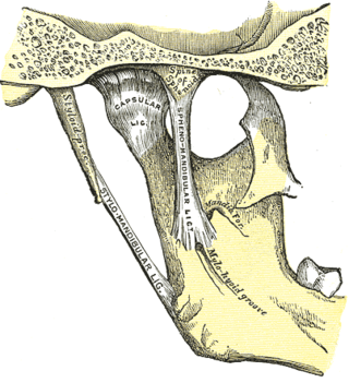
The sphenomandibular ligament is one of the three ligaments of the temporomandibular joint. It is situated medially to - and generally separate from - the articular capsule of the joint. Superiorly, it is attached to the spine of the sphenoid bone; inferiorly, it is attached to the lingula of mandible. The SML acts to limit inferior-ward movement of the mandible.

<span class="mw-page-title-main">Meckel's cartilage</span>

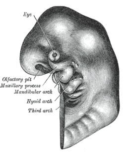
In humans, the cartilaginous bar of the mandibular arch is formed by what are known as Meckel's cartilages also known as Meckelian cartilages; above this the incus and malleus are developed. Meckel's cartilage arises from the first pharyngeal arch.

<span class="mw-page-title-main">Anterior triangle of the neck</span>

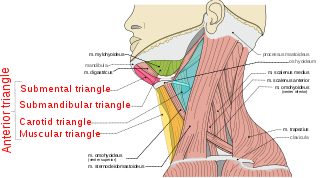
The anterior triangle is a region of the neck.

<span class="mw-page-title-main">Carotid triangle</span>

The carotid triangle is a portion of the anterior triangle of the neck.

<span class="mw-page-title-main">Cervical lymph nodes</span> Lymph nodes found in the neck

Cervical lymph nodes are lymph nodes found in the neck. Of the 800 lymph nodes in the human body, 300 are in the neck. Cervical lymph nodes are subject to a number of different pathological conditions including tumours, infection and inflammation.

<span class="mw-page-title-main">Outline of human anatomy</span> Overview of and topical guide to human anatomy

The following outline is provided as an overview of and topical guide to human anatomy:

<span class="mw-page-title-main">Mandible</span> Lower jaw bone

In jawed vertebrates, the mandible, lower jaw, or jawbone is a bone that makes up the lower – and typically more mobile – component of the mouth.

The face and neck development of the human embryo refers to the development of the structures from the third to eighth week that give rise to the future head and neck. They consist of three layers, the ectoderm, mesoderm and endoderm, which form the mesenchyme, neural crest and neural placodes. The paraxial mesoderm forms structures named somites and somitomeres that contribute to the development of the floor of the brain and voluntary muscles of the craniofacial region. The lateral plate mesoderm consists of the laryngeal cartilages. The three tissue layers give rise to the pharyngeal apparatus, formed by six pairs of pharyngeal arches, a set of pharyngeal pouches and pharyngeal grooves, which are the most typical feature in development of the head and neck. The formation of each region of the face and neck is due to the migration of the neural crest cells which come from the ectoderm. These cells determine the future structure to develop in each pharyngeal arch. Eventually, they also form the neurectoderm, which forms the forebrain, midbrain and hindbrain, cartilage, bone, dentin, tendon, dermis, pia mater and arachnoid mater, sensory neurons, and glandular stroma.

References

  1. 1 2 3 Graham A (2003). "Development of the pharyngeal arches". Am J Med Genet A. 119A (3): 251–256. doi:10.1002/ajmg.a.10980. PMID   12784288. S2CID   28318053.
  2. Graham A, Smith A (2001). "Patterning the pharyngeal arches". BioEssays. 23 (1): 54–61. doi:10.1002/1521-1878(200101)23:1<54::AID-BIES1007>3.0.CO;2-5. PMID   11135309. S2CID   10792335.
  3. Kardong KV (2003). "Vertebrates: Comparative Anatomy, Function, Evolution". Third Edition. New York (McGraw Hill).
  4. Depew MJ, Lufkin T, Rubenstein JL (2002). "Specification of jaw subdivisions by Dlx genes". Science. 298 (5592): 381–385. doi: 10.1126/science.1075703 . PMID   12193642. S2CID   10274300.
  5. Fraser GJ, Hulsey D, Bloomquist RF, Uyesugi K, Manley NR, Streelman T (2009). Jernvall J (ed.). "An Ancient Gene Network Is Co-opted for Teeth on Old and New Jaws". PLOS Biology. 7 (2): 0233–0247. doi: 10.1371/journal.pbio.1000031 . PMC   2637924 . PMID   19215146.
  6. 1 2 Larsen, William J. (1993). Human embryology. New York: Churchill Livingstone. pp. 318–323. ISBN   0-443-08724-5.
  7. McKenzie, James C. "Lecture 24. Branchial Apparatus". Howard University. Archived from the original on 2003-05-02. Retrieved 2007-09-09.
  8. Marino, Thomas A. "Text for Pharyngeal Arch Development". Temple University. Archived from the original on 2007-09-09. Retrieved 2007-09-09.
  9. 1 2 William J. Larsen (2001). Human embryology. Edinburgh: Churchill Livingstone. ISBN   0-443-06583-7
  10. Higashiyama, Hiroki; Koyabu, Daisuke; Hirasawa, Tatsuya; Werneburg, Ingmar; Kuratani, Shigeru; Kurihara, Hiroki (November 2, 2021). "Mammalian face as an evolutionary novelty". PNAS. 118 (44): e2111876118. Bibcode:2021PNAS..11811876H. doi: 10.1073/pnas.2111876118 . PMC   8673075 . PMID   34716275.
  11. Inderbir Sing, G.P Pal-Human Embryology
  12. 1 2 McMinn, R., 1994. Last's anatomy: Regional and applied (9th ed).
  13. Sudhir, Sant, 2008.Embryology for Medical Students 2nd edition
  14. Rodríguez-Vázquez JF (2008). "Morphogenesis of the second pharyngeal arch cartilage (Reichert's cartilage) in human embryos". J. Anat. 208 (2): 179–189. doi:10.1111/j.1469-7580.2006.00524.x. PMC   2100189 . PMID   16441562.
  15. 1 2 3 4 5 Sadler, Thomas W. (February 2009). Langman's Medical Embryology. Lippincott Williams & Wilkins. pp. 366–369. ISBN   978-0781790697.
  16. Graham, Anthony; Poopalasundaram, Subathra (11 Aug 2019). "A reappraisal and revision of the numbering of the pharyngeal arches". J. Anat. 235 (6): 1019–1023. doi:10.1111/joa.13067. PMC   6875933 . PMID   31402457.
  17. "marshall.edu". Archived from the original on 2009-02-27. Retrieved 2007-09-09.
  18. Sadler, Thomas W. (February 2009). Langman's Medical Embryology. Lippincott Williams & Wilkins. pp. 366–372. ISBN   978-0781790697.
  19. Higashiyama H, Kuratani S (2014). "On the maxillary nerve". Journal of Morphology. 275 (1): 17–38. doi:10.1002/jmor.20193. PMID   24151219. S2CID   32707087.
  20. 1 2 Netter, Frank H.; Cochard, Larry R. (2002). Netter's Atlas of human embryology. Teterboro, N.J: Icon Learning Systems. p. 227. ISBN   0-914168-99-1.
  21. 1 2 Kyung Won Chung (2005). Gross Anatomy (Board Review). Hagerstown, Maryland: Lippincott Williams & Wilkins. ISBN   0-7817-5309-0.