Germ layer

Last updated

A germ layer is a primary layer of cells that forms during embryonic development. [1] The three germ layers in vertebrates are particularly pronounced; however, all eumetazoans (animals that are sister taxa to the sponges) produce two or three primary germ layers. Some animals, like cnidarians, produce two germ layers (the ectoderm and endoderm) making them diploblastic. Other animals such as bilaterians produce a third layer (the mesoderm) between these two layers, making them triploblastic. Germ layers eventually give rise to all of an animal's tissues and organs through the process of organogenesis.

Contents

History

Cleavage and division of the cell of an egg of a vertebrate (Remak, 1855). Remak "Untersuchungen uber die...", 1855 Wellcome L0014201.jpg
Cleavage and division of the cell of an egg of a vertebrate (Remak, 1855).

Caspar Friedrich Wolff observed organization of the early embryo in leaf-like layers. In 1817, Heinz Christian Pander discovered three primordial germ layers while studying chick embryos. Between 1850 and 1855, Robert Remak had further refined the germ cell layer (Keimblatt) concept, stating that the external, internal and middle layers form respectively the epidermis, the gut, and the intervening musculature and vasculature. [2] [3] [4] The term "mesoderm" was introduced into English by Huxley in 1871, and "ectoderm" and "endoderm" by Lankester in 1873.

Evolution

Gastrulation of a diploblast: The formation of germ layers from a (1) blastula to a (2) gastrula. Some of the ectoderm cells (orange) move inward forming the endoderm (red). Gastrulation.png
Gastrulation of a diploblast: The formation of germ layers from a (1) blastula to a (2) gastrula. Some of the ectoderm cells (orange) move inward forming the endoderm (red).

Among animals, sponges show the least amount of compartmentalization, having a single germ layer. Although they have differentiated cells (e.g. collar cells), they lack true tissue coordination. Diploblastic animals, Cnidaria and Ctenophora, show an increase in compartmentalization, having two germ layers, the endoderm and ectoderm. Diploblastic animals are organized into recognisable tissues. All bilaterian animals (from flatworms to humans) are triploblastic, possessing a mesoderm in addition to the germ layers found in Diploblasts. Triploblastic animals develop recognizable organs.

Development

Fertilization leads to the formation of a zygote. During the next stage, cleavage, mitotic cell divisions transform the zygote into a hollow ball of cells, a blastula. This early embryonic form undergoes gastrulation, forming a gastrula with either two or three layers (the germ layers). In all vertebrates, these progenitor cells differentiate into all adult tissues and organs. [5]

In the human embryo, after about three days, the zygote forms a solid mass of cells by mitotic division, called a morula. This then changes to a blastocyst, consisting of an outer layer called a trophoblast, and an inner cell mass called the embryoblast. Filled with uterine fluid, the blastocyst breaks out of the zona pellucida and undergoes implantation. The inner cell mass initially has two layers: the hypoblast and epiblast. At the end of the second week, a primitive streak appears. The epiblast in this region moves towards the primitive streak, dives down into it, and forms a new layer, called the endoderm, pushing the hypoblast out of the way (this goes on to form the amnion.) The epiblast keeps moving and forms a second layer, the mesoderm. The top layer is now called the ectoderm. [6]

Gastrulation occurs in reference to the primary body axis. Germ layer formation is linked to the primary body axis as well, however it is less reliant on it than gastrulation is. Hydractinia shows that germ layer formation that transpires as a mixed delamination. [7]

In mice, germ layer differentiation is controlled by two transcription factors: Sox2 and Oct4 proteins. These transcription factors cause the pluripotent mouse embryonic stem cells to select a germ layer fate. Sox2 promotes ectodermal differentiation, while Oct4 promotes mesendodermal differentiation. Each gene inhibits what the other promotes. Amounts of each protein are different throughout the genome, causing the embryonic stem cells to select their fate. [8]

The germ layers

Endoderm

The endoderm produces tissue within the lungs, thyroid, and pancreas. Endoderm2.png
The endoderm produces tissue within the lungs, thyroid, and pancreas.

The endoderm is one of the germ layers formed during animal embryonic development. Cells migrating inward along the archenteron form the inner layer of the gastrula, which develops into the endoderm.

The endoderm consists at first of flattened cells, which subsequently become columnar. It forms the epithelial lining of the whole of the digestive tract except part of the mouth and pharynx and the terminal part of the rectum (which are lined by involutions of the ectoderm). It also forms the lining cells of all the glands which open into the digestive tract, including those of the liver and pancreas; the epithelium of the auditory tube and tympanic cavity; the trachea, bronchi, and alveoli of the lungs; the bladder and part of the urethra; and the follicle lining of the thyroid gland and thymus.

The endoderm forms: the pharynx, the esophagus, the stomach, the small intestine, the colon, the liver, the pancreas, the bladder, the epithelial parts of the trachea and bronchi, the lungs, the thyroid, and the parathyroid.

Mesoderm

The mesoderm aids in the production of cardiac muscle, skeletal muscle, smooth muscle, tissues within the kidneys, and red blood cells. Mesoderm.png
The mesoderm aids in the production of cardiac muscle, skeletal muscle, smooth muscle, tissues within the kidneys, and red blood cells.

The mesoderm germ layer forms in the embryos of triploblastic animals. During gastrulation, some of the cells migrating inward contribute to the mesoderm, an additional layer between the endoderm and the ectoderm. [9] The formation of a mesoderm leads to the development of a coelom. Organs formed inside a coelom can freely move, grow, and develop independently of the body wall while fluid cushions protects them from shocks. [10]

The mesoderm has several components which develop into tissues: intermediate mesoderm, paraxial mesoderm, lateral plate mesoderm, and chorda-mesoderm. The chorda-mesoderm develops into the notochord. The intermediate mesoderm develops into kidneys and gonads. The paraxial mesoderm develops into cartilage, skeletal muscle, and dermis. The lateral plate mesoderm develops into the circulatory system (including the heart and spleen), the wall of the gut, and wall of the human body. [11]

Through cell signaling cascades and interactions with the ectodermal and endodermal cells, the mesodermal cells begin the process of differentiation. [12]

The mesoderm forms: muscle (smooth and striated), bone, cartilage, connective tissue, adipose tissue, circulatory system, lymphatic system, dermis, Dentine of teeth, genitourinary system, serous membranes, spleen and notochord.

Ectoderm

The ectoderm produces tissues within the epidermis, aids in the formation of neurons within the brain, and constructs melanocytes. Ectoderm.png
The ectoderm produces tissues within the epidermis, aids in the formation of neurons within the brain, and constructs melanocytes.

The ectoderm generates the outer layer of the embryo, and it forms from the embryo's epiblast. [13] The ectoderm develops into the surface ectoderm, neural crest, and the neural tube. [14]

The surface ectoderm develops into: epidermis, hair, nails, lens of the eye, sebaceous glands, cornea, tooth enamel, the epithelium of the mouth and nose.

The neural crest of the ectoderm develops into: peripheral nervous system, adrenal medulla, melanocytes, facial cartilage.

The neural tube of the ectoderm develops into: brain, spinal cord, posterior pituitary, motor neurons, retina.

Note: The anterior pituitary develops from the ectodermal tissue of Rathke's pouch.

Neural crest

Because of its great importance, the neural crest is sometimes considered a fourth germ layer. [15] It is, however, derived from the ectoderm.

See also

Micrograph of a teratoma, a tumour that characteristically has tissue from all three germ layers. The image shows tissue derived from the mesoderm (immature cartilage - left-upper corner of image), endoderm (gastrointestinal glands - center-bottom of image) and ectoderm (epidermis - right of image). H&E stain. Teratoma 2 low mag.jpg
Micrograph of a teratoma, a tumour that characteristically has tissue from all three germ layers. The image shows tissue derived from the mesoderm (immature cartilage - left-upper corner of image), endoderm (gastrointestinal glands - center-bottom of image) and ectoderm (epidermis - right of image). H&E stain.

Related Research Articles

<span class="mw-page-title-main">Ontogeny</span> Origination and development of an organism

Ontogeny is the origination and development of an organism, usually from the time of fertilization of the egg to adult. The term can also be used to refer to the study of the entirety of an organism's lifespan.

<span class="mw-page-title-main">Embryo</span> Multicellular diploid eukaryote in its earliest stage of development

An embryo is the initial stage of development for a multicellular organism. In organisms that reproduce sexually, embryonic development is the part of the life cycle that begins just after fertilization of the female egg cell by the male sperm cell. The resulting fusion of these two cells produces a single-celled zygote that undergoes many cell divisions that produce cells known as blastomeres. The blastomeres are arranged as a solid ball that when reaching a certain size, called a morula, takes in fluid to create a cavity called a blastocoel. The structure is then termed a blastula, or a blastocyst in mammals.

<span class="mw-page-title-main">Mesoderm</span> Middle germ layer of embryonic development

The mesoderm is the middle layer of the three germ layers that develops during gastrulation in the very early development of the embryo of most animals. The outer layer is the ectoderm, and the inner layer is the endoderm.

<span class="mw-page-title-main">Gastrulation</span> Stage in embryonic development in which germ layers form

Gastrulation is the stage in the early embryonic development of most animals, during which the blastula, or in mammals the blastocyst, is reorganized into a two-layered or three-layered embryo known as the gastrula. Before gastrulation, the embryo is a continuous epithelial sheet of cells; by the end of gastrulation, the embryo has begun differentiation to establish distinct cell lineages, set up the basic axes of the body, and internalized one or more cell types including the prospective gut.

<span class="mw-page-title-main">Ectoderm</span> Outer germ layer of embryonic development

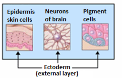
The ectoderm is one of the three primary germ layers formed in early embryonic development. It is the outermost layer, and is superficial to the mesoderm and endoderm. It emerges and originates from the outer layer of germ cells. The word ectoderm comes from the Greek ektos meaning "outside", and derma meaning "skin".

<span class="mw-page-title-main">Endoderm</span> Inner germ layer of embryonic development

Endoderm is the innermost of the three primary germ layers in the very early embryo. The other two layers are the ectoderm and mesoderm. Cells migrating inward along the archenteron form the inner layer of the gastrula, which develops into the endoderm.

<span class="mw-page-title-main">Invagination</span> Process in embryonic development

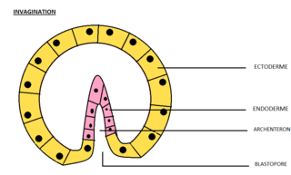
Invagination is the process of a surface folding in on itself to form a cavity, pouch or tube. In developmental biology, invagination is a mechanism that takes place during gastrulation. This mechanism or cell movement happens mostly in the vegetal pole. Invagination consists of the folding of an area of the exterior sheet of cells towards the inside of the blastula. In each organism, the complexity will be different depending on the number of cells. Invagination can be referenced as one of the steps of the establishment of the body plan. The term, originally used in embryology, has been adopted in other disciplines as well.

Organogenesis is the phase of embryonic development that starts at the end of gastrulation and continues until birth. During organogenesis, the three germ layers formed from gastrulation form the internal organs of the organism.

<span class="mw-page-title-main">Animal embryonic development</span> Process by which the embryo forms and develops

In developmental biology, animal embryonic development, also known as animal embryogenesis, is the developmental stage of an animal embryo. Embryonic development starts with the fertilization of an egg cell (ovum) by a sperm cell, (spermatozoon). Once fertilized, the ovum becomes a single diploid cell known as a zygote. The zygote undergoes mitotic divisions with no significant growth and cellular differentiation, leading to development of a multicellular embryo after passing through an organizational checkpoint during mid-embryogenesis. In mammals, the term refers chiefly to the early stages of prenatal development, whereas the terms fetus and fetal development describe later stages.

<span class="mw-page-title-main">Neurula</span> Embryo at the early stage of development in which neurulation occurs

A neurula is a vertebrate embryo at the early stage of development in which neurulation occurs. The neurula stage is preceded by the gastrula stage; consequentially, neurulation is preceded by gastrulation. Neurulation marks the beginning of the process of organogenesis.

Histogenesis is the formation of different tissues from undifferentiated cells. These cells are constituents of three primary germ layers, the endoderm, mesoderm, and ectoderm. The science of the microscopic structures of the tissues formed within histogenesis is termed histology.

The primitive node is the organizer for gastrulation in most amniote embryos. In birds it is known as Hensen's node, and in amphibians it is known as the Spemann-Mangold organizer. It is induced by the Nieuwkoop center in amphibians, or by the posterior marginal zone in amniotes including birds.

A trilaminar embryonic disc, trilaminary blastoderm, or trilaminar germ disk is an early stage in the development of triploblastic organisms, which include humans and many other animals. It is the next stage from the earlier bilaminar embryonic disc.

<span class="mw-page-title-main">Epiblast</span> Embryonic inner cell mass tissue that forms the embryo itself, through the three germ layers

In amniote embryonic development, the epiblast is one of two distinct cell layers arising from the inner cell mass in the mammalian blastocyst, or from the blastula in reptiles and birds, the other layer is the hypoblast. It drives the embryo proper through its differentiation into the three primary germ layers, ectoderm, mesoderm and endoderm, during gastrulation. The amniotic ectoderm and extraembryonic mesoderm also originate from the epiblast.

<span class="mw-page-title-main">Mesenchyme</span> Type of animal embryonic connective tissue

Mesenchyme is a type of loosely organized animal embryonic connective tissue of undifferentiated cells that give rise to most tissues, such as skin, blood or bone. The interactions between mesenchyme and epithelium help to form nearly every organ in the developing embryo.

<span class="mw-page-title-main">Bilaminar embryonic disc</span>

The bilaminar embryonic disc, bilaminar blastoderm or embryonic disc is the distinct two-layered structure of cells formed in an embryo. In the development of the human embryo this takes place by day eight. It is formed when the inner cell mass, also known as the embryoblast, forms a bilaminar disc of two layers, an upper layer called the epiblast and a lower layer called the hypoblast, which will eventually form into fetus. These two layers of cells are stretched between two fluid-filled cavities at either end: the primitive yolk sac and the amniotic sac.

<span class="mw-page-title-main">Laminar organization</span>

A laminar organization describes the way certain tissues, such as bone membrane, skin, or brain tissues, are arranged in layers.

<span class="mw-page-title-main">Fish development</span>

The development of fishes is unique in some specific aspects compared to the development of other animals.

<span class="mw-page-title-main">Human embryonic development</span> Development and formation of the human embryo

Human embryonic development or human embryogenesis is the development and formation of the human embryo. It is characterised by the processes of cell division and cellular differentiation of the embryo that occurs during the early stages of development. In biological terms, the development of the human body entails growth from a one-celled zygote to an adult human being. Fertilization occurs when the sperm cell successfully enters and fuses with an egg cell (ovum). The genetic material of the sperm and egg then combine to form the single cell zygote and the germinal stage of development commences. Embryonic development in the human, covers the first eight weeks of development; at the beginning of the ninth week the embryo is termed a fetus. The eight weeks has 23 stages.

<span class="mw-page-title-main">Koller's sickle</span>

In avian gastrulation, Koller's sickle is a local thickening of cells at the posterior edge of the upper layer of the area pellucida called the epiblast. Koller's sickle is crucial for avian development, due to its critical role in inducing the differentiation of various avian body parts. Koller's sickle induces primitive streak and Hensen's node, which are major components of avian gastrulation. Avian gastrulation is a process by which developing cells in an avian embryo move relative to one another in order to form the three germ layers.

References

  1. Gilbert, Scott F (2003). "The Epidermis and the Origin of Cutaneous Structures". Developmental Biology. Sinauer Associates.
  2. Remak, R. (1855). Untersuchungen über die Entwickelung der Wirbelthiere. Berlin: G. Reimer. link.
  3. Collins, P.; Billett, F. S. (1995). "The terminology of early development: History, concepts, and current usage". Clinical Anatomy. 8 (6): 418–425. doi:10.1002/ca.980080610. PMID   8713164. S2CID   23450709.
  4. Weyers, Wolfgang (2002). 150 Years of cell division. Dermatopathology: Practical & Conceptual, Vol. 8, No. 2. link Archived 2019-04-02 at the Wayback Machine .
  5. Gilbert, Scott F (2000). "Comparative Embryology". Developmental Biology. Sinauer Associates.
  6. Gilbert, Scott F (2000). "Early Mammalian Development". Developmental Biology. Sinauer Associates.
  7. Technau, Ulrich (September 2020). "Gastrulation and germ layer formation in the sea anemone Nematostella vectensis and other cnidarians". Mechanisms of Development. 163: 103628. doi: 10.1016/j.mod.2020.103628 . ISSN   0925-4773. PMID   32603823. S2CID   220121520.
  8. Thomson, Matt; Liu, Siyuan John; Zou, Ling-Nan; Smith, Zack; Meissner, Alexander; Ramanathan, Sharad (June 2011). "Pluripotency Factors in Embryonic Stem Cells Regulate Differentiation into Germ Layers". Cell. 145 (6): 875–889. doi:10.1016/j.cell.2011.05.017. ISSN   0092-8674. PMC   5603300 . PMID   21663792.
  9. Muhr, Jeremy; Ackerman, Kristin M. (2022), "Embryology, Gastrulation", StatPearls, Treasure Island (FL): StatPearls Publishing, PMID   32119281 , retrieved 2022-02-27
  10. "Coelom". Biology Dictionary. 2017-06-07. Retrieved 2022-02-23.
  11. Gilbert, Scott F (2003). "Paraxial and Intermediate Mesoderm". Developmental Biology. Sinauer Associates.
  12. Brand, Thomas (1 June 2003). "Heart development: molecular insights into cardiac specification and early morphogenesis". Developmental Biology. 258 (1): 1–19. doi: 10.1016/S0012-1606(03)00112-X . PMID   12781678.
  13. Gilbert, Scott F (2003). "Early Mammalian Development". Developmental Biology. Sinauer Associates.
  14. Gilbert, Scott F (2003). "The Central Nervous System and The Epidermis". Developmental Biology. Sinauer Associates.
  15. Hall BK (2000). "The neural crest as a fourth germ layer and vertebrates as quadroblastic not triploblastic". Evolution & Development. 2 (1): 3–5. doi:10.1046/j.1525-142x.2000.00032.x. PMID   11256415. S2CID   27150120.