Gastrulation

Last updated
Gastrulation
Blastula.png
Gastrulation occurs when a blastula, made up of one layer, folds inward and enlarges to create a gastrula. This diagram is color-coded: ectoderm, blue; endoderm, green; blastocoel (the yolk sac), yellow; and archenteron (the primary gut), purple.
Identifiers
MeSH D054262
Anatomical terminology

Gastrulation is the stage in the early embryonic development of most animals, during which the blastula (a single-layered hollow sphere of cells), or in mammals the blastocyst, is reorganized into a two-layered or three-layered embryo known as the gastrula. [1] Before gastrulation, the embryo is a continuous epithelial sheet of cells; by the end of gastrulation, the embryo has begun differentiation to establish distinct cell lineages, set up the basic axes of the body (e.g. dorsal–ventral, anterior–posterior), and internalized one or more cell types including the prospective gut. [2]

Contents

Gastrula layers

In triploblastic organisms, the gastrula is trilaminar (three-layered). These three germ layers are the ectoderm (outer layer), mesoderm (middle layer), and endoderm (inner layer). [3] [4] In diploblastic organisms, such as Cnidaria and Ctenophora, the gastrula has only ectoderm and endoderm. The two layers are also sometimes referred to as the hypoblast and epiblast. [5] Sponges do not go through the gastrula stage.

Gastrulation takes place after cleavage and the formation of the blastula, or blastocyst. Gastrulation is followed by organogenesis, when individual organs develop within the newly formed germ layers. [6] Each layer gives rise to specific tissues and organs in the developing embryo.

Following gastrulation, cells in the body are either organized into sheets of connected cells (as in epithelia), or as a mesh of isolated cells, such as mesenchyme. [4] [8]

Basic cell movements

Although gastrulation patterns exhibit enormous variation throughout the animal kingdom, they are unified by the five basic types of cell movements that occur during gastrulation: [2] [9]

  1. Invagination
  2. Involution
  3. Ingression
  4. Delamination
  5. Epiboly

Etymology

The terms "gastrula" and "gastrulation" were coined by Ernst Haeckel, in his 1872 work "Biology of Calcareous Sponges". [10] Gastrula (literally, "little belly") is a neo-Latin diminutive based on the Ancient Greek γαστήρgastḗr ("a belly").

Importance

Lewis Wolpert, pioneering developmental biologist in the field, has been credited for noting that "It is not birth, marriage, or death, but gastrulation which is truly the most important time in your life." [2] [11]

A description of the gastrulation process in a human embryo in three dimensions

Model systems

Gastrulation is highly variable across the animal kingdom but has underlying similarities. Gastrulation has been studied in many animals, but some models have been used for longer than others. Furthermore, it is easier to study development in animals that develop outside the mother. Model organisms whose gastrulation is understood in the greatest detail include the mollusc, sea urchin, frog, and chicken. A human model system is the gastruloid.

Protostomes versus deuterostomes

Protovsdeuterostomes.svg

The distinction between protostomes and deuterostomes is based on the direction in which the mouth (stoma) develops in relation to the blastopore. Protostome derives from the Greek word protostoma meaning "first mouth" (πρῶτος + στόμα) whereas Deuterostome's etymology is "second mouth" from the words second and mouth (δεύτερος + στόμα).[ citation needed ]

The major distinctions between deuterostomes and protostomes are found in embryonic development:

Sea urchins

Sea urchins have been important model organisms in developmental biology since the 19th century. [12] Their gastrulation is often considered the archetype for invertebrate deuterostomes. [13] Experiments along with computer simulations have been used to gain knowledge about gastrulation in the sea urchin. Recent simulations found that planar cell polarity is sufficient to drive sea urchin gastrulation. [14]

Germ layer determination

Sea urchins exhibit highly stereotyped cleavage patterns and cell fates. Maternally deposited mRNAs establish the organizing center of the sea urchin embryo. Canonical Wnt and Delta-Notch signaling progressively segregate progressive endoderm and mesoderm. [15]

Cell internalization

In sea urchins the first cells to internalize are the primary mesenchyme cells (PMCs), which have a skeletogenic fate, which ingress during the blastula stage. Gastrulation – internalization of the prospective endoderm and non-skeletogenic mesoderm – begins shortly thereafter with invagination and other cell rearrangements the vegetal pole, which contribute approximately 30% to the final archenteron length. The gut's final length depends on cell rearrangements within the archenteron. [16]

Amphibians

The frog genus Xenopus has been used as a model organism for the study of gastrulation. [17]

Symmetry breaking

The sperm contributes one of the two mitotic asters needed to complete first cleavage. The sperm can enter anywhere in the animal half of the egg but its exact point of entry will break the egg's radial symmetry by organizing the cytoskeleton. Prior to first cleavage, the egg's cortex rotates relative to the internal cytoplasm by the coordinated action of microtubules, in a process known as cortical rotation. This displacement brings maternally loaded determinants of cell fate from the equatorial cytoplasm and vegetal cortex into contact, and together these determinants set up the organizer. Thus, the area on the vegetal side opposite the sperm entry point will become the organizer. [18] Hilde Mangold, working in the lab of Hans Spemann, demonstrated that this special "organizer" of the embryo is necessary and sufficient to induce gastrulation. [19] [20] [21]

Germ layer determination

Specification of endoderm depends on rearrangement of maternally deposited determinants, leading to nuclearization of Beta-catenin. Mesoderm is induced by signaling from the presumptive endoderm to cells that would otherwise become ectoderm. [18]

Cell internalization

The dorsal lip of the blastopore is the mechanical driver of gastrulation. The first sign of invagination seen in the frog is the dorsal lip.[ citation needed ]

Cell signaling

In the frog, Xenopus, one of the signals is retinoic acid (RA). [22] RA signaling in this organism can affect the formation of the endoderm and depending on the timing of the signaling, it can determine the fate whether its pancreatic, intestinal, or respiratory. Other signals such as Wnt and BMP also play a role in respiratory fate of the Xenopus by activating cell lineage tracers. [22]

Amniotes

Overview

In amniotes (reptiles, birds and mammals), gastrulation involves the creation of the blastopore, an opening into the archenteron. Note that the blastopore is not an opening into the blastocoel, the space within the blastula, but represents a new inpocketing that pushes the existing surfaces of the blastula together. In amniotes, gastrulation occurs in the following sequence: (1) the embryo becomes asymmetric; (2) the primitive streak forms; (3) cells from the epiblast at the primitive streak undergo an epithelial to mesenchymal transition and ingress at the primitive streak to form the germ layers. [7]

Symmetry breaking

In preparation for gastrulation, the embryo must become asymmetric along both the proximal-distal axis and the anteroposterior axis. The proximal-distal axis is formed when the cells of the embryo form the "egg cylinder", which consists of the extraembryonic tissues, which give rise to structures like the placenta, at the proximal end and the epiblast at the distal end. Many signaling pathways contribute to this reorganization, including BMP, FGF, nodal, and Wnt. Visceral endoderm surrounds the epiblast. The distal visceral endoderm (DVE) migrates to the anterior portion of the embryo, forming the anterior visceral endoderm (AVE). This breaks anterior-posterior symmetry and is regulated by nodal signaling. [7]

Epithelial-mesenchymal transition - loss of cell adhesion leads to constriction and extrusion of newly formed mesenchymal cell. Epithelial-mesenchymal transition scheme.png
Epithelial–mesenchymal transition – loss of cell adhesion leads to constriction and extrusion of newly formed mesenchymal cell.

Germ layer determination

The primitive streak is formed at the beginning of gastrulation and is found at the junction between the extraembryonic tissue and the epiblast on the posterior side of the embryo and the site of ingression. [23] Formation of the primitive streak is reliant upon nodal signaling [7] in the Koller's sickle within the cells contributing to the primitive streak and BMP4 signaling from the extraembryonic tissue. [23] [24] Furthermore, Cer1 and Lefty1 restrict the primitive streak to the appropriate location by antagonizing nodal signaling. [25] The region defined as the primitive streak continues to grow towards the distal tip. [7]

During the early stages of development, the primitive streak is the structure that will establish bilateral symmetry, determine the site of gastrulation and initiate germ layer formation. [26] To form the streak, reptiles, birds and mammals arrange mesenchymal cells along the prospective midline, establishing the first embryonic axis, as well as the place where cells will ingress and migrate during the process of gastrulation and germ layer formation. [27] The primitive streak extends through this midline and creates the antero-posterior body axis, [28] becoming the first symmetry-breaking event in the embryo, and marks the beginning of gastrulation. [29] This process involves the ingression of mesoderm and endoderm progenitors and their migration to their ultimate position, [28] [30] where they will differentiate into the three germ layers. [27] The localization of the cell adhesion and signaling molecule beta-catenin is critical to the proper formation of the organizer region that is responsible for initiating gastrulation.

Cell internalization

In order for the cells to move from the epithelium of the epiblast through the primitive streak to form a new layer, the cells must undergo an epithelial to mesenchymal transition (EMT) to lose their epithelial characteristics, such as cell–cell adhesion. FGF signaling is necessary for proper EMT. FGFR1 is needed for the up regulation of SNAI1, which down regulates E-cadherin, causing a loss of cell adhesion. Following the EMT, the cells ingress through the primitive streak and spread out to form a new layer of cells or join existing layers. FGF8 is implicated in the process of this dispersal from the primitive streak. [25]

Cell signaling

There are certain signals that play a role in determination and formation of the three germ layers, such as FGF, RA, and Wnt. [22] In mammals such as mice, RA signaling can play a role in lung formation. If there is not enough RA, there will be an error in the lung production. RA also regulates the respiratory competence in this mouse model.[ citation needed ]

Cell signaling driving gastrulation

During gastrulation, the cells are differentiated into the ectoderm or mesendoderm, which then separates into the mesoderm and endoderm. [22] The endoderm and mesoderm form due to the nodal signaling. Nodal signaling uses ligands that are part of TGFβ family. These ligands will signal transmembrane serine/threonine kinase receptors, and this will then phosphorylate Smad2 and Smad3. This protein will then attach itself to Smad4 and relocate to the nucleus where the mesendoderm genes will begin to be transcribed. The Wnt pathway along with β-catenin plays a key role in nodal signaling and endoderm formation. [31] Fibroblast growth factors (FGF), canonical Wnt pathway, bone morphogenetic protein (BMP), and retinoic acid (RA) are all important in the formation and development of the endoderm. [22] FGF are important in producing the homeobox gene which regulates early anatomical development. BMP signaling plays a role in the liver and promotes hepatic fate. RA signaling also induce homeobox genes such as Hoxb1 and Hoxa5. In mice, if there is a lack in RA signaling the mouse will not develop lungs. [22] RA signaling also has multiple uses in organ formation of the pharyngeal arches, the foregut, and hindgut. [22]

Gastrulation in vitro

There have been a number of attempts to understand the processes of gastrulation using in vitro techniques in parallel and complementary to studies in embryos, usually though the use of 2D [32] [33] [34] and 3D cell (Embryonic organoids) culture techniques [35] [36] [37] [38] using embryonic stem cells (ESCs) or induced pluripotent stem cells (iPSCs). These are associated with number of clear advantages in using tissue-culture based protocols, some of which include reducing the cost of associated in vivo work (thereby reducing, replacing and refining the use of animals in experiments; the 3Rs), being able to accurately apply agonists/antagonists in spatially and temporally specific manner [36] [37] which may be technically difficult to perform during Gastrulation. However, it is important to relate the observations in culture to the processes occurring in the embryo for context.

To illustrate this, the guided differentiation of mouse ESCs has resulted in generating primitive streak–like cells that display many of the characteristics of epiblast cells that traverse through the primitive streak [32] (e.g. transient brachyury up regulation and the cellular changes associated with an epithelial to mesenchymal transition [32] ), and human ESCs cultured on micro patterns, treated with BMP4, can generate spatial differentiation pattern similar to the arrangement of the germ layers in the human embryo. [33] [34] Finally, using 3D embryoid body- and organoid-based techniques, small aggregates of mouse ESCs (Embryonic Organoids, or Gastruloids) are able to show a number of processes of early mammalian embryo development such as symmetry-breaking, polarisation of gene expression, gastrulation-like movements, axial elongation and the generation of all three embryonic axes (anteroposterior, dorsoventral and left-right axes). [35] [36] [37] [39]

In vitro fertilization occurs in a laboratory. The process of in vitro fertilization is when mature eggs are removed from the ovaries and are placed in a cultured medium where they are fertilized by sperm. In the culture the embryo will form. [40] 14 days after fertilization the primitive streak forms. The formation of the primitive streak has been known to some countries as "human individuality". [41] This means that the embryo is now a being itself, it is its own entity. The countries that believe this have created a 14-day rule in which it is illegal to study or experiment on a human embryo after the 14-day period in vitro. Research has been conducted on the first 14 days of an embryo, but no known studies have been done after the 14 days. [42] With the rule in place, mice embryos are used understand the development after 14 days; however, there are differences in the development between mice and humans.

See also

Related Research Articles

<span class="mw-page-title-main">Mesoderm</span> Middle germ layer of embryonic development

The mesoderm is the middle layer of the three germ layers that develops during gastrulation in the very early development of the embryo of most animals. The outer layer is the ectoderm, and the inner layer is the endoderm.

<span class="mw-page-title-main">Invagination</span> Process in embryonic development

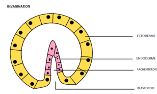
Invagination is the process of a surface folding in on itself to form a cavity, pouch or tube. In developmental biology, invagination is a mechanism that takes place during gastrulation. This mechanism or cell movement happens mostly in the vegetal pole. Invagination consists of the folding of an area of the exterior sheet of cells towards the inside of the blastula. In each organism, the complexity will be different depending on the number of cells. Invagination can be referenced as one of the steps of the establishment of the body plan. The term, originally used in embryology, has been adopted in other disciplines as well.

<span class="mw-page-title-main">Blastocoel</span> Fluid-filled or yolk-filled cavity that forms in the blastula

The blastocoel, also spelled blastocoele and blastocele, and also called cleavage cavity, or segmentation cavity is a fluid-filled or yolk-filled cavity that forms in the blastula during very early embryonic development. At this stage in mammals the blastula develops into the blastocyst containing an inner cell mass, and outer trophectoderm.

A germ layer is a primary layer of cells that forms during embryonic development. The three germ layers in vertebrates are particularly pronounced; however, all eumetazoans produce two or three primary germ layers. Some animals, like cnidarians, produce two germ layers making them diploblastic. Other animals such as bilaterians produce a third layer between these two layers, making them triploblastic. Germ layers eventually give rise to all of an animal's tissues and organs through the process of organogenesis.

<span class="mw-page-title-main">Animal embryonic development</span> Process by which the embryo forms and develops

In developmental biology, animal embryonic development, also known as animal embryogenesis, is the developmental stage of an animal embryo. Embryonic development starts with the fertilization of an egg cell (ovum) by a sperm cell, (spermatozoon). Once fertilized, the ovum becomes a single diploid cell known as a zygote. The zygote undergoes mitotic divisions with no significant growth and cellular differentiation, leading to development of a multicellular embryo after passing through an organizational checkpoint during mid-embryogenesis. In mammals, the term refers chiefly to the early stages of prenatal development, whereas the terms fetus and fetal development describe later stages.

<span class="mw-page-title-main">Neurula</span> Embryo at the early stage of development in which neurulation occurs

A neurula is a vertebrate embryo at the early stage of development in which neurulation occurs. The neurula stage is preceded by the gastrula stage; consequentially, neurulation is preceded by gastrulation. Neurulation marks the beginning of the process of organogenesis.

The primitive node is the organizer for gastrulation in most amniote embryos. In birds it is known as Hensen's node, and in amphibians it is known as the Spemann-Mangold organizer. It is induced by the Nieuwkoop center in amphibians, or by the posterior marginal zone in amniotes including birds.

<span class="mw-page-title-main">Primitive streak</span> Structure in early amniote embryogenesis

The primitive streak is a structure that forms in the early embryo in amniotes. In amphibians, the equivalent structure is the blastopore. During early embryonic development, the embryonic disc becomes oval shaped, and then pear-shaped with the broad end towards the anterior, and the narrower region projected to the posterior. The primitive streak forms a longitudinal midline structure in the narrower posterior (caudal) region of the developing embryo on its dorsal side. At first formation, the primitive streak extends for half the length of the embryo. In the human embryo, this appears by stage 6, about 17 days.

<span class="mw-page-title-main">Epiblast</span> Embryonic inner cell mass tissue that forms the embryo itself, through the three germ layers

In amniote embryonic development, the epiblast is one of two distinct cell layers arising from the inner cell mass in the mammalian blastocyst, or from the blastula in reptiles and birds, the other layer is the hypoblast. It drives the embryo proper through its differentiation into the three primary germ layers, ectoderm, mesoderm and endoderm, during gastrulation. The amniotic ectoderm and extraembryonic mesoderm also originate from the epiblast.

<span class="mw-page-title-main">Mesenchyme</span> Type of animal embryonic connective tissue

Mesenchyme is a type of loosely organized animal embryonic connective tissue of undifferentiated cells that give rise to most tissues, such as skin, blood or bone. The interactions between mesenchyme and epithelium help to form nearly every organ in the developing embryo.

<span class="mw-page-title-main">Bilaminar embryonic disc</span>

The bilaminar embryonic disc, bilaminar blastoderm or embryonic disc is the distinct two-layered structure of cells formed in an embryo. In the development of the human embryo this takes place by day eight. It is formed when the inner cell mass, also known as the embryoblast, forms a bilaminar disc of two layers, an upper layer called the epiblast and a lower layer called the hypoblast, which will eventually form into fetus. These two layers of cells are stretched between two fluid-filled cavities at either end: the primitive yolk sac and the amniotic sac.

In the field of developmental biology, regional differentiation is the process by which different areas are identified in the development of the early embryo. The process by which the cells become specified differs between organisms.

<span class="mw-page-title-main">Fish development</span>

The development of fishes is unique in some specific aspects compared to the development of other animals.

<span class="mw-page-title-main">Cerberus (protein)</span> Protein found in humans

Cerberus is a protein that in humans is encoded by the CER1 gene. Cerberus is a signaling molecule which contributes to the formation of the head, heart and left-right asymmetry of internal organs. This gene varies slightly from species to species but its overall functions seem to be similar.

<span class="mw-page-title-main">Human embryonic development</span> Development and formation of the human embryo

Human embryonic development or human embryogenesis is the development and formation of the human embryo. It is characterised by the processes of cell division and cellular differentiation of the embryo that occurs during the early stages of development. In biological terms, the development of the human body entails growth from a one-celled zygote to an adult human being. Fertilization occurs when the sperm cell successfully enters and fuses with an egg cell (ovum). The genetic material of the sperm and egg then combine to form the single cell zygote and the germinal stage of development commences. Embryonic development in the human, covers the first eight weeks of development; at the beginning of the ninth week the embryo is termed a fetus. The eight weeks has 23 stages.

<span class="mw-page-title-main">Hypoblast</span> Embryonic inner cell mass tissue that forms the yolk sac and, later, chorion

In amniote embryology, the hypoblast is one of two distinct layers arising from the inner cell mass in the mammalian blastocyst, or from the blastodisc in reptiles and birds. The hypoblast gives rise to the yolk sac, which in turn gives rise to the chorion.

<span class="mw-page-title-main">Koller's sickle</span>

In avian gastrulation, Koller's sickle is a local thickening of cells at the posterior edge of the upper layer of the area pellucida called the epiblast. Koller's sickle is crucial for avian development, due to its critical role in inducing the differentiation of various avian body parts. Koller's sickle induces primitive streak and Hensen's node, which are major components of avian gastrulation. Avian gastrulation is a process by which developing cells in an avian embryo move relative to one another in order to form the three germ layers.

The Nodal signaling pathway is a signal transduction pathway important in regional and cellular differentiation during embryonic development.

Embryogenesis in living creatures occurs in different ways depending on class and species. One of the most basic criteria of such development is independence from a water habitat.

This glossary of developmental biology is a list of definitions of terms and concepts commonly used in the study of developmental biology and related disciplines in biology, including embryology and reproductive biology, primarily as they pertain to vertebrate animals and particularly to humans and other mammals. The developmental biology of invertebrates, plants, fungi, and other organisms is treated in other articles; e.g terms relating to the reproduction and development of insects are listed in Glossary of entomology, and those relating to plants are listed in Glossary of botany.

References

Notes

  1. Urry, Lisa (2016). Campbell Biology (11th ed.). Pearson. p. 1047. ISBN   978-0134093413.
  2. 1 2 3 4 5 6 Gilbert, Scott F.; Michael J. F. Barresi (2016). Developmental biology (Eleventh ed.). Sunderland, Massachusetts: Sinauer. ISBN   978-1-60535-470-5. OCLC   945169933.
  3. Mundlos 2009: p. 422
  4. 1 2 McGeady, 2004: p. 34
  5. Jonathon M.W., Slack (2013). Essential Developmental Biology. West Sussex, UK: Wiley-Blackwell. p. 122. ISBN   978-0-470-92351-1.
  6. Hall, 1998: pp. 132-134
  7. 1 2 3 4 5 Arnold & Robinson, 2009
  8. Hall, 1998: p. 177
  9. Gilbert, Scott F. (2000). "Figure 8.6, [Types of cell movements during...]". www.ncbi.nlm.nih.gov. Retrieved 11 May 2022.
  10. Ereskovsky 2010: p. 236
  11. Wolpert L (2008) The triumph of the embryo. Courier Corporation, page 12. ISBN   978-0-486-46929-4
  12. Laubichler, M.D. and Davidson, E. H. (2008). "Boveri's long experiment: sea urchin merogones and the establishment of the role of nuclear chromosomes in development". Developmental Biology. 314(1):1–11. doi : 10.1016/j.ydbio.2007.11.024.
  13. McClay, David R.; Gross, J.M.; Range, Ryan; Peterson, R.E.; Bradham, Cynthia (2004). "Chapter 9: Sea Urchin Gastrulation". In Stern, Claudio D. (ed.). Gastrulation: From Cells to Embryos. Cold Spring Harbor Laboratory Press. pp. 123–137. ISBN   978-0-87969-707-5.
  14. Nielsen, Bjarke Frost; Nissen, Silas Boye; Sneppen, Kim; Mathiesen, Joachim; Trusina, Ala (February 21, 2020). "Model to Link Cell Shape and Polarity with Organogenesis". iScience. 23 (2): 100830. Bibcode:2020iSci...23j0830N. doi:10.1016/j.isci.2020.100830. PMC   6994644 . PMID   31986479. S2CID   210934521.
  15. McClay, D. R. 2009. Cleavage and Gastrulation in Sea Urchin. eLS. doi : 10.1002/9780470015902.a0001073.pub2
  16. Hardin J D (1990). "Context-sensitive cell behaviors during gastrulation" (PDF). Semin. Dev. Biol. 1: 335–345.
  17. Blum, Martin; Beyer, Tina; Weber, Thomas; Vick, Philipp; Andre, Philipp; Bitzer, Eva; Schweickert, Axel (June 2009). "Xenopus , an ideal model system to study vertebrate left-right asymmetry". Developmental Dynamics. 238 (6): 1215–1225. doi: 10.1002/dvdy.21855 . PMID   19208433. S2CID   39348233.
  18. 1 2 Gilbert, Scott F. (2000). "Axis Formation in Amphibians: The Phenomenon of the Organizer, The Progressive Determination of the Amphibian Axes". Developmental Biology. Sinauer Associates.
  19. Gilbert, Scott F. (2000). "Figure 10.20, [Organization of a secondary axis...]". www.ncbi.nlm.nih.gov. Retrieved 1 June 2020.
  20. Spemann H., Mangold H. (1924). "Über Induktion von Embryonanlagen durch Implantation artfremder Organisatoren". Roux' Arch. F. Entw. Mech. 100 (3–4): 599–638. doi:10.1007/bf02108133. S2CID   12605303.
  21. De Robertis Edward (2006). "Spemann's organizer and self-regulation in amphibian embryos". Nature Reviews Molecular Cell Biology. 7 (4): 296–302. doi:10.1038/nrm1855. PMC   2464568 . PMID   16482093.
  22. 1 2 3 4 5 6 7 Zorn A, Wells J (2009). "Vertebrate Endoderm Development and Organ Formation". Annu Rev Cell Dev Biol. 25: 221–251. doi:10.1146/annurev.cellbio.042308.113344. PMC   2861293 . PMID   19575677.
  23. 1 2 Tam & Behringer, 1997
  24. Catala, 2005: p. 1535
  25. 1 2 Tam, P.P.; Loebel, D.A (2007). "Gene function in mouse embryogenesis: get set for gastrulation". Nat Rev Genet. 8 (5): 368–81. doi:10.1038/nrg2084. PMID   17387317. S2CID   138874.
  26. Sheng, Guojun; Arias, Alfonso Martinez; Sutherland, Ann (2021-12-03). "The primitive streak and cellular principles of building an amniote body through gastrulation". Science. 374 (6572): abg1727. doi:10.1126/science.abg1727. PMID   34855481. S2CID   244841366.
  27. 1 2 Mikawa T, Poh AM, Kelly KA, Ishii Y, Reese DE (2004). "Induction and patterning of the primitive streak, an organizing center of gastrulation in the amniote". Dev Dyn. 229 (3): 422–32. doi: 10.1002/dvdy.10458 . PMID   14991697. S2CID   758473.
  28. 1 2 Downs KM. (2009). "The enigmatic primitive streak: prevailing notions and challenges concerning the body axis of mammals". BioEssays. 31 (8): 892–902. doi:10.1002/bies.200900038. PMC   2949267 . PMID   19609969.
  29. Chuai M, Zeng W, Yang X, Boychenko V, Glazier JA, Weijer CJ (2006). "Cell movement during chick primitive streak formation". Dev. Biol. 296 (1): 137–49. doi:10.1016/j.ydbio.2006.04.451. PMC   2556955 . PMID   16725136.
  30. Chuai M, Weijer CJ (2008). "The mechanisms underlying primitive streak formation in the chick embryo.". Current Topics in Developmental Biology. Vol. 81. pp. 135–56. doi:10.1016/S0070-2153(07)81004-0. ISBN   978-0-12-374253-7. PMID   18023726.
  31. Grapin-Botton, A.; Constam, D. (2007). "Evolution of the mechanisms and molecular control of endoderm formation". Mechanisms of Development. 124 (4): 253–78. doi:10.1016/j.mod.2007.01.001. PMID   17307341. S2CID   16552755.
  32. 1 2 3 Turner, David A.; Rué, Pau; Mackenzie, Jonathan P.; Davies, Eleanor; Martinez Arias, Alfonso (2014-01-01). "Brachyury cooperates with Wnt/β-catenin signalling to elicit primitive-streak-like behaviour in differentiating mouse embryonic stem cells". BMC Biology. 12: 63. doi: 10.1186/s12915-014-0063-7 . ISSN   1741-7007. PMC   4171571 . PMID   25115237.
  33. 1 2 Warmflash, Aryeh; Sorre, Benoit; Etoc, Fred; Siggia, Eric D; Brivanlou, Ali H (2014). "A method to recapitulate early embryonic spatial patterning in human embryonic stem cells". Nature Methods. 11 (8): 847–854. doi:10.1038/nmeth.3016. PMC   4341966 . PMID   24973948.
  34. 1 2 Etoc, Fred; Metzger, Jakob; Ruzo, Albert; Kirst, Christoph; Yoney, Anna; Ozair, M. Zeeshan; Brivanlou, Ali H.; Siggia, Eric D. (2016). "A Balance between Secreted Inhibitors and Edge Sensing Controls Gastruloid Self-Organization". Developmental Cell. 39 (3): 302–315. doi:10.1016/j.devcel.2016.09.016. PMC   5113147 . PMID   27746044.
  35. 1 2 Brink, Susanne C. van den; Baillie-Johnson, Peter; Balayo, Tina; Hadjantonakis, Anna-Katerina; Nowotschin, Sonja; Turner, David A.; Arias, Alfonso Martinez (2014-11-15). "Symmetry breaking, germ layer specification and axial organisation in aggregates of mouse embryonic stem cells". Development. 141 (22): 4231–4242. doi:10.1242/dev.113001. ISSN   0950-1991. PMC   4302915 . PMID   25371360.
  36. 1 2 3 Turner, David Andrew; Glodowski, Cherise R.; Luz, Alonso-Crisostomo; Baillie-Johnson, Peter; Hayward, Penny C.; Collignon, Jérôme; Gustavsen, Carsten; Serup, Palle; Schröter, Christian (2016-05-13). "Interactions between Nodal and Wnt signalling Drive Robust Symmetry Breaking and Axial Organisation in Gastruloids (Embryonic Organoids)". bioRxiv   10.1101/051722 .
  37. 1 2 3 Turner, David; Alonso-Crisostomo, Luz; Girgin, Mehmet; Baillie-Johnson, Peter; Glodowski, Cherise R.; Hayward, Penelope C.; Collignon, Jérôme; Gustavsen, Carsten; Serup, Palle (2017-01-31). "Gastruloids develop the three body axes in the absence of extraembryonic tissues and spatially localised signalling". bioRxiv   10.1101/104539 .
  38. Beccari, Leonardo; Moris, Naomi; Girgin, Mehmet; Turner, David A.; Baillie-Johnson, Peter; Cossy, Anne-Catherine; Lutolf, Matthias P.; Duboule, Denis; Arias, Alfonso Martinez (October 2018). "Multi-axial self-organization properties of mouse embryonic stem cells into gastruloids". Nature. 562 (7726): 272–276. Bibcode:2018Natur.562..272B. doi:10.1038/s41586-018-0578-0. ISSN   0028-0836. PMID   30283134. S2CID   52915553.
  39. Turner, David A.; Girgin, Mehmet; Alonso-Crisostomo, Luz; Trivedi, Vikas; Baillie-Johnson, Peter; Glodowski, Cherise R.; Hayward, Penelope C.; Collignon, Jérôme; Gustavsen, Carsten (2017-11-01). "Anteroposterior polarity and elongation in the absence of extra-embryonic tissues and of spatially localised signalling in gastruloids: mammalian embryonic organoids". Development. 144 (21): 3894–3906. doi:10.1242/dev.150391. ISSN   0950-1991. PMC   5702072 . PMID   28951435.
  40. "In vitro fertilization (IVF) - Mayo Clinic". www.mayoclinic.org. Retrieved 2022-04-11.
  41. Asplund, Kjell (2020). "Use of in vitro fertilization—ethical issues". Upsala Journal of Medical Sciences. 125 (2): 192–199. doi:10.1080/03009734.2019.1684405. ISSN   2000-1967. PMC   7721055 . PMID   31686575. S2CID   207896932.
  42. Davis, Caitlin (2019-03-01). "The Boundaries of Embryo Research: Extending the Fourteen-Day Rule". Journal of Bioethical Inquiry. 16 (1): 133–140. doi:10.1007/s11673-018-09895-w. ISSN   1872-4353. PMID   30635823. S2CID   58643344.

Bibliography

Further reading