Neural tube

Last updated
Neural tube
Gray19 with color.png
Transverse section of half of a chick embryo of forty-five hours' incubation. The dorsal (back) surface of the embryo is toward the top of this page, while the ventral (front) surface is toward the bottom. (Neural tube is in green.)
Gray18.png
Chick embryo of thirty-three hours' incubation, viewed from the dorsal aspect (30x magnification)
Details
Carnegie stage 10
Precursor Neural groove
Gives rise to Central nervous system (brain and spinal cord)
Identifiers
Latin tubus neuralis, tuba neuralis
MeSH D054259
TE tube_by_E5.14.1.0.0.0.1 E5.14.1.0.0.0.1
Anatomical terminology

In the developing chordate (including vertebrates), the neural tube is the embryonic precursor to the central nervous system, which is made up of the brain and spinal cord. The neural groove gradually deepens as the neural fold become elevated, and ultimately the folds meet and coalesce in the middle line and convert the groove into the closed neural tube. In humans, neural tube closure usually occurs by the fourth week of pregnancy (the 28th day after conception).

Contents

Stages of neural tube formation. Neural crest.svg
Stages of neural tube formation.

Development

The neural tube develops in two ways: primary neurulation and secondary neurulation.

Primary neurulation divides the ectoderm into three cell types:

  1. Primary neurulation begins after the neural plate forms. The edges of the neural plate start to thicken and lift upward, forming the neural folds. The center of the neural plate remains grounded, allowing a U-shaped neural groove to form. This neural groove sets the boundary between the right and left sides of the embryo. The neural folds pinch in towards the midline of the embryo and fuse together to form the neural tube. [1]
  2. In secondary neurulation, the cells of the neural plate form a cord-like structure that migrates inside the embryo and hollows to form the tube.

Each organism uses primary and secondary neurulation to varying degrees.

Mammalian neural tubes close in the head in the opposite order that they close in the trunk.

  1. Neural crest cells migrate
  2. Neural tube closes
  3. Overlying ectoderm closes
  1. Overlying ectoderm closes
  2. Neural tube closes
  3. Neural crest cells migrate

Structure

Stages of development of brain vesicles 1302 Brain Vesicle DevN.jpg
Stages of development of brain vesicles

Four neural tube subdivisions each eventually develop into distinct regions of the central nervous system by the division of neuroepithelial cells: the forebrain (prosencephalon), the midbrain (mesencephalon), the hindbrain (rhombencephalon) and the spinal cord.

For a short time, the neural tube is open both cranially and caudally. These openings, called neuropores, close during the fourth week in humans. Improper closure of the neuropores can result in neural tube defects such as anencephaly or spina bifida.

The dorsal part of the neural tube contains the alar plate, which is associated primarily with sensation. The ventral part of the neural tube contains the basal plate, which is primarily associated with motor (i.e., muscle) control.

The spinal cord develops from the posterior neural tube. As the spinal cord develops, the cells making up the wall of the neural tube proliferate and differentiate into the neurons and glia of the spinal cord. The dorsal tissues will be associated with sensory functions, and the ventral tissues will be associated with motor functions. [2]

Dorsal-ventral patterning

The neural tube patterns along the dorsal-ventral axis to establish defined compartments of neural progenitor cells that lead to distinct classes of neurons. [3] According to the French flag model of morphogenesis, this patterning occurs early in development and results from the activity of several secreted signaling molecules. Sonic hedgehog (Shh) is a key player in patterning the ventral axis, while bone morphogenic proteins (BMPs) and Wnt family members play an important role in patterning the dorsal axis. [4] Other factors shown to provide positional information to the neural progenitor cells include fibroblast growth factors (FGFs) and retinoic acid. Retinoic acid is required ventrally along with Shh to induce Pax6 and Olig2 during differentiation of motor neurons. [5]

Three main ventral cell types are established during early neural tube development: the floor plate cells, which form at the ventral midline during the neural fold stage; as well as the more dorsally located motor neurons and interneurons. [3] These cell types are specified by the secretion of the Shh from the notochord (located ventrally to the neural tube), and later from the floor plate cells. [6] Shh acts as a morphogen, meaning that it acts in a concentration-dependent manner to specify cell types as it moves further from its source. [7]

The following is a proposed mechanism for how Shh patterns the ventral neural tube: A gradient of Shh that controls the expression of a group of homeodomain (HD) and basic Helix-Loop-Helix (bHLH) transcription factors is created. These transcription factors are grouped into two protein classes based on how Shh affects them. Class I is inhibited by Shh, whereas Class II is activated by Shh. These two classes of proteins then cross-regulate each other to create more defined boundaries of expression. The different combinations of expression of these transcription factors along the dorsal-ventral axis of the neural tube are responsible for creating the identity of the neuronal progenitor cells. [4] Five molecularly distinct groups of ventral neurons form from these neuronal progenitor cells in vitro. Also, the position at which these neuronal groups are generated in vivo can be predicted by the concentration of Shh required for their induction in vitro. [8] Studies have shown that neural progenitors can evoke different responses based on the length of exposure to Shh, with a longer exposure time resulting in more ventral cell types. [9] [10]

At the dorsal end of the neural tube, BMPs are responsible for neuronal patterning. BMP is initially secreted from the overlying ectoderm. A secondary signaling center is then established in the roof plate, the dorsal most structure of the neural tube. [1] BMP from the dorsal end of the neural tube seems to act in the same concentration-dependent manner as Shh in the ventral end. [11] This was shown using zebrafish mutants that had varying amounts of BMP signaling activity. Researchers observed changes in dorsal-ventral patterning, for example, zebrafish deficient in certain BMPs showed a loss of dorsal sensory neurons and an expansion of interneurons. [12]

Shh secreted from the floor plate creates a gradient along the ventral neural tube. Shh functions in a concentration-dependent manner to specify ventral neuronal fates. V0-V3 represent four different classes of ventral interneurons, and MN indicates motor neurons. Shh gradient in the neural tube.jpg
Shh secreted from the floor plate creates a gradient along the ventral neural tube. Shh functions in a concentration-dependent manner to specify ventral neuronal fates. V0-V3 represent four different classes of ventral interneurons, and MN indicates motor neurons.

See also

Related Research Articles

<span class="mw-page-title-main">Thalamus</span> Structure within the brain

The thalamus is a large mass of gray matter located in the dorsal part of the diencephalon. Nerve fibers project out of the thalamus to the cerebral cortex in all directions, known as the thalamocortical radiations, allowing hub-like exchanges of information. It has several functions, such as the relaying of sensory and motor signals to the cerebral cortex and the regulation of consciousness, sleep, and alertness.

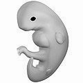
The development of the nervous system, or neural development (neurodevelopment), refers to the processes that generate, shape, and reshape the nervous system of animals, from the earliest stages of embryonic development to adulthood. The field of neural development draws on both neuroscience and developmental biology to describe and provide insight into the cellular and molecular mechanisms by which complex nervous systems develop, from nematodes and fruit flies to mammals.

<span class="mw-page-title-main">Sonic hedgehog protein</span> Signaling molecule in animals

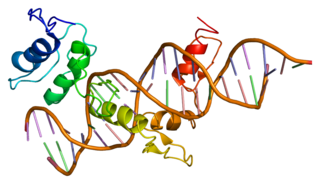
Sonic hedgehog protein (SHH) is encoded for by the SHH gene. The protein is named after the character Sonic the Hedgehog.

<span class="mw-page-title-main">Notochord</span> Flexible rod-shaped structure in all chordates

In zoology and developmental anatomy, the notochord is an elastic, rod-like anatomical structure found in many deuterostomal animals. A notochord is one of five synapomorphies used to define a species as a chordate.

<span class="mw-page-title-main">Neurulation</span> Embryological process forming the neural tube

Neurulation refers to the folding process in vertebrate embryos, which includes the transformation of the neural plate into the neural tube. The embryo at this stage is termed the neurula.

In the vertebrate embryo, a rhombomere is a transiently divided segment of the developing neural tube, within the hindbrain region in the area that will eventually become the rhombencephalon. The rhombomeres appear as a series of slightly constricted swellings in the neural tube, caudal to the cephalic flexure. In human embryonic development, the rhombomeres are present by day 29.

<span class="mw-page-title-main">Neuromere</span>

Neuromeres are distinct groups of neural crest cells, forming segments in the neural tube of the early embryonic development of the brain. There are three classes of neuromeres in the central nervous system – prosomeres, mesomeres and rhombomeres that will develop the forebrain, midbrain, and hindbrain respectively.

<span class="mw-page-title-main">Neural plate</span>

The neural plate is a key developmental structure that serves as the basis for the nervous system. Cranial to the primitive node of the embryonic primitive streak, ectodermal tissue thickens and flattens to become the neural plate. The region anterior to the primitive node can be generally referred to as the neural plate. Cells take on a columnar appearance in the process as they continue to lengthen and narrow. The ends of the neural plate, known as the neural folds, push the ends of the plate up and together, folding into the neural tube, a structure critical to brain and spinal cord development. This process as a whole is termed primary neurulation.

<span class="mw-page-title-main">Neurula</span> Embryo at the early stage of development in which neurulation occurs

A neurula is a vertebrate embryo at the early stage of development in which neurulation occurs. The neurula stage is preceded by the gastrula stage; consequentially, neurulation is preceded by gastrulation. Neurulation marks the beginning of the process of organogenesis.

<span class="mw-page-title-main">Neural crest</span> Pluripotent embyronic cell group giving rise to diverse cell lineages

Neural crest cells are a temporary group of cells that arise from the embryonic ectoderm germ layer, and in turn give rise to a diverse cell lineage—including melanocytes, craniofacial cartilage and bone, smooth muscle, peripheral and enteric neurons and glia.

<span class="mw-page-title-main">GLI1</span> Protein-coding gene in humans

Zinc finger protein GLI1 also known as glioma-associated oncogene is a protein that in humans is encoded by the GLI1 gene. It was originally isolated from human glioblastoma cells.

<span class="mw-page-title-main">Neural fold</span> Structure arising during embryonic development of birds and mammals

The neural fold is a structure that arises during neurulation in the embryonic development of both birds and mammals among other organisms. This structure is associated with primary neurulation, meaning that it forms by the coming together of tissue layers, rather than a clustering, and subsequent hollowing out, of individual cells. In humans, the neural folds are responsible for the formation of the anterior end of the neural tube. The neural folds are derived from the neural plate, a preliminary structure consisting of elongated ectoderm cells. The folds give rise to neural crest cells, as well as bringing about the formation of the neural tube.

<span class="mw-page-title-main">Floor plate</span> Embryonic structure

The floor plate is a structure integral to the developing nervous system of vertebrate organisms. Located on the ventral midline of the embryonic neural tube, the floor plate is a specialized glial structure that spans the anteroposterior axis from the midbrain to the tail regions. It has been shown that the floor plate is conserved among vertebrates, such as zebrafish and mice, with homologous structures in invertebrates such as the fruit fly Drosophila and the nematode C. elegans. Functionally, the structure serves as an organizer to ventralize tissues in the embryo as well as to guide neuronal positioning and differentiation along the dorsoventral axis of the neural tube.

<span class="mw-page-title-main">Zona limitans intrathalamica</span>

The zona limitans intrathalamica (ZLI) is a lineage-restriction compartment and primary developmental boundary in the vertebrate forebrain that serves as a signaling center and a restrictive border between the thalamus and the prethalamus.

<span class="mw-page-title-main">Limb development</span>

Limb development in vertebrates is an area of active research in both developmental and evolutionary biology, with much of the latter work focused on the transition from fin to limb.

<span class="mw-page-title-main">Fish development</span>

The development of fishes is unique in some specific aspects compared to the development of other animals.

<span class="mw-page-title-main">RAB23</span> Protein-coding gene in the species Homo sapiens

Ras-related protein Rab-23 is a protein that in humans is encoded by the RAB23 gene. Alternative splicing occurs at this gene locus and two transcript variants encoding the same protein have been identified.

<span class="mw-page-title-main">Zone of polarizing activity</span>

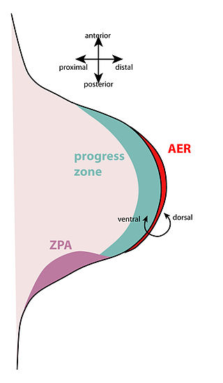
The zone of polarizing activity (ZPA) is an area of mesenchyme that contains signals which instruct the developing limb bud to form along the anterior/posterior axis. Limb bud is undifferentiated mesenchyme enclosed by an ectoderm covering. Eventually, the limb bud develops into bones, tendons, muscles and joints. Limb bud development relies not only on the ZPA, but also many different genes, signals, and a unique region of ectoderm called the apical ectodermal ridge (AER). Research by Saunders and Gasseling in 1948 identified the AER and its subsequent involvement in proximal distal outgrowth. Twenty years later, the same group did transplantation studies in chick limb bud and identified the ZPA. It wasn't until 1993 that Todt and Fallon showed that the AER and ZPA are dependent on each other.

<span class="mw-page-title-main">Spinal cord</span> Long, tubular central nervous system structure in the vertebral column

The spinal cord is a long, thin, tubular structure made up of nervous tissue that extends from the medulla oblongata in the brainstem to the lumbar region of the vertebral column (backbone) of vertebrate animals. The center of the spinal cord is hollow and contains a structure called central canal, which contains cerebrospinal fluid. The spinal cord is also covered by meninges and enclosed by the neural arches. Together, the brain and spinal cord make up the central nervous system.

The development of the cerebral cortex, known as corticogenesis is the process during which the cerebral cortex of the brain is formed as part of the development of the nervous system of mammals including its development in humans. The cortex is the outer layer of the brain and is composed of up to six layers. Neurons formed in the ventricular zone migrate to their final locations in one of the six layers of the cortex. The process occurs from embryonic day 10 to 17 in mice and between gestational weeks seven to 18 in humans.

References

PD-icon.svgThis article incorporates text in the public domain from page 50 of the 20th edition of Gray's Anatomy (1918)

  1. 1 2 Gilbert, Scott F. Developmental Biology Eighth Edition. Sunderland, Massachusetts: Sinauer Associates, Inc., 2006.
  2. Creative Commons by small.svg  This article incorporates text available under the CC BY 4.0 license.Betts, J Gordon; Desaix, Peter; Johnson, Eddie; Johnson, Jody E; Korol, Oksana; Kruse, Dean; Poe, Brandon; Wise, James; Womble, Mark D; Young, Kelly A (June 8, 2023). Anatomy & Physiology. Houston: OpenStax CNX. 13.1 Embryologic perspective. ISBN   978-1-947172-04-3.
  3. 1 2 Jessell TM (2000). "Neuronal specification in the spinal cord: inductive signals and transcriptional codes". Nat Rev Genet. 1 (1): 20–9. doi:10.1038/35049541. PMID   11262869. S2CID   205012382.
  4. 1 2 Ulloa F, Marti E (2010). "Wnt won the war: Antagonistic role of Wnt over Shh controls dorso-ventral patterning of the vertebrate neural tube". Developmental Dynamics. 239 (1): 69–76. doi: 10.1002/dvdy.22058 . PMID   19681160. S2CID   205766310.
  5. Duester G (2008). "Retinoic acid synthesis and signaling during early organogenesis". Cell. 134 (6): 921–931. doi:10.1016/j.cell.2008.09.002. PMC   2632951 . PMID   18805086.
  6. Patten I, Placzek M (2000). "The role of Sonic hedgehog in neural tube patterning". Cell Mol Life Sci. 57 (12): 1695–708. doi:10.1007/pl00000652. PMID   11130176. S2CID   20950575.
  7. Dessaud E, McMahon AP, Briscoe J (2008). "Pattern formation in the vertebrate neural tube: a sonic hedgehog morphogen-regulated transcriptional network". Development. 135 (15): 2489–2503. doi: 10.1242/dev.009324 . PMID   18621990.
  8. Ericson J, Briscoe J, Rashbass P, van Heyningen V, Jessell TM (1997). "Graded Sonic hedgehog signaling and the specification of cell fate in the ventral neural tube". Cold Spring Harb Symp Quant Biol. 62: 451–466. doi:10.1101/SQB.1997.062.01.053. PMID   9598380.
  9. Stamataki D, Ulloa F, Tsoni SV, Mynett A, Briscoe J (2005). "A gradient of Gli activity mediates graded Sonic hedgehog signaling in the neural tube". Genes Dev. 19 (5): 626–641. doi:10.1101/gad.325905. PMC   551582 . PMID   15741323.
  10. Dessaud E, Yang LL, Hill K, et al. (November 2007). "Interpretation of the sonic hedgehog morphogen gradient by a temporal adaptation mechanism" (PDF). Nature. 450 (7170): 717–20. Bibcode:2007Natur.450..717D. doi:10.1038/nature06347. hdl: 2027.42/62511 . PMID   18046410. S2CID   4419025.
  11. Wilson L, Maden M (2005). "The mechanisms of dorsoventral patterning in the vertebrate neural tube". Developmental Biology. 282 (1): 1–13. doi: 10.1016/j.ydbio.2005.02.027 . PMID   15936325.
  12. Nguyen VH, Trout J, Connors SA, Andermann P, Weinberg E, Mullins MC (2000). "Dorsal and intermediate neuronal cell types of the spinal cord are established by a BMP signaling pathway". Development. 127 (6): 1209–1220. doi: 10.1242/dev.127.6.1209 . PMID   10683174.