Hemostasis

Last updated

In biology, hemostasis or haemostasis is a process to prevent and stop bleeding, meaning to keep blood within a damaged blood vessel (the opposite of hemostasis is hemorrhage). It is the first stage of wound healing. Hemostasis involves three major steps:

Contents

Coagulation, the changing of blood from a liquid to a gel which forms the fibrin clots, is essential to hemostasis. Intact blood vessels moderate blood's tendency to form clots. The endothelial cells of intact vessels prevent blood clotting with a heparin-like molecule and thrombomodulin, and prevent platelet aggregation with nitric oxide and prostacyclin. When endothelium of a blood vessel is damaged, the endothelial cells stop secretion of coagulation and aggregation inhibitors and instead secrete von Willebrand factor, which initiates the maintenance of hemostasis after injury. These processes seal the injury or hole until tissues are healed.

Etymology and pronunciation

The word hemostasis ( /ˌhmˈstsɪs/ , [1] [2] sometimes /ˌhˈmɒstəsɪs/ ) uses the combining forms hemo- and -stasis , Neo-Latin from Ancient Greek αἱμο-haimo- (similar to αἷμαhaîma), meaning "blood", and στάσιςstásis, meaning "stasis", yielding "motionlessness or stopping of blood".

Steps of mechanism

Aggregation of thrombocytes (platelets). Platelet-rich human blood plasma (left vial) is a turbid liquid. Upon addition of ADP, platelets are activated and start to aggregate, forming white flakes (right vial) Thrombocyteaggregation.jpg
Aggregation of thrombocytes (platelets). Platelet-rich human blood plasma (left vial) is a turbid liquid. Upon addition of ADP, platelets are activated and start to aggregate, forming white flakes (right vial)

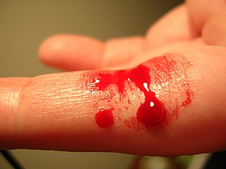
Hemostasis occurs when blood is present outside of the body or blood vessels. It is the innate response for the body to stop bleeding and loss of blood. During hemostasis three steps occur in a rapid sequence. Vascular spasm is the first response as the blood vessels constrict to allow less blood to be lost. In the second step, platelet plug formation, platelets stick together to form a temporary seal to cover the break in the vessel wall. The third and last step is called coagulation or blood clotting. Coagulation reinforces the platelet plug with fibrin threads that act as a "molecular glue". [3] Platelets are a large factor in the hemostatic process. They allow for the creation of the "platelet plug" that forms almost directly after a blood vessel has been ruptured. Within seconds of a blood vessel's epithelial wall being disrupted, platelets begin to adhere to the sub-endothelium surface. It takes approximately sixty seconds until the first fibrin strands begin to intersperse among the wound. After several minutes the platelet plug is completely formed by fibrin. [4] Hemostasis is maintained in the body via three mechanisms:

  1. Vascular spasm: Vasoconstriction is produced by vascular smooth muscle cells, and is the blood vessel's first response to injury. The smooth muscle cells are controlled by vascular endothelium, which releases intravascular signals to control the contracting properties. When a blood vessel is damaged, there is an immediate reflex, initiated by local sympathetic pain receptors, which helps promote vasoconstriction. The damaged vessels will constrict (vasoconstrict) which reduces the amount of blood flow through the area and limits the amount of blood loss. Collagen is exposed at the site of injury, the collagen promotes platelets to adhere to the injury site. Platelets release cytoplasmic granules which contain serotonin, ADP and thromboxane A2, all of which increase the effect of vasoconstriction. The spasm response becomes more effective as the amount of damage is increased. Vascular spasm is much more effective in smaller blood vessels. [5] [6]
  2. Platelet plug formation: Platelets adhere to damaged endothelium to form a platelet plug (primary hemostasis) and then degranulate. This process is regulated through thromboregulation. Plug formation is activated by a glycoprotein called von Willebrand factor (vWF), which is found in plasma. Platelets play one of major roles in the hemostatic process. When platelets come across the injured endothelium cells, they change shape, release granules and ultimately become ‘sticky’. Platelets express certain receptors, some of which are used for the adhesion of platelets to collagen. When platelets are activated, they express glycoprotein receptors that interact with other platelets, producing aggregation and adhesion. Platelets release cytoplasmic granules such as adenosine diphosphate (ADP), serotonin and thromboxane A2. Adenosine diphosphate (ADP) attracts more platelets to the affected area, serotonin is a vasoconstrictor and thromboxane A2 assists in platelet aggregation, vasoconstriction and degranulation. As more chemicals are released more platelets stick and release their chemicals; creating a platelet plug and continuing the process in a positive feedback loop. Platelets alone are responsible for stopping the bleeding of unnoticed wear and tear of our skin on a daily basis. This is referred to as primary hemostasis. [5] [7]
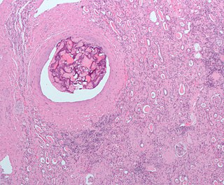
  3. Clot formation: Once the platelet plug has been formed by the platelets, the clotting factors (a dozen proteins that travel along the blood plasma in an inactive state) are activated in a sequence of events known as 'coagulation cascade' which leads to the formation of fibrin from inactive fibrinogen plasma protein. Thus, a fibrin mesh is produced all around the platelet plug to hold it in place; this step is called secondary hemostasis. During this process some red and white blood cells are trapped in the mesh which causes the primary hemostasis plug to become harder: the resultant plug is called a thrombus or blood clot. The blood clot contains the secondary hemostasis plug with blood cells trapped in it. This is a necessary step for wound healing, but it has the ability to cause severe health problems if the thrombus becomes detached from the vessel wall and travels through the circulatory system; If it reaches the brain, heart or lungs it could lead to stroke, heart attack, or pulmonary embolism respectively. [3]

Types

Hemostasis can be achieved in various other ways if the body cannot do it naturally (or needs help) during surgery or medical treatment. When the body is under shock and stress, hemostasis is harder to achieve. Though natural hemostasis is most desired, having other means of achieving this is vital for survival in many emergency settings. Without the ability to stimulate hemostasis the risk of hemorrhaging is great. During surgical procedures, the types of hemostasis listed below can be used to control bleeding while avoiding and reducing the risk of tissue destruction. Hemostasis can be achieved by chemical agent as well as mechanical or physical agents. Which hemostasis type used is determined based on the situation. [8]

Developmental Haemostasis refers to the differences in the haemostatic system between children and adults.

In emergency medicine

Debates by physicians and medical practitioners still continue to arise on the subject of hemostasis and how to handle situations with large injuries. If an individual acquires a large injury resulting in extreme blood loss, then a hemostatic agent alone would not be very effective. Medical professionals continue to debate on what the best ways are to assist a patient in a chronic state; however, it is universally accepted that hemostatic agents are the primary tool for smaller bleeding injuries. [8]

Some main types of hemostasis used in emergency medicine include:

Disorders

The body's hemostasis system requires careful regulation in order to work properly. If the blood does not clot sufficiently, it may be due to bleeding disorders such as hemophilia or immune thrombocytopenia; this requires careful investigation. Over-active clotting can also cause problems; thrombosis, where blood clots form abnormally, can potentially cause embolisms, where blood clots break off and subsequently become lodged in a vein or artery.[ citation needed ]

Hemostasis disorders can develop for many different reasons. They may be congenital, due to a deficiency or defect in an individual's platelets or clotting factors. A number of disorders can be acquired as well, such as in HELLP syndrome, which is due to pregnancy, or Hemolytic-uremic syndrome (HUS), which is due to E. coli toxins.

History of artificial hemostasis

The process of preventing blood loss from a vessel or organ of the body is referred to as hemostasis. The term comes from the Ancient Greek roots "heme" meaning blood, and "stasis" meaning halting; Put together means the "halting of the blood". [3] The origin of hemostasis dates back as far as ancient Greece; first referenced to being used in the Battle of Troy. It started with the realization that excessive bleeding inevitably equaled death. Vegetable and mineral styptics were used on large wounds by the Greeks and Romans until the takeover of Egypt around 332BC by Greece. At this time many more advances in the general medical field were developed through the study of Egyptian mummification practice, which led to greater knowledge of the hemostatic process. It was during this time that many of the veins and arteries running throughout the human body were found and the directions in which they traveled. Doctors of this time realized if these were plugged, blood could not continue to flow out of the body. Nevertheless, it took until the invention of the printing press during the fifteenth century for medical notes and ideas to travel westward, allowing for the idea and practice of hemostasis to be expanded. [13]

Research

There is currently a great deal of research being conducted on hemostasis. The most current research is based on genetic factors of hemostasis and how it can be altered to reduce the cause of genetic disorders that alter the natural process hemostasis. [14]

Von Willebrand disease is associated with a defect in the ability of the body to create the platelet plug and the fibrin mesh that ultimately stops the bleeding. New research is concluding that the von Willebrand disease is much more common in adolescence. This disease negatively hinders the natural process of Hemostasis causing excessive bleeding to be a concern in patients with this disease. There are complex treatments that can be done including a combination of therapies, estrogen-progesterone preparations, desmopressin, and Von Willebrand factor concentrates. Current research is trying to find better ways to deal with this disease; however, much more research is needed in order to find out the effectiveness of the current treatments and if there are more operative ways to treat this disease. [15]

See also

Related Research Articles

<span class="mw-page-title-main">Bleeding</span> Loss of blood escaping from the circulatory system

Bleeding, hemorrhage, haemorrhage or blood loss is blood escaping from the circulatory system from damaged blood vessels. Bleeding can occur internally, or externally either through a natural opening such as the mouth, nose, ear, urethra, vagina or anus, or through a puncture in the skin. Hypovolemia is a massive decrease in blood volume, and death by excessive loss of blood is referred to as exsanguination. Typically, a healthy person can endure a loss of 10–15% of the total blood volume without serious medical difficulties. The stopping or controlling of bleeding is called hemostasis and is an important part of both first aid and surgery.

<span class="mw-page-title-main">Thrombus</span> Blood clot

A thrombus, colloquially called a blood clot, is the final product of the blood coagulation step in hemostasis. There are two components to a thrombus: aggregated platelets and red blood cells that form a plug, and a mesh of cross-linked fibrin protein. The substance making up a thrombus is sometimes called cruor. A thrombus is a healthy response to injury intended to stop and prevent further bleeding, but can be harmful in thrombosis, when a clot obstructs blood flow through healthy blood vessels in the circulatory system.

<span class="mw-page-title-main">Platelet</span> Component of blood aiding in coagulation

Platelets or thrombocytes are a component of blood whose function is to react to bleeding from blood vessel injury by clumping, thereby initiating a blood clot. Platelets have no cell nucleus; they are fragments of cytoplasm derived from the megakaryocytes of the bone marrow or lung, which then enter the circulation. Platelets are found only in mammals, whereas in other vertebrates, thrombocytes circulate as intact mononuclear cells.

<span class="mw-page-title-main">Coagulation</span> Process of formation of blood clots

Coagulation, also known as clotting, is the process by which blood changes from a liquid to a gel, forming a blood clot. It results in hemostasis, the cessation of blood loss from a damaged vessel, followed by repair. The process of coagulation involves activation, adhesion and aggregation of platelets, as well as deposition and maturation of fibrin.

<span class="mw-page-title-main">Disseminated intravascular coagulation</span> Medical condition where blood clots block small blood vessels

Disseminated intravascular coagulation (DIC) is a condition in which blood clots form throughout the body, blocking small blood vessels. Symptoms may include chest pain, shortness of breath, leg pain, problems speaking, or problems moving parts of the body. As clotting factors and platelets are used up, bleeding may occur. This may include blood in the urine, blood in the stool, or bleeding into the skin. Complications may include organ failure.

<span class="mw-page-title-main">Fibrin</span> Fibrous protein involved in blood coagulation

Fibrin is a fibrous, non-globular protein involved in the clotting of blood. It is formed by the action of the protease thrombin on fibrinogen, which causes it to polymerize. The polymerized fibrin, together with platelets, forms a hemostatic plug or clot over a wound site.

<span class="mw-page-title-main">Fibrinogen</span> Soluble protein complex in blood plasma and involved in clot formation

Fibrinogen is a glycoprotein complex, produced in the liver, that circulates in the blood of all vertebrates. During tissue and vascular injury, it is converted enzymatically by thrombin to fibrin and then to a fibrin-based blood clot. Fibrin clots function primarily to occlude blood vessels to stop bleeding. Fibrin also binds and reduces the activity of thrombin. This activity, sometimes referred to as antithrombin I, limits clotting. Fibrin also mediates blood platelet and endothelial cell spreading, tissue fibroblast proliferation, capillary tube formation, and angiogenesis and thereby promotes revascularization and wound healing.

<span class="mw-page-title-main">Thrombin</span> Enzyme involved in blood coagulation in humans

Thrombin is a serine protease, an enzyme that, in humans, is encoded by the F2 gene.

Fibrinolysis is a process that prevents blood clots from growing and becoming problematic. Primary fibrinolysis is a normal body process, while secondary fibrinolysis is the breakdown of clots due to a medicine, a medical disorder, or some other cause.

von Willebrand disease Medical condition

Von Willebrand disease (VWD) is the most common hereditary blood-clotting disorder in humans. An acquired form can sometimes result from other medical conditions. It arises from a deficiency in the quality or quantity of von Willebrand factor (VWF), a multimeric protein that is required for platelet adhesion. It is known to affect several breeds of dogs as well as humans. The three forms of VWD are hereditary, acquired, and pseudo or platelet type. The three types of hereditary VWD are VWD type 1, VWD type 2, and VWD type 3. Type 2 contains various subtypes. Platelet type VWD is also an inherited condition.

<span class="mw-page-title-main">Endothelium</span> Layer of cells that lining inner surface of blood vessels

The endothelium is a single layer of squamous endothelial cells that line the interior surface of blood vessels and lymphatic vessels. The endothelium forms an interface between circulating blood or lymph in the lumen and the rest of the vessel wall. Endothelial cells form the barrier between vessels and tissue and control the flow of substances and fluid into and out of a tissue.

von Willebrand factor Mammalian protein involved in blood clotting

Von Willebrand factor (VWF) is a blood glycoprotein that promotes hemostasis, specifically, platelet adhesion. It is deficient and/or defective in von Willebrand disease and is involved in many other diseases, including thrombotic thrombocytopenic purpura, Heyde's syndrome, and possibly hemolytic–uremic syndrome. Increased plasma levels in many cardiovascular, neoplastic, metabolic, and connective tissue diseases are presumed to arise from adverse changes to the endothelium, and may predict an increased risk of thrombosis.

Weibel–Palade bodies (WPBs) are the storage granules of endothelial cells, the cells that form the inner lining of the blood vessels and heart. They manufacture, store and release two principal molecules, von Willebrand factor and P-selectin, and thus play a dual role in hemostasis and inflammation.

<span class="mw-page-title-main">Hypovolemic shock</span> Medical condition

Hypovolemic shock is a form of shock caused by severe hypovolemia. It could be the result of severe dehydration through a variety of mechanisms or blood loss. Hypovolemic shock is a medical emergency; if left untreated, the insufficient blood flow can cause damage to organs, leading to multiple organ failure.

<span class="mw-page-title-main">Purpura</span> Hemorrhagic lesion of the skin caused by bleeding underneath the skin

Purpura is a condition of red or purple discolored spots on the skin that do not blanch on applying pressure. The spots are caused by bleeding underneath the skin secondary to platelet disorders, vascular disorders, coagulation disorders, or other causes. They measure 3–10 mm, whereas petechiae measure less than 3 mm, and ecchymoses greater than 1 cm.

<span class="mw-page-title-main">Embolization</span> Passage and lodging of an embolus within the bloodstream

Embolization refers to the passage and lodging of an embolus within the bloodstream. It may be of natural origin (pathological), in which sense it is also called embolism, for example a pulmonary embolism; or it may be artificially induced (therapeutic), as a hemostatic treatment for bleeding or as a treatment for some types of cancer by deliberately blocking blood vessels to starve the tumor cells.

<span class="mw-page-title-main">Schistocyte</span> Fragmented portion of a red blood cell

A schistocyte or schizocyte is a fragmented part of a red blood cell. Schistocytes are typically irregularly shaped, jagged, and have two pointed ends.

An antihemorrhagic agent is a substance that promotes hemostasis. It may also be known as a hemostatic agent.

The dysfibrinogenemias consist of three types of fibrinogen disorders in which a critical blood clotting factor, fibrinogen, circulates at normal levels but is dysfunctional. Congenital dysfibrinogenemia is an inherited disorder in which one of the parental genes produces an abnormal fibrinogen. This fibrinogen interferes with normal blood clotting and/or lysis of blood clots. The condition therefore may cause pathological bleeding and/or thrombosis. Acquired dysfibrinogenemia is a non-hereditary disorder in which fibrinogen is dysfunctional due to the presence of liver disease, autoimmune disease, a plasma cell dyscrasias, or certain cancers. It is associated primarily with pathological bleeding. Hereditary fibrinogen Aα-Chain amyloidosis is a sub-category of congenital dysfibrinogenemia in which the dysfunctional fibrinogen does not cause bleeding or thrombosis but rather gradually accumulates in, and disrupts the function of, the kidney.

The platelet plug, also known as the hemostatic plug or platelet thrombus, is an aggregation of platelets formed during early stages of hemostasis in response to one or more injuries to blood vessel walls. After platelets are recruited and begin to accumulate around the breakage, their “sticky” nature allows them to adhere to each other. This forms a platelet plug, which prevents more blood from leaving the body as well as any outside contaminants from getting in. The plug provides a temporary blockage of the break in the vasculature. As such, platelet plug formation occurs after vasoconstriction of the blood vessels but before the creation of the fibrin mesh clot, which is the more permanent solution to the injury. The result of the platelet plug formation is the coagulation of blood. It can also be referred to as primary hemostasis.

References

  1. "hemostasis". Merriam-Webster.com Dictionary . Retrieved 2016-01-21.
  2. "hemostasis". Lexico UK English Dictionary. Oxford University Press. Archived from the original on 2020-03-22.
  3. 1 2 3 Marieb, Elaine Nicpon; Hoehn, Katja (2010). Human Anatomy & Physiology (8th ed.). San Francisco: Benjamin Cummings. pp. 649–650.
  4. Boon, G. D. "An Overview of Hemostasis." Toxicologic Pathology 21.2 (1993): 170–179.
  5. 1 2 Alturi, Pavan (2005). The Surgical Review: An Integrated Basic and Clinical Science Study Guide. Philadelphia: Lippincott Williams & Wilkins. p. 300.
  6. Zdanowicz, M (2003). Essentials of pathophysiology for pharmacy . Florida: CRC Press. p.  23. ISBN   9781587160363.
  7. Li, Zhenyu (11 Nov 2010). "Signaling during platelet adhesion and activation". Arteriosclerosis, Thrombosis, and Vascular Biology. 30 (12): 2341–2349. doi:10.1161/ATVBAHA.110.207522. PMC   3085271 . PMID   21071698.
  8. 1 2 Kulkarni Roshni (2004). "Alternative and Topical Approaches to Treating the Massicely Bleeding Patient" (PDF). Advances in Hematology. 2 (7): 428–431. Archived from the original (PDF) on 2009-01-06. Retrieved 2012-04-26.
  9. Aldo Moraci, et al. "The Use Of Local Agents: Bone Wax, Gelatin, Collagen, Oxidized Cellulose." European Spine Journal 2004; 13.: S89–S96.
  10. Smith Shondra L.; Belmont John M.; Casparian J. Michael (1999). "Analysis Of Pressure Achieved By Various Materials Used For Pressure Dressings". Dermatologic Surgery. 25 (12): 931–934. doi:10.1046/j.1524-4725.1999.99151.x. PMID   10594624.
  11. Kozak Orhan; et al. (2010). "A New Method For Hepatic Resection And Hemostasis: Absorbable Plaque And Suture". Eurasian Journal of Medicine. 41: 1–4.
  12. Tahriri Mohammadreza; et al. (2011). "Preparation And Characterization Of Absorbable Hemostat Crosslinked Gelatin Sponges For Surgical Applications". Current Applied Physics. 11 (3): 457–461. Bibcode:2011CAP....11..457K. doi:10.1016/j.cap.2010.08.031.
  13. Wies, C. H. (1929). "Wies, C. H. "The History of Hemostasis." Yale Journal of Biology and Medicine". The Yale Journal of Biology and Medicine. 2 (2): 167–168. PMC   2606227 .
  14. Rosen, Elliot D.; Xuei, Xiaoling; Suckow, Mark; Edenberg, Howard (2006). "Searching for hemostatic modifier genes affecting the phenotype of mice with very low levels of FVII". Blood Cells, Molecules and Diseases. 36 (2): 131–134. doi:10.1016/j.bcmd.2005.12.037. PMID   16524747.
  15. Mikhail, Sameh; Kouides, Peter (December 2010). "von Willebrand Disease in the Pediatric and Adolescent Population". Journal of Pediatric and Adolescent Gynecology. 23 (6): S3–S10. doi:10.1016/j.jpag.2010.08.005. PMID   20934894.