Lipid

Last updated

Structures of some common lipids. At the top are cholesterol and oleic acid. The middle structure is a triglyceride composed of oleoyl, stearoyl, and palmitoyl chains attached to a glycerol backbone. At the bottom is the common phospholipid phosphatidylcholine. Common lipids lmaps.png
Structures of some common lipids. At the top are cholesterol and oleic acid. The middle structure is a triglyceride composed of oleoyl, stearoyl, and palmitoyl chains attached to a glycerol backbone. At the bottom is the common phospholipid phosphatidylcholine.

Lipids are a broad group of organic compounds which include fats, waxes, sterols, fat-soluble vitamins (such as vitamins A, D, E and K), monoglycerides, diglycerides, phospholipids, and others. The functions of lipids include storing energy, signaling, and acting as structural components of cell membranes. [3] [4] Lipids have applications in the cosmetic and food industries, and in nanotechnology. [5]

Contents

Lipids may be broadly defined as hydrophobic or amphiphilic small molecules; the amphiphilic nature of some lipids allows them to form structures such as vesicles, multilamellar/unilamellar liposomes, or membranes in an aqueous environment. Biological lipids originate entirely or in part from two distinct types of biochemical subunits or "building-blocks": ketoacyl and isoprene groups. [3] Using this approach, lipids may be divided into eight categories: fatty acyls, glycerolipids, glycerophospholipids, sphingolipids, saccharolipids, and polyketides (derived from condensation of ketoacyl subunits); and sterol lipids and prenol lipids (derived from condensation of isoprene subunits). [3]

Although the term "lipid" is sometimes used as a synonym for fats, fats are a subgroup of lipids called triglycerides. Lipids also encompass molecules such as fatty acids and their derivatives (including tri-, di-, monoglycerides, and phospholipids), as well as other sterol-containing metabolites such as cholesterol. [6] Although humans and other mammals use various biosynthetic pathways both to break down and to synthesize lipids, some essential lipids cannot be made this way and must be obtained from the diet.

History

In 1815, Henri Braconnot classified lipids (graisses) in two categories, suifs (solid greases or tallow) and huiles (fluid oils). [7] In 1823, Michel Eugène Chevreul developed a more detailed classification, including oils, greases, tallow, waxes, resins, balsams and volatile oils (or essential oils). [8] [9] [10]

The first synthetic triglyceride was reported by Théophile-Jules Pelouze in 1844, when he produced tributyrin by treating butyric acid with glycerin in the presence of concentrated sulfuric acid. [11] Several years later, Marcellin Berthelot, one of Pelouze's students, synthesized tristearin and tripalmitin by reaction of the analogous fatty acids with glycerin in the presence of gaseous hydrogen chloride at high temperature. [12]

In 1827, William Prout recognized fat ("oily" alimentary matters), along with protein ("albuminous") and carbohydrate ("saccharine"), as an important nutrient for humans and animals. [13] [14]

For a century, chemists regarded "fats" as only simple lipids made of fatty acids and glycerol (glycerides), but new forms were described later. Theodore Gobley (1847) discovered phospholipids in mammalian brain and hen egg, called by him as "lecithins". Thudichum discovered in human brain some phospholipids (cephalin), glycolipids (cerebroside) and sphingolipids (sphingomyelin). [9]

The terms lipoid, lipin, lipide and lipid have been used with varied meanings from author to author. [15] In 1912, Rosenbloom and Gies proposed the substitution of "lipoid" by "lipin". [16] In 1920, Bloor introduced a new classification for "lipoids": simple lipoids (greases and waxes), compound lipoids (phospholipoids and glycolipoids), and the derived lipoids (fatty acids, alcohols, sterols). [17] [18]

The word lipide, which stems etymologically from Greek λίπος, lipos 'fat', was introduced in 1923 by the French pharmacologist Gabriel Bertrand. [19] Bertrand included in the concept not only the traditional fats (glycerides), but also the "lipoids", with a complex constitution. [9] The word lipide was unanimously approved by the international commission of the Société de Chimie Biologique during the plenary session on July 3, 1923. The word lipide was later anglicized as lipid because of its pronunciation ('lɪpɪd). In French, the suffix -ide, from Ancient Greek -ίδης (meaning 'son of' or 'descendant of'), is always pronounced (ɪd).

In 1947, T. P. Hilditch defined "simple lipids" as greases and waxes (true waxes, sterols, alcohols). [20] [ page needed ]

Categories

Lipids have been classified into eight categories by the Lipid MAPS consortium [3] as follows:

Fatty acyls

I2 - Prostacyclin (an example of a prostaglandin, an eicosanoid fatty acid) Prostacyclin-2D-skeletal.png
I2 – Prostacyclin (an example of a prostaglandin, an eicosanoid fatty acid)
LTB4 (an example of a leukotriene, an eicosanoid fatty acid) Leukotriene B4.svg
LTB4 (an example of a leukotriene, an eicosanoid fatty acid)

Fatty acyls, a generic term for describing fatty acids, their conjugates and derivatives, are a diverse group of molecules synthesized by chain-elongation of an acetyl-CoA primer with malonyl-CoA or methylmalonyl-CoA groups in a process called fatty acid synthesis. [21] [22] They are made of a hydrocarbon chain that terminates with a carboxylic acid group; this arrangement confers the molecule with a polar, hydrophilic end, and a nonpolar, hydrophobic end that is insoluble in water. The fatty acid structure is one of the most fundamental categories of biological lipids and is commonly used as a building-block of more structurally complex lipids. The carbon chain, typically between four and 24 carbons long, [23] may be saturated or unsaturated, and may be attached to functional groups containing oxygen, halogens, nitrogen, and sulfur. If a fatty acid contains a double bond, there is the possibility of either a cis or trans geometric isomerism, which significantly affects the molecule's configuration. Cis-double bonds cause the fatty acid chain to bend, an effect that is compounded with more double bonds in the chain. Three double bonds in 18-carbon linolenic acid , the most abundant fatty-acyl chains of plant thylakoid membranes, render these membranes highly fluid despite environmental low-temperatures, [24] and also makes linolenic acid give dominating sharp peaks in high resolution 13-C NMR spectra of chloroplasts. This in turn plays an important role in the structure and function of cell membranes. [25] :193–5 Most naturally occurring fatty acids are of the cis configuration, although the trans form does exist in some natural and partially hydrogenated fats and oils. [26]

Examples of biologically important fatty acids include the eicosanoids, derived primarily from arachidonic acid and eicosapentaenoic acid, that include prostaglandins, leukotrienes, and thromboxanes. Docosahexaenoic acid is also important in biological systems, particularly with respect to sight. [27] [28] Other major lipid classes in the fatty acid category are the fatty esters and fatty amides. Fatty esters include important biochemical intermediates such as wax esters, fatty acid thioester coenzyme A derivatives, fatty acid thioester ACP derivatives and fatty acid carnitines. The fatty amides include N-acyl ethanolamines, such as the cannabinoid neurotransmitter anandamide. [29]

Glycerolipids

Example of an unsaturated fat triglyceride (C55H98O6). Left part: glycerol; right part, from top to bottom: palmitic acid, oleic acid, alpha-linolenic acid. Fat triglyceride shorthand formula.PNG
Example of an unsaturated fat triglyceride (C55H98O6). Left part: glycerol; right part, from top to bottom: palmitic acid, oleic acid, alpha-linolenic acid.

Glycerolipids are composed of mono-, di-, and tri-substituted glycerols, [30] the best-known being the fatty acid triesters of glycerol, called triglycerides. The word "triacylglycerol" is sometimes used synonymously with "triglyceride". In these compounds, the three hydroxyl groups of glycerol are each esterified, typically by different fatty acids. Because they function as an energy store, these lipids comprise the bulk of storage fat in animal tissues. The hydrolysis of the ester bonds of triglycerides and the release of glycerol and fatty acids from adipose tissue are the initial steps in metabolizing fat. [31] :630–1

Additional subclasses of glycerolipids are represented by glycosylglycerols, which are characterized by the presence of one or more sugar residues attached to glycerol via a glycosidic linkage. Examples of structures in this category are the digalactosyldiacylglycerols found in plant membranes [32] and seminolipid from mammalian sperm cells. [33]

Glycerophospholipids

Phosphatidylethanolamine Phosphatidyl-ethanolamine.svg
Phosphatidylethanolamine

Glycerophospholipids, usually referred to as phospholipids (though sphingomyelins are also classified as phospholipids), are ubiquitous in nature and are key components of the lipid bilayer of cells, [34] as well as being involved in metabolism and cell signaling. [35] Neural tissue (including the brain) contains relatively high amounts of glycerophospholipids, and alterations in their composition has been implicated in various neurological disorders. [36] Glycerophospholipids may be subdivided into distinct classes, based on the nature of the polar headgroup at the sn-3 position of the glycerol backbone in eukaryotes and eubacteria, or the sn-1 position in the case of archaebacteria. [37]

Examples of glycerophospholipids found in biological membranes are phosphatidylcholine (also known as PC, GPCho or lecithin), phosphatidylethanolamine (PE or GPEtn) and phosphatidylserine (PS or GPSer). In addition to serving as a primary component of cellular membranes and binding sites for intra- and intercellular proteins, some glycerophospholipids in eukaryotic cells, such as phosphatidylinositols and phosphatidic acids are either precursors of or, themselves, membrane-derived second messengers. [31] :844 Typically, one or both of these hydroxyl groups are acylated with long-chain fatty acids, but there are also alkyl-linked and 1Z-alkenyl-linked (plasmalogen) glycerophospholipids, as well as dialkylether variants in archaebacteria. [38]

Sphingolipids

Sphingomyelin Sphingomyelin-horizontal-2D-skeletal.png
Sphingomyelin

Sphingolipids are a complicated family of compounds [39] that share a common structural feature, a sphingoid base backbone that is synthesized de novo from the amino acid serine and a long-chain fatty acyl CoA, then converted into ceramides, phosphosphingolipids, glycosphingolipids and other compounds. The major sphingoid base of mammals is commonly referred to as sphingosine. Ceramides (N-acyl-sphingoid bases) are a major subclass of sphingoid base derivatives with an amide-linked fatty acid. The fatty acids are typically saturated or mono-unsaturated with chain lengths from 16 to 26 carbon atoms. [25] :421–2

The major phosphosphingolipids of mammals are sphingomyelins (ceramide phosphocholines), [40] whereas insects contain mainly ceramide phosphoethanolamines [41] and fungi have phytoceramide phosphoinositols and mannose-containing headgroups. [42] The glycosphingolipids are a diverse family of molecules composed of one or more sugar residues linked via a glycosidic bond to the sphingoid base. Examples of these are the simple and complex glycosphingolipids such as cerebrosides and gangliosides.

Sterols

Chemical structure of cholesterol Cholesterol.svg
Chemical structure of cholesterol

Sterols, such as cholesterol and its derivatives, are an important component of membrane lipids, [43] along with the glycerophospholipids and sphingomyelins. Other examples of sterols are the bile acids and their conjugates, [44] which in mammals are oxidized derivatives of cholesterol and are synthesized in the liver. The plant equivalents are the phytosterols, such as β-sitosterol, stigmasterol, and brassicasterol; the latter compound is also used as a biomarker for algal growth. [45] The predominant sterol in fungal cell membranes is ergosterol. [46]

Sterols are steroids in which one of the hydrogen atoms is substituted with a hydroxyl group, at position 3 in the carbon chain. They have in common with steroids the same fused four-ring core structure. Steroids have different biological roles as hormones and signaling molecules. The eighteen-carbon (C18) steroids include the estrogen family whereas the C19 steroids comprise the androgens such as testosterone and androsterone. The C21 subclass includes the progestogens as well as the glucocorticoids and mineralocorticoids. [2] :749 The secosteroids, comprising various forms of vitamin D, are characterized by cleavage of the B ring of the core structure. [47]

Prenols

Prenol lipid (2E-geraniol) Geraniol structure.png
Prenol lipid (2E-geraniol)

Prenol lipids are synthesized from the five-carbon-unit precursors isopentenyl diphosphate and dimethylallyl diphosphate, which are produced mainly via the mevalonic acid (MVA) pathway. [48] The simple isoprenoids (linear alcohols, diphosphates, etc.) are formed by the successive addition of C5 units, and are classified according to number of these terpene units. Structures containing greater than 40 carbons are known as polyterpenes. Carotenoids are important simple isoprenoids that function as antioxidants and as precursors of vitamin A. [49] Another biologically important class of molecules is exemplified by the quinones and hydroquinones, which contain an isoprenoid tail attached to a quinonoid core of non-isoprenoid origin. [50] Vitamin E and vitamin K, as well as the ubiquinones, are examples of this class. Prokaryotes synthesize polyprenols (called bactoprenols) in which the terminal isoprenoid unit attached to oxygen remains unsaturated, whereas in animal polyprenols (dolichols) the terminal isoprenoid is reduced. [51]

Saccharolipids

Structure of the saccharolipid Kdo2-lipid A. Glucosamine residues in blue, Kdo residues in red, acyl chains in black and phosphate groups in green. Kdo2-lipidA.png
Structure of the saccharolipid Kdo2-lipid A. Glucosamine residues in blue, Kdo residues in red, acyl chains in black and phosphate groups in green.

Saccharolipids describe compounds in which fatty acids are linked to a sugar backbone, forming structures that are compatible with membrane bilayers. In the saccharolipids, a monosaccharide substitutes for the glycerol backbone present in glycerolipids and glycerophospholipids. The most familiar saccharolipids are the acylated glucosamine precursors of the Lipid A component of the lipopolysaccharides in Gram-negative bacteria. Typical lipid A molecules are disaccharides of glucosamine, which are derivatized with as many as seven fatty-acyl chains. The minimal lipopolysaccharide required for growth in E. coli is Kdo2-Lipid A, a hexa-acylated disaccharide of glucosamine that is glycosylated with two 3-deoxy-D-manno-octulosonic acid (Kdo) residues. [52]

Polyketides

Polyketides are synthesized by polymerization of acetyl and propionyl subunits by classic enzymes as well as iterative and multimodular enzymes that share mechanistic features with the fatty acid synthases. They comprise many secondary metabolites and natural products from animal, plant, bacterial, fungal and marine sources, and have great structural diversity. [53] [54] Many polyketides are cyclic molecules whose backbones are often further modified by glycosylation, methylation, hydroxylation, oxidation, or other processes. Many commonly used antimicrobial, antiparasitic, and anticancer agents are polyketides or polyketide derivatives, such as erythromycins, tetracyclines, avermectins, and antitumor epothilones. [55]

Biological functions

Component of biological membranes

Eukaryotic cells feature the compartmentalized membrane-bound organelles that carry out different biological functions. The glycerophospholipids are the main structural component of biological membranes, as the cellular plasma membrane and the intracellular membranes of organelles; in animal cells, the plasma membrane physically separates the intracellular components from the extracellular environment.[ citation needed ] The glycerophospholipids are amphipathic molecules (containing both hydrophobic and hydrophilic regions) that contain a glycerol core linked to two fatty acid-derived "tails" by ester linkages and to one "head" group by a phosphate ester linkage.[ citation needed ] While glycerophospholipids are the major component of biological membranes, other non-glyceride lipid components such as sphingomyelin and sterols (mainly cholesterol in animal cell membranes) are also found in biological membranes. [56] [2] :329–331 In plants and algae, the galactosyldiacylglycerols, [57] and sulfoquinovosyldiacylglycerol, [32] which lack a phosphate group, are important components of membranes of chloroplasts and related organelles and are among the most abundant lipids in photosynthetic tissues, including those of higher plants, algae and certain bacteria. [58]

Plant thylakoid membranes have the largest lipid component of a non-bilayer forming monogalactosyl diglyceride (MGDG), and little phospholipids; despite this unique lipid composition, chloroplast thylakoid membranes have been shown to contain a dynamic lipid-bilayer matrix as revealed by magnetic resonance and electron microscope studies. [59]

Self-organization of phospholipids: a spherical liposome, a micelle, and a lipid bilayer. Phospholipids aqueous solution structures.svg
Self-organization of phospholipids: a spherical liposome, a micelle, and a lipid bilayer.

A biological membrane is a form of lamellar phase lipid bilayer. The formation of lipid bilayers is an energetically preferred process when the glycerophospholipids described above are in an aqueous environment. [2] :333–4 This is known as the hydrophobic effect. In an aqueous system, the polar heads of lipids align towards the polar, aqueous environment, while the hydrophobic tails minimize their contact with water and tend to cluster together, forming a vesicle; depending on the concentration of the lipid, this biophysical interaction may result in the formation of micelles, liposomes, or lipid bilayers. Other aggregations are also observed and form part of the polymorphism of amphiphile (lipid) behavior. Phase behavior is an area of study within biophysics. [60] [61] Micelles and bilayers form in the polar medium by a process known as the hydrophobic effect. [62] When dissolving a lipophilic or amphiphilic substance in a polar environment, the polar molecules (i.e., water in an aqueous solution) become more ordered around the dissolved lipophilic substance, since the polar molecules cannot form hydrogen bonds to the lipophilic areas of the amphiphile. So in an aqueous environment, the water molecules form an ordered "clathrate" cage around the dissolved lipophilic molecule. [63]

The formation of lipids into protocell membranes represents a key step in models of abiogenesis, the origin of life. [64]

Energy storage

Triglycerides, stored in adipose tissue, are a major form of energy storage both in animals and plants. They are a major source of energy in aerobic respiration. The complete oxidation of fatty acids releases about 38 kJ/g (9  kcal/g), compared with only 17 kJ/g (4 kcal/g) for the oxidative breakdown of carbohydrates and proteins. The adipocyte, or fat cell, is designed for continuous synthesis and breakdown of triglycerides in animals, with breakdown controlled mainly by the activation of hormone-sensitive enzyme lipase. [65] Migratory birds that must fly long distances without eating use triglycerides to fuel their flights. [2] :619

Signaling

Evidence has emerged showing that lipid signaling is a vital part of the cell signaling. [66] [67] [68] [69] Lipid signaling may occur via activation of G protein-coupled or nuclear receptors, and members of several different lipid categories have been identified as signaling molecules and cellular messengers. [70] These include sphingosine-1-phosphate, a sphingolipid derived from ceramide that is a potent messenger molecule involved in regulating calcium mobilization, [71] cell growth, and apoptosis; [72] diacylglycerol and the phosphatidylinositol phosphates (PIPs), involved in calcium-mediated activation of protein kinase C; [73] the prostaglandins, which are one type of fatty-acid derived eicosanoid involved in inflammation and immunity; [74] the steroid hormones such as estrogen, testosterone and cortisol, which modulate a host of functions such as reproduction, metabolism and blood pressure; and the oxysterols such as 25-hydroxy-cholesterol that are liver X receptor agonists. [75] Phosphatidylserine lipids are known to be involved in signaling for the phagocytosis of apoptotic cells or pieces of cells. They accomplish this by being exposed to the extracellular face of the cell membrane after the inactivation of flippases which place them exclusively on the cytosolic side and the activation of scramblases, which scramble the orientation of the phospholipids. After this occurs, other cells recognize the phosphatidylserines and phagocytosize the cells or cell fragments exposing them. [76]

Other functions

The "fat-soluble" vitamins (A, D, E and K) – which are isoprene-based lipids – are essential nutrients stored in the liver and fatty tissues, with a diverse range of functions. Acyl-carnitines are involved in the transport and metabolism of fatty acids in and out of mitochondria, where they undergo beta oxidation. [77] Polyprenols and their phosphorylated derivatives also play important transport roles, in this case the transport of oligosaccharides across membranes. Polyprenol phosphate sugars and polyprenol diphosphate sugars function in extra-cytoplasmic glycosylation reactions, in extracellular polysaccharide biosynthesis (for instance, peptidoglycan polymerization in bacteria), and in eukaryotic protein N-glycosylation. [78] [79] Cardiolipins are a subclass of glycerophospholipids containing four acyl chains and three glycerol groups that are particularly abundant in the inner mitochondrial membrane. [80] [81] They are believed to activate enzymes involved with oxidative phosphorylation. [82] Lipids also form the basis of steroid hormones. [83]

Metabolism

The major dietary lipids for humans and other animals are animal and plant triglycerides, sterols, and membrane phospholipids. The process of lipid metabolism synthesizes and degrades the lipid stores and produces the structural and functional lipids characteristic of individual tissues.

Biosynthesis

In animals, when there is an oversupply of dietary carbohydrate, the excess carbohydrate is converted to triglycerides. This involves the synthesis of fatty acids from acetyl-CoA and the esterification of fatty acids in the production of triglycerides, a process called lipogenesis. [2] :634 Fatty acids are made by fatty acid synthases that polymerize and then reduce acetyl-CoA units. The acyl chains in the fatty acids are extended by a cycle of reactions that add the acetyl group, reduce it to an alcohol, dehydrate it to an alkene group and then reduce it again to an alkane group. The enzymes of fatty acid biosynthesis are divided into two groups, in animals and fungi all these fatty acid synthase reactions are carried out by a single multifunctional protein, [84] while in plant plastids and bacteria separate enzymes perform each step in the pathway. [85] [86] The fatty acids may be subsequently converted to triglycerides that are packaged in lipoproteins and secreted from the liver.

The synthesis of unsaturated fatty acids involves a desaturation reaction, whereby a double bond is introduced into the fatty acyl chain. For example, in humans, the desaturation of stearic acid by stearoyl-CoA desaturase-1 produces oleic acid. The doubly unsaturated fatty acid linoleic acid as well as the triply unsaturated α-linolenic acid cannot be synthesized in mammalian tissues, and are therefore essential fatty acids and must be obtained from the diet. [2] :643

Triglyceride synthesis takes place in the endoplasmic reticulum by metabolic pathways in which acyl groups in fatty acyl-CoAs are transferred to the hydroxyl groups of glycerol-3-phosphate and diacylglycerol. [2] :733–9

Terpenes and isoprenoids, including the carotenoids, are made by the assembly and modification of isoprene units donated from the reactive precursors isopentenyl pyrophosphate and dimethylallyl pyrophosphate. [48] These precursors can be made in different ways. In animals and archaea, the mevalonate pathway produces these compounds from acetyl-CoA, [87] while in plants and bacteria the non-mevalonate pathway uses pyruvate and glyceraldehyde 3-phosphate as substrates. [48] [88] One important reaction that uses these activated isoprene donors is steroid biosynthesis. Here, the isoprene units are joined together to make squalene and then folded up and formed into a set of rings to make lanosterol. [89] Lanosterol can then be converted into other steroids such as cholesterol and ergosterol. [89] [90]

Degradation

Beta oxidation is the metabolic process by which fatty acids are broken down in the mitochondria or in peroxisomes to generate acetyl-CoA. For the most part, fatty acids are oxidized by a mechanism that is similar to, but not identical with, a reversal of the process of fatty acid synthesis. That is, two-carbon fragments are removed sequentially from the carboxyl end of the acid after steps of dehydrogenation, hydration, and oxidation to form a beta-keto acid, which is split by thiolysis. The acetyl-CoA is then ultimately converted into adenosine triphosphate (ATP), CO2, and H2O using the citric acid cycle and the electron transport chain. Hence the citric acid cycle can start at acetyl-CoA when fat is being broken down for energy if there is little or no glucose available. The energy yield of the complete oxidation of the fatty acid palmitate is 106 ATP. [2] :625–6 Unsaturated and odd-chain fatty acids require additional enzymatic steps for degradation.

Nutrition and health

Most of the fat found in food is in the form of triglycerides, cholesterol, and phospholipids. Some dietary fat is necessary to facilitate absorption of fat-soluble vitamins (A, D, E, and K) and carotenoids. [91] :903 Humans and other mammals have a dietary requirement for certain essential fatty acids, such as linoleic acid (an omega-6 fatty acid) and alpha-linolenic acid (an omega-3 fatty acid) because they cannot be synthesized from simple precursors in the diet. [2] :643 Both of these fatty acids are 18-carbon polyunsaturated fatty acids differing in the number and position of the double bonds. Most vegetable oils are rich in linoleic acid (safflower, sunflower, and corn oils). Alpha-linolenic acid is found in the green leaves of plants and in some seeds, nuts, and legumes (in particular flax, rapeseed, walnut, and soy). [92] Fish oils are particularly rich in the longer-chain omega-3 fatty acids eicosapentaenoic acid and docosahexaenoic acid. [91] :388 Many studies have shown positive health benefits associated with consumption of omega-3 fatty acids on infant development, cancer, cardiovascular diseases, and various mental illnesses (such as depression, attention-deficit hyperactivity disorder, and dementia). [93] [94]

In contrast, it is now well-established that consumption of trans fats, such as those present in partially hydrogenated vegetable oils, are a risk factor for cardiovascular disease. Fats that are good for one may be turned into trans fats by improper cooking methods that result in overcooking the lipids. [95] [96] [97]

A few studies have suggested that total dietary fat intake is linked to an increased risk of obesity. [98] [99] and diabetes; [100] Others, including the Women's Health Initiative Dietary Modification Trial, an eight-year study of 49,000 women, the Nurses' Health Study, and the Health Professionals Follow-up Study, revealed no such links. [101] [102] None of these studies suggested any connection between percentage of calories from fat and risk of cancer, heart disease, or weight gain. The Nutrition Source, [103] a website maintained by the department of nutrition at the T. H. Chan School of Public Health at Harvard University, summarizes the current evidence on the effect of dietary fat: "Detailed research—much of it done at Harvard—shows that the total amount of fat in the diet isn't really linked with weight or disease." [104]

See also

Related Research Articles

<span class="mw-page-title-main">Cholesterol</span> Sterol biosynthesized by all animal cells

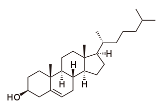
Cholesterol is the principal sterol of all higher animals, distributed in body tissues, especially the brain and spinal cord, and in animal fats and oils.

<span class="mw-page-title-main">Fatty acid</span> Carboxylic acid

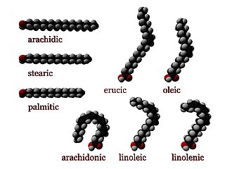
In chemistry, particularly in biochemistry, a fatty acid is a carboxylic acid with an aliphatic chain, which is either saturated or unsaturated. Most naturally occurring fatty acids have an unbranched chain of an even number of carbon atoms, from 4 to 28. Fatty acids are a major component of the lipids in some species such as microalgae but in some other organisms are not found in their standalone form, but instead exist as three main classes of esters: triglycerides, phospholipids, and cholesteryl esters. In any of these forms, fatty acids are both important dietary sources of fuel for animals and important structural components for cells.

<span class="mw-page-title-main">Phospholipid</span> Class of lipids

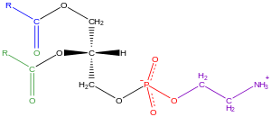
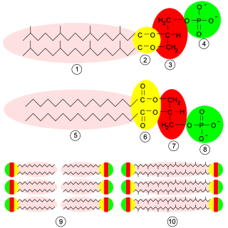
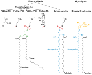
Phospholipids are a class of lipids whose molecule has a hydrophilic "head" containing a phosphate group and two hydrophobic "tails" derived from fatty acids, joined by an alcohol residue. Marine phospholipids typically have omega-3 fatty acids EPA and DHA integrated as part of the phospholipid molecule. The phosphate group can be modified with simple organic molecules such as choline, ethanolamine or serine.

<span class="mw-page-title-main">Lipoprotein</span> Biochemical assembly whose purpose is to transport hydrophobic lipid molecules

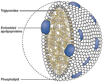
A lipoprotein is a biochemical assembly whose primary function is to transport hydrophobic lipid molecules in water, as in blood plasma or other extracellular fluids. They consist of a triglyceride and cholesterol center, surrounded by a phospholipid outer shell, with the hydrophilic portions oriented outward toward the surrounding water and lipophilic portions oriented inward toward the lipid center. A special kind of protein, called apolipoprotein, is embedded in the outer shell, both stabilising the complex and giving it a functional identity that determines its role.

<span class="mw-page-title-main">Acetyl-CoA</span> Chemical compound

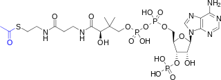
Acetyl-CoA is a molecule that participates in many biochemical reactions in protein, carbohydrate and lipid metabolism. Its main function is to deliver the acetyl group to the citric acid cycle to be oxidized for energy production.

<span class="mw-page-title-main">Chylomicron</span> One of the five major groups of lipoprotein

Chylomicra, also known as ultra low-density lipoproteins (ULDL), are lipoprotein particles that consist of triglycerides (85–92%), phospholipids (6–12%), cholesterol (1–3%), and proteins (1–2%). They transport dietary lipids from the intestines to other locations in the body. ULDLs are one of the five major groups of lipoproteins that enable fats and cholesterol to move within the water-based solution of the bloodstream. A protein specific to chylomicra is ApoB48.

<span class="mw-page-title-main">Sphingomyelin</span> Class of chemical compounds

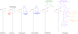
Sphingomyelin is a type of sphingolipid found in animal cell membranes, especially in the membranous myelin sheath that surrounds some nerve cell axons. It usually consists of phosphocholine and ceramide, or a phosphoethanolamine head group; therefore, sphingomyelins can also be classified as sphingophospholipids. In humans, SPH represents ~85% of all sphingolipids, and typically make up 10–20 mol % of plasma membrane lipids.

Fatty acid metabolism consists of various metabolic processes involving or closely related to fatty acids, a family of molecules classified within the lipid macronutrient category. These processes can mainly be divided into (1) catabolic processes that generate energy and (2) anabolic processes where they serve as building blocks for other compounds.

<span class="mw-page-title-main">Glycerophospholipid</span> Class of lipids

Glycerophospholipids or phosphoglycerides are glycerol-based phospholipids. They are the main component of biological membranes in eukaryotic cells. They are a type of lipid, of which its composition affects membrane structure and properties. Two major classes are known: those for bacteria and eukaryotes and a separate family for archaea.

Cardiolipin is an important component of the inner mitochondrial membrane, where it constitutes about 20% of the total lipid composition. It can also be found in the membranes of most bacteria. The name "cardiolipin" is derived from the fact that it was first found in animal hearts. It was first isolated from the beef heart in the early 1940s by Mary C. Pangborn. In mammalian cells, but also in plant cells, cardiolipin (CL) is found almost exclusively in the inner mitochondrial membrane, where it is essential for the optimal function of numerous enzymes that are involved in mitochondrial energy metabolism.

<span class="mw-page-title-main">Ether lipid</span>

In biochemistry, an ether lipid refers to any lipid in which the lipid "tail" group is attached to the glycerol backbone via an ether bond at any position. In contrast, conventional glycerophospholipids and triglycerides are triesters. Structural types include:

In biochemistry, lipogenesis is the conversion of fatty acids and glycerol into fats, or a metabolic process through which acetyl-CoA is converted to triglyceride for storage in fat. Lipogenesis encompasses both fatty acid and triglyceride synthesis, with the latter being the process by which fatty acids are esterified to glycerol before being packaged into very-low-density lipoprotein (VLDL). Fatty acids are produced in the cytoplasm of cells by repeatedly adding two-carbon units to acetyl-CoA. Triacylglycerol synthesis, on the other hand, occurs in the endoplasmic reticulum membrane of cells by bonding three fatty acid molecules to a glycerol molecule. Both processes take place mainly in liver and adipose tissue. Nevertheless, it also occurs to some extent in other tissues such as the gut and kidney. A review on lipogenesis in the brain was published in 2008 by Lopez and Vidal-Puig. After being packaged into VLDL in the liver, the resulting lipoprotein is then secreted directly into the blood for delivery to peripheral tissues.

In chemistry, de novo synthesis is the synthesis of complex molecules from simple molecules such as sugars or amino acids, as opposed to recycling after partial degradation. For example, nucleotides are not needed in the diet as they can be constructed from small precursor molecules such as formate and aspartate. Methionine, on the other hand, is needed in the diet because while it can be degraded to and then regenerated from homocysteine, it cannot be synthesized de novo.

Lipid metabolism is the synthesis and degradation of lipids in cells, involving the breakdown and storage of fats for energy and the synthesis of structural and functional lipids, such as those involved in the construction of cell membranes. In animals, these fats are obtained from food and are synthesized by the liver. Lipogenesis is the process of synthesizing these fats. The majority of lipids found in the human body from ingesting food are triglycerides and cholesterol. Other types of lipids found in the body are fatty acids and membrane lipids. Lipid metabolism is often considered the digestion and absorption process of dietary fat; however, there are two sources of fats that organisms can use to obtain energy: from consumed dietary fats and from stored fat. Vertebrates use both sources of fat to produce energy for organs such as the heart to function. Since lipids are hydrophobic molecules, they need to be solubilized before their metabolism can begin. Lipid metabolism often begins with hydrolysis, which occurs with the help of various enzymes in the digestive system. Lipid metabolism also occurs in plants, though the processes differ in some ways when compared to animals. The second step after the hydrolysis is the absorption of the fatty acids into the epithelial cells of the intestinal wall. In the epithelial cells, fatty acids are packaged and transported to the rest of the body.

<span class="mw-page-title-main">Membrane lipid</span> Lipid molecules on cell membrane

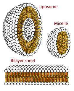
Membrane lipids are a group of compounds which form the lipid bilayer of the cell membrane. The three major classes of membrane lipids are phospholipids, glycolipids, and cholesterol. Lipids are amphiphilic: they have one end that is soluble in water ('polar') and an ending that is soluble in fat ('nonpolar'). By forming a double layer with the polar ends pointing outwards and the nonpolar ends pointing inwards membrane lipids can form a 'lipid bilayer' which keeps the watery interior of the cell separate from the watery exterior. The arrangements of lipids and various proteins, acting as receptors and channel pores in the membrane, control the entry and exit of other molecules and ions as part of the cell's metabolism. In order to perform physiological functions, membrane proteins are facilitated to rotate and diffuse laterally in two dimensional expanse of lipid bilayer by the presence of a shell of lipids closely attached to protein surface, called annular lipid shell.

Sterol O-acyltransferase is an intracellular protein located in the endoplasmic reticulum that forms cholesteryl esters from cholesterol.

<span class="mw-page-title-main">Lipase</span> Class of enzymes which cleave fats via hydrolysis

In biochemistry, lipase refers to a class of enzymes that catalyzes the hydrolysis of fats. Some lipases display broad substrate scope including esters of cholesterol, phospholipids, and of lipid-soluble vitamins and sphingomyelinases; however, these are usually treated separately from "conventional" lipases. Unlike esterases, which function in water, lipases "are activated only when adsorbed to an oil–water interface". Lipases perform essential roles in digestion, transport and processing of dietary lipids in most, if not all, organisms.

Lipid droplets, also referred to as lipid bodies, oil bodies or adiposomes, are lipid-rich cellular organelles that regulate the storage and hydrolysis of neutral lipids and are found largely in the adipose tissue. They also serve as a reservoir for cholesterol and acyl-glycerols for membrane formation and maintenance. Lipid droplets are found in all eukaryotic organisms and store a large portion of lipids in mammalian adipocytes. Initially, these lipid droplets were considered to merely serve as fat depots, but since the discovery in the 1990s of proteins in the lipid droplet coat that regulate lipid droplet dynamics and lipid metabolism, lipid droplets are seen as highly dynamic organelles that play a very important role in the regulation of intracellular lipid storage and lipid metabolism. The role of lipid droplets outside of lipid and cholesterol storage has recently begun to be elucidated and includes a close association to inflammatory responses through the synthesis and metabolism of eicosanoids and to metabolic disorders such as obesity, cancer, and atherosclerosis. In non-adipocytes, lipid droplets are known to play a role in protection from lipotoxicity by storage of fatty acids in the form of neutral triacylglycerol, which consists of three fatty acids bound to glycerol. Alternatively, fatty acids can be converted to lipid intermediates like diacylglycerol (DAG), ceramides and fatty acyl-CoAs. These lipid intermediates can impair insulin signaling, which is referred to as lipid-induced insulin resistance and lipotoxicity. Lipid droplets also serve as platforms for protein binding and degradation. Finally, lipid droplets are known to be exploited by pathogens such as the hepatitis C virus, the dengue virus and Chlamydia trachomatis among others.

<span class="mw-page-title-main">Diglyceride</span> Type of fat derived from glycerol and two fatty acids

A diglyceride, or diacylglycerol (DAG), is a glyceride consisting of two fatty acid chains covalently bonded to a glycerol molecule through ester linkages. Two possible forms exist, 1,2-diacylglycerols and 1,3-diacylglycerols. Diglycerides are natural components of food fats, though minor in comparison to triglycerides. DAGs can act as surfactants and are commonly used as emulsifiers in processed foods. DAG-enriched oil has been investigated extensively as a fat substitute due to its ability to suppress the accumulation of body fat; with total annual sales of approximately USD 200 million in Japan since its introduction in the late 1990s till 2009.

<span class="mw-page-title-main">Laurdan</span> Chemical compound

Laurdan is an organic compound which is used as a fluorescent dye when applied to fluorescence microscopy. It is used to investigate membrane qualities of the phospholipid bilayers of cell membranes. One of its most important characteristics is its sensitivity to membrane phase transitions as well as other alterations to membrane fluidity such as the penetration of water.

References

  1. Maitland J Jr (1998). Organic Chemistry. W W Norton & Co Inc (Np). p. 139. ISBN   978-0-393-97378-5.
  2. 1 2 3 4 5 6 7 8 9 10 Stryer L, Berg JM, Tymoczko JL (2007). Biochemistry (6th ed.). San Francisco: W.H. Freeman. ISBN   978-0-7167-8724-2.
  3. 1 2 3 4 Fahy E, Subramaniam S, Murphy RC, Nishijima M, Raetz CR, Shimizu T, Spener F, van Meer G, Wakelam MJ, Dennis EA (April 2009). "Update of the LIPID MAPS comprehensive classification system for lipids". Journal of Lipid Research. 50 (S1): S9–14. doi: 10.1194/jlr.R800095-JLR200 . PMC   2674711 . PMID   19098281.
  4. Subramaniam S, Fahy E, Gupta S, Sud M, Byrnes RW, Cotter D, Dinasarapu AR, Maurya MR (October 2011). "Bioinformatics and systems biology of the lipidome". Chemical Reviews. 111 (10): 6452–6490. doi:10.1021/cr200295k. PMC   3383319 . PMID   21939287.
  5. Mashaghi S, Jadidi T, Koenderink G, Mashaghi A (February 2013). "Lipid nanotechnology". International Journal of Molecular Sciences. 14 (2): 4242–4282. doi: 10.3390/ijms14024242 . PMC   3588097 . PMID   23429269. Open Access logo PLoS transparent.svg
  6. Michelle A, Hopkins J, McLaughlin CW, Johnson S, Warner MQ, LaHart D, Wright JD (1993). Human Biology and Health. Englewood Cliffs, New Jersey: Prentice Hall. ISBN   978-0-13-981176-0.
  7. Braconnot H (31 March 1815). "Sur la nature des corps gras". Annales de chimie. 2 (XCIII): 225–277.
  8. Chevreul ME (1823). Recherches sur les corps gras d'origine animale. Paris: Levrault.
  9. 1 2 3 Leray C (2012). Introduction to Lipidomics. Boca Raton: CRC Press. ISBN   978-1466551466.
  10. Leray C (2015). "Introduction, History and Evolution.". Lipids. Nutrition and health. Boca Raton: CRC Press. ISBN   978-1482242317.
  11. Pelouze TJ, Gélis A (1844). "Mémoire sur l'acide butyrique". Annales de Chimie et de Physique. 10: 434.
  12. Comptes rendus hebdomadaires des séances de l'Académie des Sciences, Paris, 1853, 36, 27; Annales de Chimie et de Physique 1854, 41, 216
  13. Leray C. "Chronological history of lipid center". Cyberlipid Center. Archived from the original on 13 October 2017. Retrieved 1 December 2017.
  14. Prout W (1827). "On the ultimate composition of simple alimentary substances, with some preliminary remarks on the analysis of organised bodies in general". Phil. Trans.: 355–388.
  15. Culling CF (1974). "Lipids. (Fats, Lipoids. Lipins).". Handbook of Histopathological Techniques (3rd ed.). London: Butterworths. pp. 351–376. ISBN   978-1483164793.
  16. Rosenbloom J, Gies WJ (1911). "Suggestion to teachers of biochemistry. I. A proposed chemical classification of lipins, with a note on the intimate relation between cholesterols and bile salts". Biochem. Bull. 1: 51–56.
  17. Bloor WR (1920). "Outline of a classication of the lipids". Proc. Soc. Exp. Biol. Med. 17 (6): 138–140. doi:10.3181/00379727-17-75. S2CID   75844378.
  18. Christie WW, Han X (2010). Lipid Analysis: Isolation, Separation, Identification and Lipidomic Analysis. Bridgwater, England: The Oily Press. ISBN   978-0857097866.
  19. Bertrand G (1923). "Projet de reforme de la nomenclature de Chimie biologique". Bulletin de la Société de Chimie Biologique. 5: 96–109.
  20. Hilditch, Thomas Percy (1956). The Chemical Constitution of Natural Fats. Wiley.
  21. Vance JE, Vance DE (2002). Biochemistry of Lipids, Lipoproteins and Membranes. Amsterdam: Elsevier. ISBN   978-0-444-51139-3.
  22. Brown HA, ed. (2007). Lipodomics and Bioactive Lipids: Mass Spectrometry Based Lipid Analysis. Methods in Enzymology. Vol. 423. Boston: Academic Press. ISBN   978-0-12-373895-0.
  23. Hunt SM, Groff JL, Gropper SA (1995). Advanced Nutrition and Human Metabolism. Belmont, California: West Pub. Co. p.  98. ISBN   978-0-314-04467-9.
  24. Yashroy RC (1987). "13C NMR studies of lipid fatty acyl chains of chloroplast membranes". Indian Journal of Biochemistry and Biophysics. 24 (6): 177–178. doi:10.1016/0165-022X(91)90019-S. PMID   3428918.
  25. 1 2 Devlin TM (1997). Textbook of Biochemistry: With Clinical Correlations (4th ed.). Chichester: John Wiley & Sons. ISBN   978-0-471-17053-2.
  26. Hunter JE (November 2006). "Dietary trans fatty acids: review of recent human studies and food industry responses". Lipids. 41 (11): 967–992. doi:10.1007/s11745-006-5049-y. PMID   17263298. S2CID   1625062.
  27. Furse S (2 December 2011). "A Long Lipid, a Long Name: Docosahexaenoic Acid". The Lipid Chronicles.
  28. "DHA for Optimal Brain and Visual Functioning". DHA/EPA Omega-3 Institute.
  29. Fezza F, De Simone C, Amadio D, Maccarrone M (2008). "Fatty Acid Amide Hydrolase: A Gate-Keeper of the Endocannabinoid System". Lipids in Health and Disease. Subcellular Biochemistry. Vol. 49. pp. 101–132. doi:10.1007/978-1-4020-8831-5_4. ISBN   978-1-4020-8830-8. PMID   18751909.
  30. Coleman RA, Lee DP (March 2004). "Enzymes of triacylglycerol synthesis and their regulation". Progress in Lipid Research. 43 (2): 134–176. doi:10.1016/S0163-7827(03)00051-1. PMID   14654091.
  31. 1 2 van Holde KE, Mathews CK (1996). Biochemistry (2nd ed.). Menlo Park, California: Benjamin/Cummings Pub. Co. ISBN   978-0-8053-3931-4.
  32. 1 2 Hölzl G, Dörmann P (September 2007). "Structure and function of glycoglycerolipids in plants and bacteria". Progress in Lipid Research. 46 (5): 225–243. doi:10.1016/j.plipres.2007.05.001. PMID   17599463.
  33. Honke K, Zhang Y, Cheng X, Kotani N, Taniguchi N (2004). "Biological roles of sulfoglycolipids and pathophysiology of their deficiency". Glycoconjugate Journal. 21 (1–2): 59–62. doi:10.1023/B:GLYC.0000043749.06556.3d. PMID   15467400. S2CID   2678053.
  34. "The Structure of a Membrane". The Lipid Chronicles. 5 November 2011. Retrieved 31 December 2011.
  35. Berridge MJ, Irvine RF (September 1989). "Inositol phosphates and cell signalling". Nature. 341 (6239): 197–205. Bibcode:1989Natur.341..197B. doi:10.1038/341197a0. PMID   2550825. S2CID   26822092.
  36. Farooqui AA, Horrocks LA, Farooqui T (June 2000). "Glycerophospholipids in brain: their metabolism, incorporation into membranes, functions, and involvement in neurological disorders". Chemistry and Physics of Lipids. 106 (1): 1–29. doi:10.1016/S0009-3084(00)00128-6. PMID   10878232.
  37. Ivanova PT, Milne SB, Byrne MO, Xiang Y, Brown HA (2007). "Glycerophospholipid identification and quantitation by electrospray ionization mass spectrometry". Lipidomics and Bioactive Lipids: Mass-Spectrometry–Based Lipid Analysis. Methods in Enzymology. Vol. 432. pp. 21–57. doi:10.1016/S0076-6879(07)32002-8. ISBN   978-0-12-373895-0. PMID   17954212.
  38. Paltauf F (December 1994). "Ether lipids in biomembranes". Chemistry and Physics of Lipids. 74 (2): 101–139. doi:10.1016/0009-3084(94)90054-X. PMID   7859340.
  39. Merrill AH, Sandoff K (2002). "Chapter 14: Sphingolipids: Metabolism and Cell Signaling" (PDF). In Vance JE, Vance EE (eds.). Biochemistry of Lipids, Lipoproteins and Membranes (4th ed.). Amsterdam: Elsevier. pp. 373–407. ISBN   978-0-444-51138-6.
  40. Hori T, Sugita M (1993). "Sphingolipids in lower animals". Progress in Lipid Research. 32 (1): 25–45. doi:10.1016/0163-7827(93)90003-F. PMID   8415797.
  41. Wiegandt H (January 1992). "Insect glycolipids". Biochimica et Biophysica Acta (BBA) - Lipids and Lipid Metabolism. 1123 (2): 117–126. doi:10.1016/0005-2760(92)90101-Z. PMID   1739742.
  42. Guan X, Wenk MR (May 2008). "Biochemistry of inositol lipids". Frontiers in Bioscience. 13 (13): 3239–3251. doi: 10.2741/2923 . PMID   18508430.
  43. Bach D, Wachtel E (March 2003). "Phospholipid/cholesterol model membranes: formation of cholesterol crystallites". Biochimica et Biophysica Acta (BBA) - Biomembranes. 1610 (2): 187–197. doi: 10.1016/S0005-2736(03)00017-8 . PMID   12648773.
  44. Russell DW (2003). "The enzymes, regulation, and genetics of bile acid synthesis". Annual Review of Biochemistry. 72: 137–174. doi:10.1146/annurev.biochem.72.121801.161712. PMID   12543708.
  45. Villinski JC, Hayes JM, Brassell SC, Riggert VL, Dunbar R (2008). "Sedimentary sterols as biogeochemical indicators in the Southern Ocean". Organic Geochemistry. 39 (5): 567–588. Bibcode:2008OrGeo..39..567V. doi:10.1016/j.orggeochem.2008.01.009.
  46. Deacon J (2005). Fungal Biology. Cambridge, Massachusetts: Blackwell Publishers. p. 342. ISBN   978-1-4051-3066-0.
  47. Bouillon R, Verstuyf A, Mathieu C, Van Cromphaut S, Masuyama R, Dehaes P, Carmeliet G (December 2006). "Vitamin D resistance". Best Practice & Research. Clinical Endocrinology & Metabolism. 20 (4): 627–645. doi:10.1016/j.beem.2006.09.008. PMID   17161336.
  48. 1 2 3 Kuzuyama T, Seto H (April 2003). "Diversity of the biosynthesis of the isoprene units". Natural Product Reports. 20 (2): 171–183. doi:10.1039/b109860h. PMID   12735695.
  49. Rao AV, Rao LG (March 2007). "Carotenoids and human health". Pharmacological Research. 55 (3): 207–216. doi:10.1016/j.phrs.2007.01.012. PMID   17349800.
  50. Brunmark A, Cadenas E (1989). "Redox and addition chemistry of quinoid compounds and its biological implications". Free Radical Biology & Medicine. 7 (4): 435–477. doi:10.1016/0891-5849(89)90126-3. PMID   2691341.
  51. Swiezewska E, Danikiewicz W (July 2005). "Polyisoprenoids: structure, biosynthesis and function". Progress in Lipid Research. 44 (4): 235–258. doi:10.1016/j.plipres.2005.05.002. PMID   16019076.
  52. 1 2 Raetz CR, Garrett TA, Reynolds CM, Shaw WA, Moore JD, Smith DC, et al. (May 2006). "Kdo2-Lipid A of Escherichia coli, a defined endotoxin that activates macrophages via TLR-4". Journal of Lipid Research. 47 (5): 1097–1111. doi: 10.1194/jlr.M600027-JLR200 . hdl: 10919/74310 . PMID   16479018. Open Access logo PLoS transparent.svg
  53. Walsh CT (March 2004). "Polyketide and nonribosomal peptide antibiotics: modularity and versatility". Science. 303 (5665): 1805–1810. Bibcode:2004Sci...303.1805W. doi:10.1126/science.1094318. PMID   15031493. S2CID   44858908.
  54. Caffrey P, Aparicio JF, Malpartida F, Zotchev SB (2008). "Biosynthetic engineering of polyene macrolides towards generation of improved antifungal and antiparasitic agents". Current Topics in Medicinal Chemistry. 8 (8): 639–653. doi:10.2174/156802608784221479. hdl: 10197/8333 . PMID   18473889.
  55. Minto RE, Blacklock BJ (July 2008). "Biosynthesis and function of polyacetylenes and allied natural products". Progress in Lipid Research. 47 (4): 233–306. doi:10.1016/j.plipres.2008.02.002. PMC   2515280 . PMID   18387369.
  56. Coones RT, Green RJ, Frazier RA (July 2021). "Investigating lipid headgroup composition within epithelial membranes: a systematic review". Soft Matter. 17 (28): 6773–6786. Bibcode:2021SMat...17.6773C. doi: 10.1039/D1SM00703C . ISSN   1744-683X. PMID   34212942. S2CID   235708094.
  57. Heinz E. (1996). "Plant glycolipids: structure, isolation and analysis", pp. 211–332 in Advances in Lipid Methodology, Vol. 3. W.W. Christie (ed.). Oily Press, Dundee. ISBN   978-0-9514171-6-4
  58. Lyu, Jiabao; Gao, Renjun; Guo, Zheng (2021). "Galactosyldiacylglycerols: From a photosynthesis-associated apparatus to structure-defined in vitro assembling". Journal of Agricultural and Food Chemistry. 69 (32): 8910–8928. doi:10.1021/acs.jafc.1c00204. PMID   33793221. S2CID   232761961.
  59. Yashroy RC (1990). "Magnetic resonance studies of dynamic organisation of lipids in chloroplast membranes". Journal of Biosciences. 15 (4): 281–288. doi:10.1007/BF02702669. S2CID   360223.
  60. van Meer G, Voelker DR, Feigenson GW (February 2008). "Membrane lipids: where they are and how they behave". Nature Reviews Molecular Cell Biology. 9 (2): 112–124. doi:10.1038/nrm2330. PMC   2642958 . PMID   18216768.
  61. Feigenson GW (November 2006). "Phase behavior of lipid mixtures". Nature Chemical Biology. 2 (11): 560–563. doi:10.1038/nchembio1106-560. PMC   2685072 . PMID   17051225.
  62. Wiggins PM (December 1990). "Role of water in some biological processes". Microbiological Reviews. 54 (4): 432–449. doi:10.1128/MMBR.54.4.432-449.1990. PMC   372788 . PMID   2087221.
  63. Raschke TM, Levitt M (May 2005). "Nonpolar solutes enhance water structure within hydration shells while reducing interactions between them". Proceedings of the National Academy of Sciences of the United States of America. 102 (19): 6777–6782. doi: 10.1073/pnas.0500225102 . PMC   1100774 . PMID   15867152.
  64. Segré D, Ben-Eli D, Deamer DW, Lancet D (2001). "The lipid world" (PDF). Origins of Life and Evolution of the Biosphere. 31 (1–2): 119–145. Bibcode:2001OLEB...31..119S. doi:10.1023/A:1006746807104. PMID   11296516. S2CID   10959497. Archived from the original (PDF) on 11 September 2008. Retrieved 15 March 2015.
  65. Brasaemle DL (December 2007). "Thematic review series: adipocyte biology. The perilipin family of structural lipid droplet proteins: stabilization of lipid droplets and control of lipolysis". Journal of Lipid Research. 48 (12): 2547–2559. doi: 10.1194/jlr.R700014-JLR200 . PMID   17878492.
  66. Malinauskas T, Aricescu AR, Lu W, Siebold C, Jones EY (July 2011). "Modular mechanism of Wnt signaling inhibition by Wnt inhibitory factor 1". Nature Structural & Molecular Biology. 18 (8): 886–893. doi:10.1038/nsmb.2081. PMC   3430870 . PMID   21743455.
  67. Malinauskas T (March 2008). "Docking of fatty acids into the WIF domain of the human Wnt inhibitory factor-1". Lipids. 43 (3): 227–230. doi:10.1007/s11745-007-3144-3. PMID   18256869. S2CID   31357937.
  68. Wang X (June 2004). "Lipid signaling". Current Opinion in Plant Biology. 7 (3): 329–336. doi:10.1016/j.pbi.2004.03.012. PMID   15134755.
  69. Dinasarapu AR, Saunders B, Ozerlat I, Azam K, Subramaniam S (June 2011). "Signaling gateway molecule pages – a data model perspective". Bioinformatics. 27 (12): 1736–1738. doi:10.1093/bioinformatics/btr190. PMC   3106186 . PMID   21505029.
  70. Eyster KM (March 2007). "The membrane and lipids as integral participants in signal transduction: lipid signal transduction for the non-lipid biochemist". Advances in Physiology Education. 31 (1): 5–16. doi:10.1152/advan.00088.2006. PMID   17327576. S2CID   9194419.
  71. Hinkovska-Galcheva V, VanWay SM, Shanley TP, Kunkel RG (November 2008). "The role of sphingosine-1-phosphate and ceramide-1-phosphate in calcium homeostasis". Current Opinion in Investigational Drugs. 9 (11): 1192–1205. PMID   18951299.
  72. Saddoughi SA, Song P, Ogretmen B (2008). "Roles of Bioactive Sphingolipids in Cancer Biology and Therapeutics". Lipids in Health and Disease. Subcellular Biochemistry. Vol. 49. pp. 413–440. doi:10.1007/978-1-4020-8831-5_16. ISBN   978-1-4020-8830-8. PMC   2636716 . PMID   18751921.
  73. Klein C, Malviya AN (January 2008). "Mechanism of nuclear calcium signaling by inositol 1,4,5-trisphosphate produced in the nucleus, nuclear located protein kinase C and cyclic AMP-dependent protein kinase". Frontiers in Bioscience. 13 (13): 1206–1226. doi: 10.2741/2756 . PMID   17981624.
  74. Boyce JA (August 2008). "Eicosanoids in asthma, allergic inflammation, and host defense". Current Molecular Medicine. 8 (5): 335–349. doi:10.2174/156652408785160989. PMID   18691060.
  75. Bełtowski J (2008). "Liver X receptors (LXR) as therapeutic targets in dyslipidemia". Cardiovascular Therapeutics. 26 (4): 297–316. doi: 10.1111/j.1755-5922.2008.00062.x . PMID   19035881.
  76. Biermann M, Maueröder C, Brauner JM, Chaurio R, Janko C, Herrmann M, Muñoz LE (December 2013). "Surface code--biophysical signals for apoptotic cell clearance". Physical Biology. 10 (6): 065007. Bibcode:2013PhBio..10f5007B. doi:10.1088/1478-3975/10/6/065007. PMID   24305041. S2CID   23782770.
  77. Indiveri C, Tonazzi A, Palmieri F (October 1991). "Characterization of the unidirectional transport of carnitine catalyzed by the reconstituted carnitine carrier from rat liver mitochondria". Biochimica et Biophysica Acta (BBA) - Biomembranes. 1069 (1): 110–116. doi:10.1016/0005-2736(91)90110-t. PMID   1932043.
  78. Parodi AJ, Leloir LF (April 1979). "The role of lipid intermediates in the glycosylation of proteins in the eucaryotic cell". Biochimica et Biophysica Acta (BBA) - Reviews on Biomembranes. 559 (1): 1–37. doi:10.1016/0304-4157(79)90006-6. PMID   375981.
  79. Helenius A, Aebi M (March 2001). "Intracellular functions of N-linked glycans". Science. 291 (5512): 2364–2369. Bibcode:2001Sci...291.2364H. doi:10.1126/science.291.5512.2364. PMID   11269317. S2CID   7277949.
  80. Nowicki M, Müller F, Frentzen M (April 2005). "Cardiolipin synthase of Arabidopsis thaliana". FEBS Letters. 579 (10): 2161–2165. doi: 10.1016/j.febslet.2005.03.007 . PMID   15811335. S2CID   21937549.
  81. Gohil VM, Greenberg ML (February 2009). "Mitochondrial membrane biogenesis: phospholipids and proteins go hand in hand". The Journal of Cell Biology. 184 (4): 469–472. doi:10.1083/jcb.200901127. PMC   2654137 . PMID   19237595.
  82. Hoch FL (March 1992). "Cardiolipins and biomembrane function" (PDF). Biochimica et Biophysica Acta (BBA) - Reviews on Biomembranes. 1113 (1): 71–133. doi:10.1016/0304-4157(92)90035-9. hdl: 2027.42/30145 . PMID   1550861.
  83. "Steroids". Elmhurst. edu. Archived from the original on 23 October 2011. Retrieved 10 October 2013.
  84. Chirala SS, Wakil SJ (November 2004). "Structure and function of animal fatty acid synthase". Lipids. 39 (11): 1045–1053. doi:10.1007/s11745-004-1329-9. PMID   15726818. S2CID   4043407.
  85. White SW, Zheng J, Zhang YM (2005). "The structural biology of type II fatty acid biosynthesis". Annual Review of Biochemistry. 74: 791–831. doi:10.1146/annurev.biochem.74.082803.133524. PMID   15952903.
  86. Ohlrogge JB, Jaworski JG (June 1997). "Regulation of fatty acid synthesis". Annual Review of Plant Physiology and Plant Molecular Biology. 48: 109–136. doi:10.1146/annurev.arplant.48.1.109. PMID   15012259. S2CID   46348092.
  87. Grochowski LL, Xu H, White RH (May 2006). "Methanocaldococcus jannaschii uses a modified mevalonate pathway for biosynthesis of isopentenyl diphosphate". Journal of Bacteriology. 188 (9): 3192–3198. doi:10.1128/JB.188.9.3192-3198.2006. PMC   1447442 . PMID   16621811.
  88. Lichtenthaler HK (June 1999). "The 1-dideoxy-D-xylulose-5-phosphate pathway of isoprenoid biosynthesis in plants". Annual Review of Plant Physiology and Plant Molecular Biology. 50: 47–65. doi:10.1146/annurev.arplant.50.1.47. PMID   15012203.
  89. 1 2 Schroepfer GJ (1981). "Sterol biosynthesis". Annual Review of Biochemistry. 50: 585–621. doi:10.1146/annurev.bi.50.070181.003101. PMID   7023367.
  90. Lees ND, Skaggs B, Kirsch DR, Bard M (March 1995). "Cloning of the late genes in the ergosterol biosynthetic pathway of Saccharomyces cerevisiae--a review". Lipids. 30 (3): 221–226. doi:10.1007/BF02537824. PMID   7791529. S2CID   4019443.
  91. 1 2 Bhagavan NV (2002). Medical Biochemistry. San Diego: Harcourt/Academic Press. ISBN   978-0-12-095440-7.
  92. Russo GL (March 2009). "Dietary n-6 and n-3 polyunsaturated fatty acids: from biochemistry to clinical implications in cardiovascular prevention". Biochemical Pharmacology. 77 (6): 937–946. doi:10.1016/j.bcp.2008.10.020. PMID   19022225.
  93. Riediger ND, Othman RA, Suh M, Moghadasian MH (April 2009). "A systemic review of the roles of n-3 fatty acids in health and disease". Journal of the American Dietetic Association. 109 (4): 668–679. doi:10.1016/j.jada.2008.12.022. PMID   19328262.
  94. Galli C, Risé P (2009). "Fish consumption, omega 3 fatty acids and cardiovascular disease. The science and the clinical trials". Nutrition and Health. 20 (1): 11–20. doi:10.1177/026010600902000102. PMID   19326716. S2CID   20742062.
  95. Micha R, Mozaffarian D (2008). "Trans fatty acids: effects on cardiometabolic health and implications for policy". Prostaglandins, Leukotrienes, and Essential Fatty Acids. 79 (3–5): 147–152. doi:10.1016/j.plefa.2008.09.008. PMC   2639783 . PMID   18996687.
  96. Dalainas I, Ioannou HP (April 2008). "The role of trans fatty acids in atherosclerosis, cardiovascular disease and infant development". International Angiology. 27 (2): 146–156. PMID   18427401.
  97. Mozaffarian D, Willett WC (December 2007). "Trans fatty acids and cardiovascular risk: a unique cardiometabolic imprint?". Current Atherosclerosis Reports. 9 (6): 486–493. doi:10.1007/s11883-007-0065-9. PMID   18377789. S2CID   24998042.
  98. Astrup A, Dyerberg J, Selleck M, Stender S (2008), "Nutrition transition and its relationship to the development of obesity and related chronic diseases", Obes Rev, 9 (S1): 48–52, doi:10.1111/j.1467-789X.2007.00438.x, PMID   18307699, S2CID   34030743
  99. Astrup A (February 2005). "The role of dietary fat in obesity". Seminars in Vascular Medicine. 5 (1): 40–47. doi:10.1055/s-2005-871740. PMID   15968579. S2CID   260372605.
  100. Astrup A (2008). "Dietary management of obesity". Journal of Parenteral and Enteral Nutrition. 32 (5): 575–577. doi:10.1177/0148607108321707. PMID   18753397.
  101. Beresford SA, Johnson KC, Ritenbaugh C, Lasser NL, Snetselaar LG, Black HR, et al. (February 2006). "Low-fat dietary pattern and risk of colorectal cancer: the Women's Health Initiative Randomized Controlled Dietary Modification Trial". Journal of the American Medical Association. 295 (6): 643–654. doi:10.1001/jama.295.6.643. PMID   16467233. Open Access logo PLoS transparent.svg
  102. Howard BV, Manson JE, Stefanick ML, Beresford SA, Frank G, Jones B, Rodabough RJ, Snetselaar L, Thomson C, Tinker L, Vitolins M, Prentice R (January 2006). "Low-fat dietary pattern and weight change over 7 years: the Women's Health Initiative Dietary Modification Trial". Journal of the American Medical Association. 295 (1): 39–49. doi:10.1001/jama.295.1.39. PMID   16391215. Open Access logo PLoS transparent.svg
  103. "The Nutrition Source". T. H. Chan School of Public Health. Harvard University.
  104. "Fats and Cholesterol: Out with the Bad, In with the Good — What Should You Eat? – The Nutrition Source". Harvard School of Public Health.

Bibliography

Introductory

Nomenclature

Databases

General