Liposome

Last updated
Scheme of a liposome formed by phospholipids in an aqueous solution. Liposome scheme-en.svg
Scheme of a liposome formed by phospholipids in an aqueous solution.
Liposomes are composite structures made of phospholipids and may contain small amounts of other molecules. Though liposomes can vary in size from low micrometer range to tens of micrometers, unilamellar liposomes, as pictured here, are typically in the lower size range with various targeting ligands attached to their surface allowing for their surface-attachment and accumulation in pathological areas for treatment of disease. Liposome.jpg
Liposomes are composite structures made of phospholipids and may contain small amounts of other molecules. Though liposomes can vary in size from low micrometer range to tens of micrometers, unilamellar liposomes, as pictured here, are typically in the lower size range with various targeting ligands attached to their surface allowing for their surface-attachment and accumulation in pathological areas for treatment of disease.

A liposome is a small artificial vesicle, spherical in shape, having at least one lipid bilayer. [2] Due to their hydrophobicity and/or hydrophilicity, biocompatibility, particle size and many other properties, [2] liposomes can be used as drug delivery vehicles for administration of pharmaceutical drugs and nutrients, [3] such as lipid nanoparticles in mRNA vaccines, and DNA vaccines. Liposomes can be prepared by disrupting biological membranes (such as by sonication).

Contents

Liposomes are most often composed of phospholipids, [4] especially phosphatidylcholine, and cholesterol, [2] but may also include other lipids, such as those found in egg and phosphatidylethanolamine, as long as they are compatible with lipid bilayer structure. [5] A liposome design may employ surface ligands for attaching to desired cells or tissues. [1]

Based on vesicle structure, there are seven main categories for liposomes: multilamellar large (MLV), oligolamellar (OLV), small unilamellar (SUV), medium-sized unilamellar (MUV), large unilamellar (LUV), giant unilamellar (GUV) and multivesicular vesicles (MVV). [6] The major types of liposomes are the multilamellar vesicle (MLV, with several lamellar phase lipid bilayers), the small unilamellar liposome vesicle (SUV, with one lipid bilayer), the large unilamellar vesicle (LUV), and the cochleate vesicle. A less desirable form is multivesicular liposomes in which one vesicle contains one or more smaller vesicles.

Seven main categories for liposomes: multilamellar large (MLV), oligolamellar (OLV), small unilamellar (SUV), medium-sized unilamellar (MUV), large unilamellar (LUV), giant unilamellar (GUV) and multivesicular vesicles (MVV)) . 1-s2.0-S0168365921005034-gr6 lrg.jpg
Seven main categories for liposomes: multilamellar large (MLV), oligolamellar (OLV), small unilamellar (SUV), medium-sized unilamellar (MUV), large unilamellar (LUV), giant unilamellar (GUV) and multivesicular vesicles (MVV)) .

Liposomes should not be confused with lysosomes, or with micelles and reverse micelles. [8] In contrast to liposomes, micelles typically contain a monolayer of fatty acids or surfactants. [9]

Discovery

The word liposome derives from two Greek words: lipo ("fat") and soma ("body"); it is so named because its composition is primarily of phospholipid.

Liposomes were first described by British hematologist Alec Douglas Bangham [10] [11] [12] in 1961 at the Babraham Institute, in Cambridge—findings that were published 1964. The discovery came about when Bangham and R. W. Horne were testing the institute's new electron microscope by adding negative stain to dry phospholipids. The resemblance to the plasmalemma was obvious, and the microscopic pictures provided the first evidence that the cell membrane is a bilayer lipid structure. The following year, Bangham, his colleague Malcolm Standish, and Gerald Weissmann, an American physician, established the integrity of this closed, bilayer structure and its ability to release its contents following detergent treatment (structure-linked latency). [13] During a Cambridge pub discussion with Bangham, Weissmann first named the structures "liposomes" after something which laboratory had been studying, the lysosome: a simple organelle whose structure-linked latency could be disrupted by detergents and streptolysins. [14] Liposomes are readily distinguishable from micelles and hexagonal lipid phases through negative staining transmission electron microscopy. [15]

Bangham, with colleagues Jeff Watkins and Standish, wrote the 1965 paper that effectively launched what would become the liposome "industry."Around that same time, Weissmann joined Bangham at the Babraham. Later, Weissmann, then an emeritus professor at New York University School of Medicine, recalled the two of them sitting in a Cambridge pub, reflecting on the role of lipid sheets in separating the cell interior from its exterior milieu. This insight, they felt, would be to cell function what the discovery of the double helix had been to genetics. As Bangham had been calling his lipid structures "multilamellar smectic mesophases," or sometimes "Banghasomes," Weissmann proposed the more user-friendly term liposome. [16] [17]

Mechanism

A micrograph of phosphatidylcholine liposomes, which were stained with fluorochrome acridine orange. Method of fluorescence microscopy (1250-fold magnification). Phosphatidylcholine liposomes stained with acridine orange.jpg
A micrograph of phosphatidylcholine liposomes, which were stained with fluorochrome acridine orange. Method of fluorescence microscopy (1250-fold magnification).
Various types of phosphatidylcholine liposomes in suspension. Method of phase-contrast microscopy (1000-fold magnification). The following types of liposomes are visible: small monolamellar vesicles, large monolamellar vesicles, multilamellar vesicles, oligolamellar vesicles. Phosphatidylcholine liposomes at phase-contrast microscopy.jpg
Various types of phosphatidylcholine liposomes in suspension. Method of phase-contrast microscopy (1000-fold magnification). The following types of liposomes are visible: small monolamellar vesicles, large monolamellar vesicles, multilamellar vesicles, oligolamellar vesicles.

Encapsulation in liposomes

A liposome has an aqueous solution core surrounded by a hydrophobic membrane, in the form of a lipid bilayer; hydrophilic solutes dissolved in the core cannot readily pass through the bilayer. Hydrophobic chemicals associate with the bilayer. This property can be utilized to load liposomes with hydrophobic and/or hydrophilic molecules, a process known as encapsulation. [18] Typically, liposomes are prepared in a solution containing the compound to be trapped, which can either be an aqueous solution for encapsulating hydrophilic compounds like proteins, [19] [20] or solutions in organic solvents mixed with lipids for encapsulating hydrophobic molecules. Encapsulation techniques can be categorized into two types: passive, which relies on the stochastic trapping of molecules during liposome formation, and active, which relies on the presence of charged lipids or transmembrane ion gradients. [18] A crucial parameter to consider is the "encapsulation efficiency," which is defined as the amount of compound present in the liposome solution divided by the total initial amount of compound used during the preparation. [21] In more recent developments, the application of liposomes in single-molecule experiments has introduced the concept of "single entity encapsulation efficiency." This term refers to the probability of a specific liposome containing the required number of copies of the compound. [22]

Delivery

To deliver the molecules to a site of action, the lipid bilayer can fuse with other bilayers such as the cell membrane, thus delivering the liposome contents; this is a complex and non-spontaneous event, however, [23] that does not apply to nutrients and drug delivery. By preparing liposomes in a solution of DNA or drugs (which would normally be unable to diffuse through the membrane) they can be (indiscriminately) delivered past the lipid bilayer. [24] Liposomes can also be designed to deliver drugs in other ways. Liposomes that contain low (or high) pH can be constructed such that dissolved aqueous drugs will be charged in solution (i.e., the pH is outside the drug's pI range). As the pH naturally neutralizes within the liposome (protons can pass through some membranes), the drug will also be neutralized, allowing it to freely pass through a membrane. These liposomes work to deliver drug by diffusion rather than by direct cell fusion. However, the efficacy of this pH regulated passage depends on the physiochemical nature of the drug in question (e.g. pKa and having a basic or acid nature), which is very low for many drugs.

A similar approach can be exploited in the biodetoxification of drugs by injecting empty liposomes with a transmembrane pH gradient. In this case the vesicles act as sinks to scavenge the drug in the blood circulation and prevent its toxic effect. [25] Another strategy for liposome drug delivery is to target endocytosis events. Liposomes can be made in a particular size range that makes them viable targets for natural macrophage phagocytosis. These liposomes may be digested while in the macrophage's phagosome, thus releasing its drug. Liposomes can also be decorated with opsonins and ligands to activate endocytosis in other cell types.

Regarding pH-sensitive liposomes, there are three mechanisms of drug delivery intracellularly, which occurs via endocytosis [26] . This is possible because of the acidic environment within endosomes [26] . The first mechanism is through the destabilization of the liposome within the endosome, triggering pore formation on the endosomal membrane and allowing diffusion of the liposome and its contents into the cytoplasm [26] . Another is the release of the encapsulated content within the endosome, eventually diffusing out into the cytoplasm through the endosomal membrane [26] . Lastly, the membrane of the liposome and the endosome fuse together, releasing the encapsulated contents onto the cytoplasm and avoiding degradation at the lysosomal level due to minimal contact time [26] .

Certain anticancer drugs such as doxorubicin (Doxil) and daunorubicin may be administered encapsulated in liposomes. Liposomal cisplatin has received orphan drug designation for pancreatic cancer from EMEA. [27]

The use of liposomes for transformation or transfection of DNA into a host cell is known as lipofection.

In addition to gene and drug delivery applications, liposomes can be used as carriers for the delivery of dyes to textiles, [28] pesticides to plants, enzymes and nutritional supplements to foods, and cosmetics to the skin. [29]

Liposomes are also used as outer shells of some microbubble contrast agents used in contrast-enhanced ultrasound.


Dietary and nutritional supplements

Until recently, the clinical uses of liposomes were for targeted drug delivery, but new applications for the oral delivery of certain dietary and nutritional supplements are in development. [30] This new application of liposomes is in part due to the low absorption and bioavailability rates of traditional oral dietary and nutritional tablets and capsules. The low oral bioavailability and absorption of many nutrients is clinically well documented. [31] Therefore, the natural encapsulation of lypophilic and hydrophilic nutrients within liposomes would be an effective method of bypassing the destructive elements of the gastric system and small intestines allowing the encapsulated nutrient to be efficiently delivered to the cells and tissues. [32]

The term nutraceutical combines the words nutrient and pharmaceutical, originally coined by Stephen DeFelice, who defined nutraceuticals as “food or part of a food that provides medical or health benefits, including the prevention and/or treatment of a disease”. [33] However, currently, there is no conclusive definition of nutraceuticals yet, to distinguish them from other food‐derived categories, such as food (dietary) supplements, herbal products, pre‐ and probiotics, functional foods, and fortified foods. [34] Generally, this term is used to describe any product derived from food sources which is expected to provide health benefits additionally to the nutritional value of daily food. A wide range of nutrients or other substances with nutritional or physiological effects (EU Directive 2002/46/EC) might be present in these products, including vitamins, minerals, amino acids, essential fatty acids, fibres and various plants and herbal extracts. Liposomal nutraceuticals contain bioactive compounds with health-promoting effects. The encapsulation of bioactive compounds in liposomes is attractive as liposomes have been shown to be able to overcome serious hurdles bioactives would otherwise encounter in the gastrointestinal (GI) tract upon oral intake. [35]

It is important to note that certain factors have far-reaching effects on the percentage of liposome that are yielded in manufacturing, as well as the actual amount of realized liposome entrapment and the actual quality and long-term stability of the liposomes themselves. [36] They are the following: (1) The actual manufacturing method and preparation of the liposomes themselves; (2) The constitution, quality, and type of raw phospholipid used in the formulation and manufacturing of the liposomes; (3) The ability to create homogeneous liposome particle sizes that are stable and hold their encapsulated payload. These are the primary elements in developing effective liposome carriers for use in dietary and nutritional supplements.

Manufacturing

The choice of liposome preparation method depends, i.a., on the following parameters: [37] [38]

  1. the physicochemical characteristics of the material to be entrapped and those of the liposomal ingredients;
  2. the nature of the medium in which the lipid vesicles are dispersed
  3. the effective concentration of the entrapped substance and its potential toxicity;
  4. additional processes involved during application/delivery of the vesicles;
  5. optimum size, polydispersity and shelf-life of the vesicles for the intended application; and,
  6. batch-to-batch reproducibility and possibility of large-scale production of safe and efficient liposomal products

Useful liposomes rarely form spontaneously. They typically form after supplying enough energy to a dispersion of (phospho)lipids in a polar solvent, such as water, to break down multilamellar aggregates into oligo- or unilamellar bilayer vesicles. [5] [24]

Liposomes can hence be created by sonicating a dispersion of amphipatic lipids, such as phospholipids, in water. [8] Low shear rates create multilamellar liposomes. The original aggregates, which have many layers like an onion, thereby form progressively smaller and finally unilamellar liposomes (which are often unstable, owing to their small size and the sonication-created defects). Sonication is generally considered a "gross" method of preparation as it can damage the structure of the drug to be encapsulated. Newer methods such as extrusion, micromixing [39] [40] [41] and Mozafari method [42] are employed to produce materials for human use. Using lipids other than phosphatidylcholine can greatly facilitate liposome preparation. [5]

Prospect

Pictorial representation of targeted theranostics liposomal delivery A traditional nanotheranostic agent.jpg
Pictorial representation of targeted theranostics liposomal delivery

Further advances in liposome research have been able to allow liposomes to avoid detection by the body's immune system, specifically, the cells of reticuloendothelial system (RES). These liposomes are known as "stealth liposomes". They were first proposed by G. Cevc and G. Blume [43] and, independently and soon thereafter, the groups of L. Huang and Vladimir Torchilin [44] and are constructed with PEG (Polyethylene Glycol) studding the outside of the membrane. The PEG coating, which is inert in the body, allows for longer circulatory life for the drug delivery mechanism. Studies have also shown that PEGylated liposomes elicit anti-IgM antibodies, thus leading to an enhanced blood clearance of the liposomes upon re-injection, depending on lipid dose and time interval between injections. [45] [46] In addition to a PEG coating, some stealth liposomes also have some sort of biological species attached as a ligand to the liposome, to enable binding via a specific expression on the targeted drug delivery site. These targeting ligands could be monoclonal antibodies (making an immunoliposome), vitamins, or specific antigens, but must be accessible. [47] Targeted liposomes can target certain cell type in the body and deliver drugs that would otherwise be systemically delivered. Naturally toxic drugs can be much less systemically toxic if delivered only to diseased tissues. Polymersomes, morphologically related to liposomes, can also be used this way. Also morphologically related to liposomes are highly deformable vesicles, designed for non-invasive transdermal material delivery, known as transfersomes. [48]

Liposomes are used as models for artificial cells.

Liposomes can be used on their own or in combination with traditional antibiotics as neutralizing agents of bacterial toxins. Many bacterial toxins evolved to target specific lipids of the host cells membrane and can be baited and neutralized by liposomes containing those specific lipid targets. [49]

A study published in May 2018 also explored the potential use of liposomes as "nano-carriers" of fertilizing nutrients to treat malnourished or sickly plants. Results showed that these synthetic particles "soak into plant leaves more easily than naked nutrients", further validating the utilization of nanotechnology to increase crop yields. [50] [51]

Machine learning has started to contribute to liposome research. For example, deep learning was used to monitor a multistep bioassay containing sucrose-loaded and nucleotides-loaded liposomes interacting with a lipid membrane-perforating peptide. [52] Artificial neural networks were also used to optimize formulation parameters of leuprolide acetate loaded liposomes [53] and to predict the particle size and the polydispersity index of liposomes. [54]

See also

Related Research Articles

<span class="mw-page-title-main">Phospholipid</span> Class of lipids

Phospholipids are a class of lipids whose molecule has a hydrophilic "head" containing a phosphate group and two hydrophobic "tails" derived from fatty acids, joined by an alcohol residue. Marine phospholipids typically have omega-3 fatty acids EPA and DHA integrated as part of the phospholipid molecule. The phosphate group can be modified with simple organic molecules such as choline, ethanolamine or serine.

<span class="mw-page-title-main">Vesicle (biology and chemistry)</span> Any small, fluid-filled, spherical organelle enclosed by a membrane

In cell biology, a vesicle is a structure within or outside a cell, consisting of liquid or cytoplasm enclosed by a lipid bilayer. Vesicles form naturally during the processes of secretion (exocytosis), uptake (endocytosis), and the transport of materials within the plasma membrane. Alternatively, they may be prepared artificially, in which case they are called liposomes. If there is only one phospholipid bilayer, the vesicles are called unilamellar liposomes; otherwise they are called multilamellar liposomes. The membrane enclosing the vesicle is also a lamellar phase, similar to that of the plasma membrane, and intracellular vesicles can fuse with the plasma membrane to release their contents outside the cell. Vesicles can also fuse with other organelles within the cell. A vesicle released from the cell is known as an extracellular vesicle.

<span class="mw-page-title-main">Lipid bilayer</span> Membrane of two layers of lipid molecules

The lipid bilayer is a thin polar membrane made of two layers of lipid molecules. These membranes are flat sheets that form a continuous barrier around all cells. The cell membranes of almost all organisms and many viruses are made of a lipid bilayer, as are the nuclear membrane surrounding the cell nucleus, and membranes of the membrane-bound organelles in the cell. The lipid bilayer is the barrier that keeps ions, proteins and other molecules where they are needed and prevents them from diffusing into areas where they should not be. Lipid bilayers are ideally suited to this role, even though they are only a few nanometers in width, because they are impermeable to most water-soluble (hydrophilic) molecules. Bilayers are particularly impermeable to ions, which allows cells to regulate salt concentrations and pH by transporting ions across their membranes using proteins called ion pumps.

<span class="mw-page-title-main">Dipalmitoylphosphatidylcholine</span> Chemical compound

Dipalmitoylphosphatidylcholine (DPPC) is a phospholipid (and a lecithin) consisting of two C16 palmitic acid groups attached to a phosphatidylcholine head-group.

A lamella in biology refers to a thin layer, membrane or plate of tissue. This is a very broad definition, and can refer to many different structures. Any thin layer of organic tissue can be called a lamella and there is a wide array of functions an individual layer can serve. For example, an intercellular lipid lamella is formed when lamellar disks fuse to form a lamellar sheet. It is believed that these disks are formed from vesicles, giving the lamellar sheet a lipid bilayer that plays a role in water diffusion.

<span class="mw-page-title-main">Virosome</span> Drug or vaccine delivery mechanism

A virosome is a drug or vaccine delivery mechanism consisting of unilamellar phospholipid membrane vesicle incorporating virus derived proteins to allow the virosomes to fuse with target cells. Viruses are infectious agents that can replicate in their host organism, however virosomes do not replicate. The properties that virosomes share with viruses are based on their structure; virosomes are essentially safely modified viral envelopes that contain the phospholipid membrane and surface glycoproteins. As a drug or vaccine delivery mechanism they are biologically compatible with many host organisms and are also biodegradable. The use of reconstituted virally derived proteins in the formation of the virosome allows for the utilization of what would otherwise be the immunogenic properties of a live-attenuated virus, but is instead a safely killed virus. A safely killed virus can serve as a promising vector because it won't cause infection and the viral structure allows the virosome to recognize specific components of its target cells.

<span class="mw-page-title-main">Cationic liposome</span>

Cationic liposomes are spherical structures that contain positively charged lipids. Cationic liposomes can vary in size between 40 nm and 500 nm, and they can either have one lipid bilayer (monolamellar) or multiple lipid bilayers (multilamellar). The positive charge of the phospholipids allows cationic liposomes to form complexes with negatively charged nucleic acids through ionic interactions. Upon interacting with nucleic acids, cationic liposomes form clusters of aggregated vesicles. These interactions allow cationic liposomes to condense and encapsulate various therapeutic and diagnostic agents in their aqueous compartment or in their lipid bilayer. These cationic liposome-nucleic acid complexes are also referred to as lipoplexes. Due to the overall positive charge of cationic liposomes, they interact with negatively charged cell membranes more readily than classic liposomes. This positive charge can also create some issues in vivo, such as binding to plasma proteins in the bloodstream, which leads to opsonization. These issues can be reduced by optimizing the physical and chemical properties of cationic liposomes through their lipid composition. Cationic liposomes are increasingly being researched for use as delivery vectors in gene therapy due to their capability to efficiently transfect cells. A common application for cationic liposomes is cancer drug delivery.

<span class="mw-page-title-main">Niosome</span> Non-ionic surfactant-based vesicle

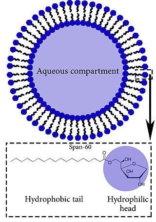
Niosomes are vesicles composed of non-ionic surfactants, incorporating cholesterol as an excipient. Niosomes are utilized for drug delivery to specific sites to achieve desired therapeutic effects. Structurally, niosomes are similar to liposomes as both consist of a lipid bilayer. However, niosomes are more stable than liposomes during formation processes and storage. Niosomes trap hydrophilic and lipophilic drugs, either in an aqueous compartment or in a vesicular membrane compartment composed of lipid material.

<span class="mw-page-title-main">Nanodisc</span> Synthetic model membrane system

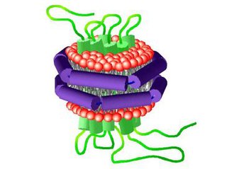
A nanodisc is a synthetic model membrane system which assists in the study of membrane proteins. Nanodiscs are discoidal proteins in which a lipid bilayer is surrounded by molecules that are amphipathic molecules including proteins, peptides, and synthetic polymers. It is composed of a lipid bilayer of phospholipids with the hydrophobic edge screened by two amphipathic proteins. These proteins are called membrane scaffolding proteins (MSP) and align in double belt formation. Nanodiscs are structurally very similar to discoidal high-density lipoproteins (HDL) and the MSPs are modified versions of apolipoprotein A1 (apoA1), the main constituent in HDL. Nanodiscs are useful in the study of membrane proteins because they can solubilise and stabilise membrane proteins and represent a more native environment than liposomes, detergent micelles, bicelles and amphipols.

<span class="mw-page-title-main">Lipid bilayer fusion</span>

In membrane biology, fusion is the process by which two initially distinct lipid bilayers merge their hydrophobic cores, resulting in one interconnected structure. If this fusion proceeds completely through both leaflets of both bilayers, an aqueous bridge is formed and the internal contents of the two structures can mix. Alternatively, if only one leaflet from each bilayer is involved in the fusion process, the bilayers are said to be hemifused. In hemifusion, the lipid constituents of the outer leaflet of the two bilayers can mix, but the inner leaflets remain distinct. The aqueous contents enclosed by each bilayer also remain separated.

A model lipid bilayer is any bilayer assembled in vitro, as opposed to the bilayer of natural cell membranes or covering various sub-cellular structures like the nucleus. They are used to study the fundamental properties of biological membranes in a simplified and well-controlled environment, and increasingly in bottom-up synthetic biology for the construction of artificial cells. A model bilayer can be made with either synthetic or natural lipids. The simplest model systems contain only a single pure synthetic lipid. More physiologically relevant model bilayers can be made with mixtures of several synthetic or natural lipids.

<span class="mw-page-title-main">Cell membrane</span> Biological membrane that separates the interior of a cell from its outside environment

The cell membrane is a biological membrane that separates and protects the interior of a cell from the outside environment. The cell membrane consists of a lipid bilayer, made up of two layers of phospholipids with cholesterols interspersed between them, maintaining appropriate membrane fluidity at various temperatures. The membrane also contains membrane proteins, including integral proteins that span the membrane and serve as membrane transporters, and peripheral proteins that loosely attach to the outer (peripheral) side of the cell membrane, acting as enzymes to facilitate interaction with the cell's environment. Glycolipids embedded in the outer lipid layer serve a similar purpose. The cell membrane controls the movement of substances in and out of a cell, being selectively permeable to ions and organic molecules. In addition, cell membranes are involved in a variety of cellular processes such as cell adhesion, ion conductivity, and cell signalling and serve as the attachment surface for several extracellular structures, including the cell wall and the carbohydrate layer called the glycocalyx, as well as the intracellular network of protein fibers called the cytoskeleton. In the field of synthetic biology, cell membranes can be artificially reassembled.

A vesosome is a multi-compartmental structure of lipidic nature used to deliver drugs. They can be considered multivesicular vesicles (MVV) and are, therefore, liposome-derived structures.

Nanoparticles for drug delivery to the brain is a method for transporting drug molecules across the blood–brain barrier (BBB) using nanoparticles. These drugs cross the BBB and deliver pharmaceuticals to the brain for therapeutic treatment of neurological disorders. These disorders include Parkinson's disease, Alzheimer's disease, schizophrenia, depression, and brain tumors. Part of the difficulty in finding cures for these central nervous system (CNS) disorders is that there is yet no truly efficient delivery method for drugs to cross the BBB. Antibiotics, antineoplastic agents, and a variety of CNS-active drugs, especially neuropeptides, are a few examples of molecules that cannot pass the BBB alone. With the aid of nanoparticle delivery systems, however, studies have shown that some drugs can now cross the BBB, and even exhibit lower toxicity and decrease adverse effects throughout the body. Toxicity is an important concept for pharmacology because high toxicity levels in the body could be detrimental to the patient by affecting other organs and disrupting their function. Further, the BBB is not the only physiological barrier for drug delivery to the brain. Other biological factors influence how drugs are transported throughout the body and how they target specific locations for action. Some of these pathophysiological factors include blood flow alterations, edema and increased intracranial pressure, metabolic perturbations, and altered gene expression and protein synthesis. Though there exist many obstacles that make developing a robust delivery system difficult, nanoparticles provide a promising mechanism for drug transport to the CNS.

A unilamellar liposome is a spherical liposome, a vesicle, bounded by a single bilayer of an amphiphilic lipid or a mixture of such lipids, containing aqueous solution inside the chamber. Unilamellar liposomes are used to study biological systems and to mimic cell membranes, and are classified into three groups based on their size: small unilamellar liposomes/vesicles (SUVs) that with a size range of 20–100 nm, large unilamellar liposomes/vesicles (LUVs) with a size range of 100–1000 nm and giant unilamellar liposomes/vesicles (GUVs) with a size range of 1–200 µm. GUVs are mostly used as models for biological membranes in research work. Animal cells are 10–30 µm and plant cells are typically 10–100 µm. Even smaller cell organelles such as mitochondria are typically 1–2 µm. Therefore, a proper model should account for the size of the specimen being studied. In addition, the size of vesicles dictates their membrane curvature which is an important factor in studying fusion proteins. SUVs have a higher membrane curvature and vesicles with high membrane curvature can promote membrane fusion faster than vesicles with lower membrane curvature such as GUVs.

<span class="mw-page-title-main">Lamellar phase</span>

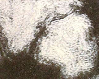
Lamellar phase refers generally to packing of polar-headed long chain nonpolar-tail molecules in an environment of bulk polar liquid, as sheets of bilayers separated by bulk liquid. In biophysics, polar lipids pack as a liquid crystalline bilayer, with hydrophobic fatty acyl long chains directed inwardly and polar headgroups of lipids aligned on the outside in contact with water, as a 2-dimensional flat sheet surface. Under transmission electron microscope (TEM), after staining with polar headgroup reactive chemical osmium tetroxide, lamellar lipid phase appears as two thin parallel dark staining lines/sheets, constituted by aligned polar headgroups of lipids. 'Sandwiched' between these two parallel lines, there exists one thicker line/sheet of non-staining closely packed layer of long lipid fatty acyl chains. This TEM-appearance became famous as Robertson's unit membrane - the basis of all biological membranes, and structure of lipid bilayer in unilamellar liposomes. In multilamellar liposomes, many such lipid bilayer sheets are layered concentrically with water layers in between.

<span class="mw-page-title-main">Liposome extruder</span> Lab equipment

A liposome extruder is a device that prepares cell membranes, exosomes and also generates nanoscale liposome formulations. The liposome extruder employs the track-etched membrane to filter huge particles and achieve sterile filtration.

<span class="mw-page-title-main">Ligand-targeted liposome</span> Ligand-targeted liposomes for use in medical applications

A ligand-targeted liposome (LTL) is a nanocarrier with specific ligands attached to its surface to enhance localization for targeted drug delivery. The targeting ability of LTLs enhances cellular localization and uptake of these liposomes for therapeutic or diagnostic purposes. LTLs have the potential to enhance drug delivery by decreasing peripheral systemic toxicity, increasing in vivo drug stability, enhancing cellular uptake, and increasing efficiency for chemotherapeutics and other applications. Liposomes are beneficial in therapeutic manufacturing because of low batch-to-batch variability, easy synthesis, favorable scalability, and strong biocompatibility. Ligand-targeting technology enhances liposomes by adding targeting properties for directed drug delivery.

<span class="mw-page-title-main">Invasomes</span> Transdermal drug delivery method

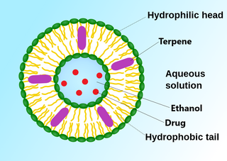
An invasome are a type of artificial vesicle nanocarrier that transport substances through the skin, the most superficial biological barrier. Vesicles are small particles surrounded by a lipid layer that can carry substances into and out of the cell. Artificial vesicles can be engineered to deliver drugs within the cell, with specific applications within transdermal drug delivery. However, the skin proves to be a barrier to effective penetration and delivery of drug therapies. Thus, invasomes are a new generation of vesicle with added structural components to assist with skin penetration.

Immunoliposome therapy is a targeted drug delivery method that involves the use of liposomes coupled with monoclonal antibodies to deliver therapeutic agents to specific sites or tissues in the body. The antibody modified liposomes target tissue through cell-specific antibodies with the release of drugs contained within the assimilated liposomes. Immunoliposome aims to improve drug stability, personalize treatments, and increased drug efficacy. This form of therapy has been used to target specific cells, protecting the encapsulated drugs from degradation in order to enhance their stability, to facilitate sustained drug release and hence to advance current traditional cancer treatment.

References

  1. 1 2 Torchilin, V (2006). "Multifunctional nanocarriers". Advanced Drug Delivery Reviews. 58 (14): 1532–55. doi:10.1016/j.addr.2006.09.009. PMID   17092599. S2CID   9464592.
  2. 1 2 3 Akbarzadeh, A.; Rezaei-Sadabady, R.; Davaran, S.; Joo, S. W.; Zarghami, N.; Hanifehpour, Y.; Samiei, M.; Kouhi, M.; Nejati-Koshki, K. (22 February 2013). "Liposome: classification, preparation, and applications". Nanoscale Research Letters . 8 (1): 102. Bibcode:2013NRL.....8..102A. doi: 10.1186/1556-276X-8-102 . ISSN   1931-7573. PMC   3599573 . PMID   23432972.
  3. "Cell Membranes - Kimball's Biology Pages". 16 August 2002. Archived from the original on 25 January 2009.
  4. Mashaghi S., et al. Lipid Nanotechnology. Int J Mol Sci. 2013 Feb; 14(2): 4242–4282.
  5. 1 2 3 Cevc, G (1993). "Rational design of new product candidates: the next generation of highly deformable bilayer vesicles for noninvasive, targeted therapy". Journal of Controlled Release. 160 (2): 135–146. doi:10.1016/j.jconrel.2012.01.005. PMID   22266051.
  6. Moghassemi, Saeid; Dadashzadeh, Arezoo; Azevedo, Ricardo Bentes; Feron, Olivier; Amorim, Christiani A. (November 2021). "Photodynamic cancer therapy using liposomes as an advanced vesicular photosensitizer delivery system". Journal of Controlled Release. 339: 75–90. doi:10.1016/j.jconrel.2021.09.024. PMID   34562540. S2CID   237636495.
  7. Moghassemi, Saeid; Dadashzadeh, Arezoo; Azevedo, Ricardo Bentes; Feron, Olivier; Amorim, Christiani A. (November 2021). "Photodynamic cancer therapy using liposomes as an advanced vesicular photosensitizer delivery system". Journal of Controlled Release. 339: 75–90. doi:10.1016/j.jconrel.2021.09.024. PMID   34562540. S2CID   237636495.
  8. 1 2 Stryer S. (1981) Biochemistry, 213
  9. Mashaghi S., et al. Lipid Nanotechnology. Int J Mol Sci. 2013 Feb; 14(2): 4242–4282.
  10. Bangham, A. D.; Horne, R. W. (1964). "Negative Staining of Phospholipids and Their Structural Modification by Surface-Active Agents As Observed in the Electron Microscope". Journal of Molecular Biology. 8 (5): 660–668. doi:10.1016/S0022-2836(64)80115-7. PMID   14187392.
  11. Horne, R. W.; Bangham, A. D.; Whittaker, V. P. (1963). "Negatively Stained Lipoprotein Membranes". Nature. 200 (4913): 1340. Bibcode:1963Natur.200.1340H. doi: 10.1038/2001340a0 . PMID   14098499. S2CID   4153775.
  12. Bangham, A. D.; Horne, R. W.; Glauert, A. M.; Dingle, J. T.; Lucy, J. A. (1962). "Action of saponin on biological cell membranes". Nature. 196 (4858): 952–955. Bibcode:1962Natur.196..952B. doi:10.1038/196952a0. PMID   13966357. S2CID   4181517.
  13. Bangham A.D.; Standish M.M.; Weissmann G. (1965). "The action of steroids and streptolysin S on the permeability of phospholipid structures to cations". J. Molecular Biol. 13 (1): 253–259. doi:10.1016/s0022-2836(65)80094-8. PMID   5859040.
  14. Sessa G.; Weissmann G. (1970). "Incorporation of lysozyme into liposomes: A model for structure-linked latency". J. Biol. Chem. 245 (13): 3295–3301. doi: 10.1016/S0021-9258(18)62994-1 . PMID   5459633.
  15. YashRoy R.C. (1990). "Lamellar dispersion and phase separation of chloroplast membrane lipids by negative staining electron microscopy" (PDF). Journal of Biosciences. 15 (2): 93–98. doi:10.1007/bf02703373. S2CID   39712301.
  16. Weissmann G.; Sessa G.; Standish M.; Bangham A. D. (1965). "ABSTRACTS". J. Clin. Invest. 44 (6): 1109–1116. doi: 10.1172/jci105203 . PMC   539946 .
  17. Geoff Watts (2010-06-12). "Alec Douglas Bangham". The Lancet. 375 (9731): 2070. doi:10.1016/S0140-6736(10)60950-6. S2CID   54382511 . Retrieved 2014-10-01.
  18. 1 2 Mayer, Lawrence D.; Bally, Marcel B.; Hope, Michael J.; Cullis, Pieter R. (1 June 1986). "Techniques for encapsulating bioactive agents into liposomes". Chemistry and Physics of Lipids. 40 (2): 333–345. doi:10.1016/0009-3084(86)90077-0. ISSN   0009-3084. PMID   3742676.
  19. Chaize, Barnabé; Colletier, Jacques-Philippe; Winterhalter, Mathias; Fournier, Didier (January 2004). "Encapsulation of Enzymes in Liposomes: High Encapsulation Efficiency and Control of Substrate Permeability". Artificial Cells, Blood Substitutes, and Biotechnology. 32 (1): 67–75. doi: 10.1081/BIO-120028669 . ISSN   1073-1199. PMID   15027802. S2CID   21897676.
  20. Colletier, Jacques-Philippe; Chaize, Barnabé; Winterhalter, Mathias; Fournier, Didier (10 May 2002). "Protein encapsulation in liposomes: efficiency depends on interactions between protein and phospholipid bilayer". BMC Biotechnology. 2 (1): 9. doi: 10.1186/1472-6750-2-9 . ISSN   1472-6750. PMC   113741 . PMID   12003642.
  21. Edwards, Katie A.; Baeumner, Antje J. (28 February 2006). "Analysis of liposomes". Talanta. 68 (5): 1432–1441. doi:10.1016/j.talanta.2005.08.031. ISSN   0039-9140. PMID   18970482.
  22. Longatte, Guillaume; Lisi, Fabio; Chen, Xueqian; Walsh, James; Wang, Wenqian; Ariotti, Nicholas; Boecking, Till; Gaus, Katharina; Gooding, J. Justin (23 November 2022). "Statistical predictions on the encapsulation of single molecule binding pairs into sized-dispersed nanocontainers". Physical Chemistry Chemical Physics. 24 (45): 28029–28039. Bibcode:2022PCCP...2428029L. doi:10.1039/D2CP03627D. ISSN   1463-9084. PMID   36373851.
  23. Cevc, G; Richardsen, H (1993). "Lipid vesicles and membrane fusion". Advanced Drug Delivery Reviews. 38 (3): 207–232. doi:10.1016/s0169-409x(99)00030-7. PMID   10837758.
  24. 1 2 Barenholz, Y; G, Cevc (2000). Physical chemistry of biological surfaces, Chapter 7: Structure and properties of membranes. New York: Marcel Dekker. pp. 171–241.
  25. Bertrand, Nicolas; Bouvet, CéLine; Moreau, Pierre; Leroux, Jean-Christophe (2010). "Transmembrane pH-Gradient Liposomes to Treat Cardiovascular Drug Intoxication". ACS Nano. 4 (12): 7552–8. doi:10.1021/nn101924a. PMID   21067150.
  26. 1 2 3 4 5 Paliwal, Shivani Rai; Paliwal, Rishi; Vyas, Suresh P. (2015-04-03). "A review of mechanistic insight and application of pH-sensitive liposomes in drug delivery". Drug Delivery. 22 (3): 231–242. doi:10.3109/10717544.2014.882469. ISSN   1071-7544.
  27. Anonymous (2018-09-17). "EU/3/07/451". European Medicines Agency. Retrieved 2020-01-10.
  28. Barani, H; Montazer, M (2008). "A review on applications of liposomes in textile processing". Journal of Liposome Research. 18 (3): 249–62. doi:10.1080/08982100802354665. PMID   18770074. S2CID   137500401.
  29. Meure, LA; Knott, R; Foster, NR; Dehghani, F (2009). "The depressurization of an expanded solution into aqueous media for the bulk production of liposomes". Langmuir: The ACS Journal of Surfaces and Colloids. 25 (1): 326–37. doi:10.1021/la802511a. PMID   19072018.
  30. Yoko Shojia; Hideki Nakashima (2004). "Nutraceutics and Delivery Systems". Journal of Drug Targeting.
  31. Williamson, G; Manach, C (2005). "Bioavailability and bioefficacy of polyphenols in humans. II. Review of 93 intervention studies". The American Journal of Clinical Nutrition. 81 (1 Suppl): 243S–255S. doi: 10.1093/ajcn/81.1.243S . PMID   15640487.
  32. Bender, David A. (2003). Nutritional Biochemistry of Vitamins. Cambridge, U.K.: Cambridge University Press. OCLC   57204737.
  33. DeFelice, Stephen L. (February 1995). "The nutraceutical revolution: its impact on food industry R&D". Trends in Food Science & Technology. 6 (2): 59–61. doi:10.1016/s0924-2244(00)88944-x. ISSN   0924-2244.
  34. Santini, Antonello; Cammarata, Silvia Miriam; Capone, Giacomo; Ianaro, Angela; Tenore, Gian Carlo; Pani, Luca; Novellino, Ettore (2018-02-14). "Nutraceuticals: opening the debate for a regulatory framework". British Journal of Clinical Pharmacology. 84 (4): 659–672. doi: 10.1111/bcp.13496 . ISSN   0306-5251. PMC   5867125 . PMID   29433155.
  35. Porter, Christopher J. H.; Trevaskis, Natalie L.; Charman, William N. (March 2007). "Lipids and lipid-based formulations: optimizing the oral delivery of lipophilic drugs". Nature Reviews Drug Discovery. 6 (3): 231–248. doi:10.1038/nrd2197. ISSN   1474-1776. PMID   17330072. S2CID   29805601.
  36. Szoka Jr, F; Papahadjopoulos, D (1980). "Comparative properties and methods of preparation of lipid vesicles (liposomes)". Annual Review of Biophysics and Bioengineering. 9: 467–508. doi:10.1146/annurev.bb.09.060180.002343. PMID   6994593.
  37. Gomezhens, A; Fernandezromero, J (2006). "Analytical methods for the control of liposomal delivery systems". TrAC Trends in Analytical Chemistry. 25 (2): 167–178. doi:10.1016/j.trac.2005.07.006.
  38. Mozafari, MR; Johnson, C; Hatziantoniou, S; Demetzos, C (2008). "Nanoliposomes and their applications in food nanotechnology". Journal of Liposome Research. 18 (4): 309–27. doi:10.1080/08982100802465941. PMID   18951288. S2CID   98836972.
  39. Jahn, Andreas; Stavis, Samuel M.; Hong, Jennifer S.; Vreeland, Wyatt N.; DeVoe, Don L.; Gaitan, Michael (2010-04-27). "Microfluidic Mixing and the Formation of Nanoscale Lipid Vesicles". ACS Nano. 4 (4): 2077–2087. doi:10.1021/nn901676x. ISSN   1936-0851. PMID   20356060.
  40. Zhigaltsev, Igor V.; Belliveau, Nathan; Hafez, Ismail; Leung, Alex K. K.; Huft, Jens; Hansen, Carl; Cullis, Pieter R. (2012-02-21). "Bottom-Up Design and Synthesis of Limit Size Lipid Nanoparticle Systems with Aqueous and Triglyceride Cores Using Millisecond Microfluidic Mixing". Langmuir. 28 (7): 3633–3640. doi:10.1021/la204833h. ISSN   0743-7463. PMID   22268499.
  41. López, Rubén R.; Ocampo, Ixchel; Sánchez, Luz-María; Alazzam, Anas; Bergeron, Karl-F.; Camacho-León, Sergio; Mounier, Catherine; Stiharu, Ion; Nerguizian, Vahé (2020-02-25). "Surface Response Based Modeling of Liposome Characteristics in a Periodic Disturbance Mixer". Micromachines. 11 (3): 235. doi: 10.3390/mi11030235 . ISSN   2072-666X. PMC   7143066 . PMID   32106424.
  42. Colas, JC; Shi, W; Rao, VS; Omri, A; Mozafari, MR; Singh, H (2007). "Microscopical investigations of nisin-loaded nanoliposomes prepared by Mozafari method and their bacterial targeting". Micron. 38 (8): 841–7. doi:10.1016/j.micron.2007.06.013. PMID   17689087.
  43. Blume, G; Cevc, G (1990). "Liposomes for the sustained drug release in vivo". Biochimica et Biophysica Acta (BBA) - Biomembranes. 1029 (1): 92–97. doi:10.1016/0005-2736(90)90440-y. PMID   2223816.
  44. Klibanov, AL; Maruyama, K; Torchilin, VP; Huang, L (1990). "Amphipathic polyethyleneglycols effectively prolong the circulation time of liposomes". FEBS Letters. 268 (1): 235–237. doi: 10.1016/0014-5793(90)81016-h . PMID   2384160. S2CID   11437990.
  45. Wang, XinYu; Ishida, Tatsuhiro; Kiwada, Hiroshi (2007-06-01). "Anti-PEG IgM elicited by injection of liposomes is involved in the enhanced blood clearance of a subsequent dose of PEGylated liposomes". Journal of Controlled Release. 119 (2): 236–244. doi:10.1016/j.jconrel.2007.02.010. ISSN   0168-3659. PMID   17399838.
  46. Dams, E.T.M.; Laverman, P.; Oyen, W.J.G.; Storm, G.; Scherphof, G.L.; Meer, J.W.M.; van der Corstens, F.H.M.; Boerman, O.C. (March 2000). "Accelerated blood clearance and altered biodistribution of repeated injections of sterically stabilized liposomes". The Journal of Pharmacology and Experimental Therapeutics. 292 (3). Amer soc pharmacology experimental therapeutics: 1071–9. PMID   10688625.
  47. Blume, G; Cevc, G; Crommelin, M D A J; Bakker-Woudenberg, I A J M; Kluft, C; Storm, G (1993). "Specific targeting with poly (ethylene glycol)-modified liposomes: coupling of homing devices to the ends of the polymeric chains combines effective target binding with long circulation times". Biochimica et Biophysica Acta (BBA) - Biomembranes. 1149 (1): 180–184. doi:10.1016/0005-2736(93)90039-3. PMID   8318529.
  48. Cevc, G (2004). "Lipid vesicles and other colloids as drug carriers on the skin". Advanced Drug Delivery Reviews. 56 (5): 675–711. doi:10.1016/j.addr.2003.10.028. PMID   15019752.
  49. Besançon, Hervé; Babiychuk, Viktoriia; Larpin, Yu; Köffel, René; Schittny, Dominik; Brockhus, Lara; Hathaway, Lucy J.; Sendi, Parham; Draeger, Annette; Babiychuk, Eduard (14 February 2021). "Tailored liposomal nanotraps for the treatment of Streptococcal infections". Journal of Nanobiotechnology. 19 (1): 46. doi: 10.1186/s12951-021-00775-x . PMC   7885208 . PMID   33588835..
  50. Karny, Avishai; Zinger, Assaf; Kajal, Ashima; Shainsky-Roitman, Janna; Schroeder, Avi (2018-05-17). "Therapeutic nanoparticles penetrate leaves and deliver nutrients to agricultural crops". Scientific Reports. 8 (1): 7589. Bibcode:2018NatSR...8.7589K. doi:10.1038/s41598-018-25197-y. ISSN   2045-2322. PMC   5958142 . PMID   29773873.
  51. Temming, Maria (2018-05-17). "Nanoparticles could help rescue malnourished crops". Science News. Retrieved 2018-05-18.
  52. John-Herpin, Aurelian; Kavungal, Deepthy; von Mücke, Lea; Altug, Hatice (2020). "Infrared Metasurface Augmented by Deep Learning for Monitoring Dynamics between All Major Classes of Biomolecules". Advanced Materials. 33 (14): e2006054. doi: 10.1002/adma.202006054 . PMID   33615570.
  53. Arulsudar, N.; Subramanian, N.; Murthy, R. S. R. (2005). "Comparison of artificial neural network and multiple linear regression in the optimization of formulation parameters of leuprolide acetate loaded liposomes". J Pharm Pharm Sci. 8 (2): 243–258. PMID   16124936.
  54. Sansare, Sameera; Duran, Tibo; Mohammadiarani, Hossein; Goyal, Manish; Yenduri, Gowtham; Costa, Antonio; Xu, Xiaoming; O'Connor, Thomas; Burgess, Diane; Chaudhuri, Bodhisattwa (2021). "Artificial neural networks in tandem with molecular descriptors as predictive tools for continuous liposome manufacturing". International Journal of Pharmaceutics. 603 (120713): 120713. doi:10.1016/j.ijpharm.2021.120713. PMID   34019974. S2CID   235093636.