Protein metabolism

Last updated

Protein metabolism denotes the various biochemical processes responsible for the synthesis of proteins and amino acids (anabolism), and the breakdown of proteins by catabolism.

Contents

The steps of protein synthesis include transcription, translation, and post translational modifications. During transcription, RNA polymerase transcribes a coding region of the DNA in a cell producing a sequence of RNA, specifically messenger RNA (mRNA). This mRNA sequence contains codons: 3 nucleotide long segments that code for a specific amino acid. Ribosomes translate the codons to their respective amino acids. [1] In humans, non-essential amino acids are synthesized from intermediates in major metabolic pathways such as the Citric Acid Cycle. [2] Essential amino acids must be consumed and are made in other organisms. The amino acids are joined by peptide bonds making a polypeptide chain. This polypeptide chain then goes through post translational modifications and is sometimes joined with other polypeptide chains to form a fully functional protein.

Dietary proteins are first broken down to individual amino acids by various enzymes and hydrochloric acid present in the gastrointestinal tract. These amino acids are absorbed into the bloodstream to be transported to the liver and onward to the rest of the body. Absorbed amino acids are typically used to create functional proteins, but may also be used to create energy. [3] They can also be converted into glucose. [4] This glucose can then be converted to triglycerides and stored in fat cells. [5]

Proteins can be broken down by enzymes known as peptidases or can break down as a result of denaturation. Proteins can denature in environmental conditions the protein is not made for. [6]

Protein synthesis

Protein anabolism is the process by which proteins are formed from amino acids. It relies on five processes: amino acid synthesis, transcription, translation, post translational modifications, and protein folding. Proteins are made from amino acids. In humans, some amino acids can be synthesized using already existing intermediates. These amino acids are known as non-essential amino acids. Essential amino acids require intermediates not present in the human body. These intermediates must be ingested, mostly from eating other organisms. [6]  

Amino Acid Synthesis

Pathways that form each amino acid [7]
Amino Acid R-group Pathway*
Glycine H- Serine + THF Glycine ( hydroxymethyltransferase)
Alanine CH3- Pyruvate Alanine ( aminotransferase)
Valine §(CH3)2-CH-Hydroxyethyl-TPP + Pyruvate → α-acetolactate → Valine
Leucine §(CH3)2-CH-CH2-Hydroxyethyl-TPP + Pyruvate → α-ketobutyrate → Leucine
Isoleucine §CH3-CH2-CH(CH3)-Hydroxyethyl-TPP + Pyruvate → α-acetolactate → Isoleucine
Methionine §CH3-S-(CH2)2- Homocysteine Methionine ( methionine synthase)
Proline -(CH2)3- Glutamic Acid Glutamate-5-semialdehydeProline (γ-glutamyl kinase)
Phenylalanine §Ph-CH2- Phosphoenolpyruvate → 2-keto-3-deoxy arabino heptulosonate-7-phosphate → ChorismatePhenylalanine
Tryptophan §Ph-NH-CH=C-CH2-Phosphoenolpyruvate → 2-keto-3-deoxy arabino heptulosonate-7-phosphate → ChorismateTryptophan
Tyrosine HO-Ph-CH2- PhenylalanineTyrosine ( phenylalanine hydroxylase )
Serine HO-CH2- 3-phosphoglycerate3-phosphohydroxypyruvate ( 3-phosphoglycerate dehydrogenase)3-phosphoserine ( aminotransferase)Serine ( phosphoserine phosphatase)
Threonine §CH3-CH(OH)- Aspartate → β-aspartate-semialdehyde → HomoserineThreonine
Cysteine HS-CH2- SerineCystathionineα-ketobutyrateCysteine
Asparagine H2N-CO-CH2- Aspartic Acid Asparagine ( asparagine synthetase)
Glutamine H2N-CO-(CH2)2- Glutamic Acid Glutamine ( glutamine synthetase)
Arginine +H2N=C(NH2)-NH-(CH2)3- Glutamate Glutamate-5-semialdehyde (γ-glutamyl kinase)Arginine
Histidine §NH-CH=N-CH=C-CH2- GlucoseGlucose-6-phosphateRibose-5-phosphateHistidine
Lysine §+H3N-(CH2)4- Aspartate → β-aspartate-semialdehyde → Homoserine + lysine
Aspartic Acid OOC-CH2- OxaloacetateAspartic Acid ( aminotransferase)
Glutamic Acid OOC-(CH2)2- α-ketoglutarate Glutamic Acid ( aminotransferase)
Shown at physiological conditions.

*Complexes that are italicized are enzymes.

§Cannot be synthesized in humans.

Polypeptide synthesis

Transcription

DNA is transcribed to mRNA which is translated into amino acids. Protein Transcription&Translation.jpg
DNA is transcribed to mRNA which is translated into amino acids.

In transcription, RNA polymerase reads a DNA strand and produces an mRNA strand that can be further translated. In order to initiate transcription, the DNA segment that is to be transcribed must be accessible (i.e. it cannot be tightly packed). Once the DNA segment is accessible, the RNA polymerase can begin to transcribe the coding DNA strand by pairing RNA nucleotides to the template DNA strand. During the initial transcription phase, the RNA polymerase searches for a promoter region on the DNA template strand. Once the RNA polymerase binds to this region, it begins to “read” the template DNA strand in the 3’ to 5’ direction. [8] RNA polymerase attaches RNA bases complementary to the template DNA strand (Uracil will be used instead of Thymine). The new nucleotide bases are bonded to each other covalently. [9] The new bases eventually dissociate from the DNA bases but stay linked to each other, forming a new mRNA strand. This mRNA strand is synthesized in the 5’ to 3’ direction. [10] Once the RNA reaches a terminator sequence, it dissociates from the DNA template strand and terminates the mRNA sequence as well.

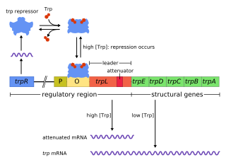
Transcription is regulated in the cell via transcription factors. Transcription factors are proteins that bind to regulatory sequences in the DNA strand such as promoter regions or operator regions. Proteins bound to these regions can either directly halt or allow RNA polymerase to read the DNA strand or can signal other proteins to halt or allow RNA polymerase reading. [11]

Translation

Formation of a dipeptide via a peptide bond. Peptidformationball.svg
Formation of a dipeptide via a peptide bond.

During translation, ribosomes convert a sequence of mRNA (messenger RNA) to an amino acid sequence. Each 3-base-pair-long segment of mRNA is a codon which corresponds to one amino acid or stop signal. [12] Amino acids can have multiple codons that correspond to them. Ribosomes do not directly attach amino acids to mRNA codons. They must utilize tRNAs (transfer RNAs) as well. Transfer RNAs can bind to amino acids and contain an anticodon which can hydrogen bind to an mRNA codon. [13] The process of bind an amino acid to a tRNA is known as tRNA charging. Here, the enzyme aminoacyl-tRNA-synthetase catalyzes two reactions. In the first one, it attaches an AMP molecule (cleaved from ATP) to the amino acid. The second reaction cleaves the aminoacyl-AMP producing the energy to join the amino acid to the tRNA molecule. [14]

Ribosomes have two subunits, one large and one small. These subunits surround the mRNA strand. The larger subunit contains three binding sites: A (aminoacyl), P (peptidyl), and E (exit). After translational initiation (which is different in prokaryotes and eukaryotes), the ribosome enters the elongation period which follows a repetitive cycle. First a tRNA with the correct amino acid enters the A site. The ribosome transfers the peptide from the tRNA in the P site to the new amino acid on the tRNA in the A site. The tRNA from the P site will be shifted into the E site where it will be ejected. This continually occurs until the ribosome reaches a stop codon or receives a signal to stop. [13] A peptide bond forms between the amino acid attached to the tRNA in the P site and the amino acid attached to a tRNA in the A site. The formation of a peptide bond requires an input of energy. The two reacting molecules are the alpha amino group of one amino acid and the alpha carboxyl group of the other amino acids. A by-product of this bond formation is the release of water (the amino group donates a proton while the carboxyl group donates a hydroxyl). [2]

Translation can be downregulated by miRNAs (microRNAs). These RNA strands can cleave mRNA strands they are complementary to and will thus stop translation. [15] Translation can also be regulated via helper proteins. For example, a protein called eukaryotic initiation factor-2 (eIF-2) can bind to the smaller subunit of the ribosome, starting translation. When elF-2 is phosphorylated, it cannot bind to the ribosome and translation is halted. [16]

Post-translational Modifications

Methylation of Lysine (amino acid) General Lysine Methylation Mechanism.svg
Methylation of Lysine (amino acid)

Once the peptide chain is synthesized, it still must be modified. Post-translational modifications can occur before protein folding or after. Common biological methods of modifying peptide chains after translation include methylation, phosphorylation, and disulfide bond formation. Methylation often occurs to arginine or lysine and involves adding a methyl group to a nitrogen (replacing a hydrogen). The R groups on these amino acids can be methylated multiple times as long as the bonds to nitrogen does not exceed 4. Methylation reduces the ability of these amino acids to form hydrogen bonds so arginine and lysine that are methylated have different properties than their standard counterparts. Phosphorylation often occurs to serine, threonine, and tyrosine and involves replacing a hydrogen on the alcohol group at the terminus of the R group with a phosphate group. This adds a negative charge on the R groups and will thus change how the amino acids behave in comparison to their standard counterparts. Disulfide bond formation is the creation of disulfide bridges (covalent bonds) between two cysteine amino acids in a chain which adds stability to the folded structure. [17]

Protein folding

A polypeptide chain in the cell does not have to stay linear; it can become branched or fold in on itself. Polypeptide chains fold in a particular manner depending on the solution they are in. The fact that all amino acids contain R groups with different properties is the main reason proteins fold. In a hydrophilic environment such as cytosol, the hydrophobic amino acids will concentrate at the core of the protein, while the hydrophilic amino acids will be on the exterior. This is entropically favorable since water molecules can move much more freely around hydrophilic amino acids than hydrophobic amino acids. In a hydrophobic environment, the hydrophilic amino acids will concentrate at the core of the protein, while the hydrophobic amino acids will be on the exterior. Since the new interactions between the hydrophilic amino acids are stronger than hydrophobic-hydrophilic interactions, this is enthalpically favorable. [18] Once a polypeptide chain is fully folded, it is called a protein. Often many subunits will combine to make a fully functional protein although physiological proteins do exist that contain only one polypeptide chain. Proteins may also incorporate other molecules such as the heme group in hemoglobin, a protein responsible for carrying oxygen in the blood. [19]

Protein breakdown

Protein catabolism is the process by which proteins are broken down to their amino acids. This is also called proteolysis and can be followed by further amino acid degradation.

Protein catabolism via enzymes

Proteases

Originally thought to only disrupt enzymatic reactions, proteases (also known as peptidases) actually help with catabolizing proteins through cleavage and creating new proteins that were not present before. Proteases also help to regulate metabolic pathways. One way they do this is to cleave enzymes in pathways that do not need to be running (i.e. gluconeogenesis when blood glucose concentrations are high). This helps to conserve as much energy as possible and to avoid futile cycles. Futile cycles occur when the catabolic and anabolic pathways are both in effect at the same time and rate for the same reaction. Since the intermediates being created are consumed, the body makes no net gain. Energy is lost through futile cycles. Proteases prevent this cycle from occurring by altering the rate of one of the pathways, or by cleaving a key enzyme, they can stop one of the pathways. Proteases are also nonspecific when binding to substrate, allowing for great amounts of diversity inside the cells and other proteins, as they can be cleaved much easier in an energy efficient manner. [20]

Possible mechanism for Aspartyl Protease cleaving a peptide bond. Only the peptide bond and active site are shown. Aspartyl protease mechanism.png
Possible mechanism for Aspartyl Protease cleaving a peptide bond. Only the peptide bond and active site are shown.

Because many proteases are nonspecific, they are highly regulated in the cell. Without regulation, proteases will destroy many proteins which are essential to physiological processes. One way the body regulates proteases is through protease inhibitors. Protease inhibitors can be other proteins, small peptides, or molecules. There are two types of protease inhibitors: reversible and irreversible. Reversible protease inhibitors form non-covalent interactions with the protease limiting its functionality. They can be competitive inhibitors, uncompetitive inhibitors, and noncompetitive inhibitors. Competitive inhibitors compete with the peptide to bind to the protease active site. Uncompetitive inhibitors bind to the protease while the peptide is bound but do not let the protease cleave the peptide bond. Noncompetitive inhibitors can do both. Irreversible protease inhibitors covalently modify the active site of the protease so it cannot cleave peptides. [21]

Exopeptidases

Exopeptidases are enzymes that can cleave the end of an amino acid side chain mostly through the addition of water. [6] Exopeptidase enzymes exist in the small intestine. These enzymes have two classes: aminopeptidases are a brush border enzyme and carboxypeptidases which is from the pancreas. Aminopeptidases are enzymes that remove amino acids from the amino terminus of protein. They are present in all lifeforms and are crucial for survival since they do many cellular tasks in order to maintain stability. This form of peptidase is a zinc metalloenzyme and it is inhibited by the transition state analog. This analog is similar to the actual transition state, so it can make the enzyme bind to it instead of the actual transition state, thus preventing substrate binding and decreasing reaction rates. [22] Carboxypeptidases cleave at the carboxyl end of the protein. While they can catabolize proteins, they are more often used in post-transcriptional modifications. [23]

Endopeptidases

Endopeptidases are enzymes that add water to an internal peptide bond in a peptide chain and break that bond. [6] Three common endopeptidases that come from the pancreas are pepsin, trypsin, and chymotrypsin. Chymotrypsin performs a hydrolysis reaction that cleaves after aromatic residues. The main amino acids involved are serine, histidine, and aspartic acid. They all play a role in cleaving the peptide bond. These three amino acids are known as the catalytic triad which means that these three must all be present in order to properly function. [6] Trypsin cleaves after long positively charged residues and has a negatively charged binding pocket at the active site. Both are produced as zymogens, meaning they are initially found in their inactive state and after cleavage though a hydrolysis reaction, they becomes activated. [2] Non-covalent interactions such as hydrogen bonding between the peptide backbone and the catalytic triad help increase reaction rates, allowing these peptidases to cleave many peptides efficiently. [6]

Protein catabolism via environmental changes

pH

Cellular proteins are held in a relatively constant pH in order to prevent changes in the protonation state of amino acids. [24] If the pH drops, some amino acids in the polypeptide chain can become protonated if the pka of their R groups is higher than the new pH. Protonation can change the charge these R groups have. If the pH raises, some amino acids in the chain can become deprotonated (if the pka of the R group is lower than the new pH). This also changes the R group charge. Since many amino acids interact with other amino acids based on electrostatic attraction, changing the charge can break these interactions. The loss of these interactions alters the proteins structure, but most importantly it alters the proteins function, which can be beneficial or detrimental. A significant change in pH may even disrupt many interactions the amino acids make and denature (unfold) the protein. [24]

Temperature

As the temperature in the environment increases, molecules move faster. Hydrogen bonds and hydrophobic interactions are important stabilizing forces in proteins. If the temperature rises and molecules containing these interactions are moving too fast, the interactions become compromised or even break. At high temperatures, these interactions cannot form, and a functional protein is denatured. [25] However, it relies on two factors; the type of protein used and the amount of heat applied. The amount of heat applied determines whether this change in protein is permanent or if it can be transformed back to its original form. [26]

Related Research Articles

<span class="mw-page-title-main">Messenger RNA</span> RNA that is read by the ribosome to produce a protein

In molecular biology, messenger ribonucleic acid (mRNA) is a single-stranded molecule of RNA that corresponds to the genetic sequence of a gene, and is read by a ribosome in the process of synthesizing a protein.

<span class="mw-page-title-main">Protein primary structure</span> Linear sequence of amino acids in a peptide or protein

Protein primary structure is the linear sequence of amino acids in a peptide or protein. By convention, the primary structure of a protein is reported starting from the amino-terminal (N) end to the carboxyl-terminal (C) end. Protein biosynthesis is most commonly performed by ribosomes in cells. Peptides can also be synthesized in the laboratory. Protein primary structures can be directly sequenced, or inferred from DNA sequences.

<span class="mw-page-title-main">Protein biosynthesis</span> Assembly of proteins inside biological cells

Protein biosynthesis is a core biological process, occurring inside cells, balancing the loss of cellular proteins through the production of new proteins. Proteins perform a number of critical functions as enzymes, structural proteins or hormones. Protein synthesis is a very similar process for both prokaryotes and eukaryotes but there are some distinct differences.

<span class="mw-page-title-main">Proteolysis</span> Breakdown of proteins into smaller polypeptides or amino acids

Proteolysis is the breakdown of proteins into smaller polypeptides or amino acids. Uncatalysed, the hydrolysis of peptide bonds is extremely slow, taking hundreds of years. Proteolysis is typically catalysed by cellular enzymes called proteases, but may also occur by intra-molecular digestion.

<span class="mw-page-title-main">Central dogma of molecular biology</span> Explanation of the flow of genetic information within a biological system

The central dogma of molecular biology is an explanation of the flow of genetic information within a biological system. It is often stated as "DNA makes RNA, and RNA makes protein", although this is not its original meaning. It was first stated by Francis Crick in 1957, then published in 1958:

The Central Dogma. This states that once "information" has passed into protein it cannot get out again. In more detail, the transfer of information from nucleic acid to nucleic acid, or from nucleic acid to protein may be possible, but transfer from protein to protein, or from protein to nucleic acid is impossible. Information here means the precise determination of sequence, either of bases in the nucleic acid or of amino acid residues in the protein.

<span class="mw-page-title-main">Active site</span> Active region of an enzyme

In biology and biochemistry, the active site is the region of an enzyme where substrate molecules bind and undergo a chemical reaction. The active site consists of amino acid residues that form temporary bonds with the substrate, the binding site, and residues that catalyse a reaction of that substrate, the catalytic site. Although the active site occupies only ~10–20% of the volume of an enzyme, it is the most important part as it directly catalyzes the chemical reaction. It usually consists of three to four amino acids, while other amino acids within the protein are required to maintain the tertiary structure of the enzymes.

<span class="mw-page-title-main">Translation (biology)</span> Cellular process of protein synthesis

In biology, translation is the process in living cells in which proteins are produced using RNA molecules as templates. The generated protein is a sequence of amino acids. This sequence is determined by the sequence of nucleotides in the RNA. The nucleotides are considered three at a time. Each such triple results in addition of one specific amino acid to the protein being generated. The matching from nucleotide triple to amino acid is called the genetic code. The translation is performed by a large complex of functional RNA and proteins called ribosomes. The entire process is called gene expression.

<span class="mw-page-title-main">Biomolecule</span> Molecule that is produced by a living organism

A biomolecule or biological molecule is a loosely used term for molecules present in organisms that are essential to one or more typically biological processes, such as cell division, morphogenesis, or development. Biomolecules include the primary metabolites which are large macromolecules such as proteins, carbohydrates, lipids, and nucleic acids, as well as small molecules such as vitamins and hormones. A more general name for this class of material is biological materials. Biomolecules are an important element of living organisms, those biomolecules are often endogenous, produced within the organism but organisms usually need exogenous biomolecules, for example certain nutrients, to survive.

<span class="mw-page-title-main">Serine protease</span> Class of enzymes

Serine proteases are enzymes that cleave peptide bonds in proteins. Serine serves as the nucleophilic amino acid at the (enzyme's) active site. They are found ubiquitously in both eukaryotes and prokaryotes. Serine proteases fall into two broad categories based on their structure: chymotrypsin-like (trypsin-like) or subtilisin-like.

In molecular biology, biosynthesis is a multi-step, enzyme-catalyzed process where substrates are converted into more complex products in living organisms. In biosynthesis, simple compounds are modified, converted into other compounds, or joined to form macromolecules. This process often consists of metabolic pathways. Some of these biosynthetic pathways are located within a single cellular organelle, while others involve enzymes that are located within multiple cellular organelles. Examples of these biosynthetic pathways include the production of lipid membrane components and nucleotides. Biosynthesis is usually synonymous with anabolism.

<span class="mw-page-title-main">Silent mutation</span> DNA mutation with no observable effect on an organisms phenotype

Silent mutations are mutations in DNA that do not have an observable effect on the organism's phenotype. They are a specific type of neutral mutation. The phrase silent mutation is often used interchangeably with the phrase synonymous mutation; however, synonymous mutations are not always silent, nor vice versa. Synonymous mutations can affect transcription, splicing, mRNA transport, and translation, any of which could alter phenotype, rendering the synonymous mutation non-silent. The substrate specificity of the tRNA to the rare codon can affect the timing of translation, and in turn the co-translational folding of the protein. This is reflected in the codon usage bias that is observed in many species. Mutations that cause the altered codon to produce an amino acid with similar functionality are often classified as silent; if the properties of the amino acid are conserved, this mutation does not usually significantly affect protein function.

The peptidyl transferase is an aminoacyltransferase as well as the primary enzymatic function of the ribosome, which forms peptide bonds between adjacent amino acids using tRNAs during the translation process of protein biosynthesis. The substrates for the peptidyl transferase reaction are two tRNA molecules, one bearing the growing peptide chain and the other bearing the amino acid that will be added to the chain. The peptidyl chain and the amino acids are attached to their respective tRNAs via ester bonds to the O atom at the CCA-3' ends of these tRNAs. Peptidyl transferase is an enzyme that catalyzes the addition of an amino acid residue in order to grow the polypeptide chain in protein synthesis. It is located in the large ribosomal subunit, where it catalyzes the peptide bond formation. It is composed entirely of RNA. The alignment between the CCA ends of the ribosome-bound peptidyl tRNA and aminoacyl tRNA in the peptidyl transferase center contribute to its ability to catalyze these reactions. This reaction occurs via nucleophilic displacement. The amino group of the aminoacyl tRNA attacks the terminal carboxyl group of the peptidyl tRNA. Peptidyl transferase activity is carried out by the ribosome. Peptidyl transferase activity is not mediated by any ribosomal proteins but by ribosomal RNA (rRNA), a ribozyme. Ribozymes are the only enzymes which are not made up of proteins, but ribonucleotides. All other enzymes are made up of proteins. This RNA relic is the most significant piece of evidence supporting the RNA World hypothesis.

Bacterial translation is the process by which messenger RNA is translated into proteins in bacteria.

Eukaryotic translation is the biological process by which messenger RNA is translated into proteins in eukaryotes. It consists of four phases: initiation, elongation, termination, and recapping.

In genetics, attenuation is a regulatory mechanism for some bacterial operons that results in premature termination of transcription. The canonical example of attenuation used in many introductory genetics textbooks, is ribosome-mediated attenuation of the trp operon. Ribosome-mediated attenuation of the trp operon relies on the fact that, in bacteria, transcription and translation proceed simultaneously. Attenuation involves a provisional stop signal (attenuator), located in the DNA segment that corresponds to the leader sequence of mRNA. During attenuation, the ribosome becomes stalled (delayed) in the attenuator region in the mRNA leader. Depending on the metabolic conditions, the attenuator either stops transcription at that point or allows read-through to the structural gene part of the mRNA and synthesis of the appropriate protein.

<i>trp</i> operon Operon that codes for the components for production of tryptophan

The trp operon is a group of genes that are transcribed together, encoding the enzymes that produce the amino acid tryptophan in bacteria. The trp operon was first characterized in Escherichia coli, and it has since been discovered in many other bacteria. The operon is regulated so that, when tryptophan is present in the environment, the genes for tryptophan synthesis are repressed.

mRNA display

mRNA display is a display technique used for in vitro protein, and/or peptide evolution to create molecules that can bind to a desired target. The process results in translated peptides or proteins that are associated with their mRNA progenitor via a puromycin linkage. The complex then binds to an immobilized target in a selection step. The mRNA-protein fusions that bind well are then reverse transcribed to cDNA and their sequence amplified via a polymerase chain reaction. The result is a nucleotide sequence that encodes a peptide with high affinity for the molecule of interest.

<span class="mw-page-title-main">Chloroplast DNA</span> DNA located in cellular organelles called chloroplasts

Chloroplast DNA (cpDNA) is the DNA located in chloroplasts, which are photosynthetic organelles located within the cells of some eukaryotic organisms. Chloroplasts, like other types of plastid, contain a genome separate from that in the cell nucleus. The existence of chloroplast DNA was identified biochemically in 1959, and confirmed by electron microscopy in 1962. The discoveries that the chloroplast contains ribosomes and performs protein synthesis revealed that the chloroplast is genetically semi-autonomous. The first complete chloroplast genome sequences were published in 1986, Nicotiana tabacum (tobacco) by Sugiura and colleagues and Marchantia polymorpha (liverwort) by Ozeki et al. Since then, a great number of chloroplast DNAs from various species have been sequenced.

This glossary of genetics is a list of definitions of terms and concepts commonly used in the study of genetics and related disciplines in biology, including molecular biology, cell biology, and evolutionary biology. It is intended as introductory material for novices; for more specific and technical detail, see the article corresponding to each term. For related terms, see Glossary of evolutionary biology.

This glossary of cell and molecular biology is a list of definitions of terms and concepts commonly used in the study of cell biology, molecular biology, and related disciplines, including genetics, microbiology, and biochemistry. It is split across two articles:

References

  1. "Transcription, Translation and Replication". www.atdbio.com. Retrieved 2019-02-12.
  2. 1 2 3 Berg JM, Tymoczko JL, Stryer L (2002). Biochemistry (5th ed.). New York: W.H. Freeman. ISBN   978-0716730514. OCLC   48055706.
  3. "Protein Metabolism". Encyclopedia.com. 7 October 2020.
  4. Nuttall FQ, Gannon MC. | Dietary protein and the blood glucose concentration. | Diabetes. 2013 May;62(5):1371-2. | doi: 10.2337/db12-1829. PMID: 23613553; PMCID: PMC3636610.
  5. Mechanism of Storage and Synthesis of Fatty Acids and Triglycerides in White Adipocytes | DOI: 10.1007/978-2-8178-0343-2_8
  6. 1 2 3 4 5 6 Voet D, Pratt CW, Voet JG (2013) [2012]. Fundamentals of biochemistry : life at the molecular level (4th ed.). Hoboken, NJ: John Wiley & Sons. pp. 712–765. ISBN   9780470547847. OCLC   782934336.
  7. "Amino Acid Synthesis". homepages.rpi.edu. Retrieved 2019-02-20.
  8. Brown TA (2002). Genomes (2nd ed.). Oxford: Bios. ISBN   978-1859962282. OCLC   50331286.
  9. "Chemistry for Biologists: Nucleic acids". www.rsc.org. Retrieved 2019-02-20.
  10. Griffiths AJ (2000). An introduction to genetic analysis (7th ed.). New York: W.H. Freeman. ISBN   978-0716735205. OCLC   42049331.
  11. Alberts B, Johnson A, Lewis J, Raff M, Roberts K, Walter P. Molecular biology of the cell (4th ed.). New York. ISBN   978-0815332183. OCLC   48122761.
  12. "National Human Genome Research Institute (NHGRI)". National Human Genome Research Institute (NHGRI). Retrieved 2019-02-20.
  13. 1 2 Cooper GM (2000). The Cell: A Molecular Approach (2nd ed.). Washington, D.C.: ASM Press. ISBN   978-0878931194. OCLC   43708665.
  14. "MolGenT - tRNA Charging". halo.umbc.edu. Retrieved 2019-03-22.
  15. "miRNA (microRNA) Introduction". Sigma-Aldrich. Retrieved 2019-03-22.
  16. Kimball SR (January 1999). "Eukaryotic initiation factor eIF2". The International Journal of Biochemistry & Cell Biology. 31 (1): 25–9. doi:10.1016/S1357-2725(98)00128-9. PMID   10216940.
  17. Green KD, Garneau-Tsodikova S (2010). "Posttranslational Modification of Proteins". Comprehensive Natural Products II. Reference Module in Chemistry, Molecular Sciences and Chemical Engineering. Vol. 5. Elsevier. pp. 433–468. doi:10.1016/b978-008045382-8.00662-6. ISBN   9780080453828.
  18. Lodish HF (2000). Molecular cell biology (4th ed.). New York: W.H. Freeman. ISBN   978-0716731368. OCLC   41266312.
  19. "Heme". PubChem. Retrieved 2019-02-20.
  20. López-Otín C, Bond JS (November 2008). "Proteases: multifunctional enzymes in life and disease". The Journal of Biological Chemistry. 283 (45): 30433–7. doi: 10.1074/jbc.R800035200 . PMC   2576539 . PMID   18650443.
  21. Geretti AM (2006). Antiretroviral resistance in clinical practice. London, England: Mediscript Ltd. ISBN   978-0955166907. OCLC   77517389.
  22. Taylor A (February 1993). "Aminopeptidases: structure and function". FASEB Journal. 7 (2): 290–8. doi:10.1096/fasebj.7.2.8440407. PMID   8440407. S2CID   23354720.
  23. "Carboxypeptidase". www.chemistry.wustl.edu. Retrieved 2019-03-23.
  24. 1 2 Nelson DL, Cox MM, Lehninger AL (2013). Lehninger principles of biochemistry (6th ed.). New York: W.H. Freeman and Company. OCLC   824794893.
  25. "Denaturation Protein". chemistry.elmhurst.edu. Retrieved 2019-02-20.
  26. Djikaev, Y. S.; Ruckenstein, Eli (2008). "Temperature effects on the nucleation mechanism of protein folding and on the barrierless thermal denaturation of a native protein". Physical Chemistry Chemical Physics. 10 (41): 6281–300. Bibcode:2008PCCP...10.6281D. doi:10.1039/b807399f. ISSN   1463-9076. PMID   18936853.