Related Research Articles

A protein kinase is a kinase which selectively modifies other proteins by covalently adding phosphates to them (phosphorylation) as opposed to kinases which modify lipids, carbohydrates, or other molecules. Phosphorylation usually results in a functional change of the target protein (substrate) by changing enzyme activity, cellular location, or association with other proteins. The human genome contains about 500 protein kinase genes and they constitute about 2% of all human genes. There are two main types of protein kinase. The great majority are serine/threonine kinases, which phosphorylate the hydroxyl groups of serines and threonines in their targets. Most of the others are tyrosine kinases, although additional types exist. Protein kinases are also found in bacteria and plants. Up to 30% of all human proteins may be modified by kinase activity, and kinases are known to regulate the majority of cellular pathways, especially those involved in signal transduction.
A protein phosphatase is a phosphatase enzyme that removes a phosphate group from the phosphorylated amino acid residue of its substrate protein. Protein phosphorylation is one of the most common forms of reversible protein posttranslational modification (PTM), with up to 30% of all proteins being phosphorylated at any given time. Protein kinases (PKs) are the effectors of phosphorylation and catalyse the transfer of a γ-phosphate from ATP to specific amino acids on proteins. Several hundred PKs exist in mammals and are classified into distinct super-families. Proteins are phosphorylated predominantly on Ser, Thr and Tyr residues, which account for 79.3, 16.9 and 3.8% respectively of the phosphoproteome, at least in mammals. In contrast, protein phosphatases (PPs) are the primary effectors of dephosphorylation and can be grouped into three main classes based on sequence, structure and catalytic function. The largest class of PPs is the phosphoprotein phosphatase (PPP) family comprising PP1, PP2A, PP2B, PP4, PP5, PP6 and PP7, and the protein phosphatase Mg2+- or Mn2+-dependent (PPM) family, composed primarily of PP2C. The protein Tyr phosphatase (PTP) super-family forms the second group, and the aspartate-based protein phosphatases the third. The protein pseudophosphatases form part of the larger phosphatase family, and in most cases are thought to be catalytically inert, instead functioning as phosphate-binding proteins, integrators of signalling or subcellular traps. Examples of membrane-spanning protein phosphatases containing both active (phosphatase) and inactive (pseudophosphatase) domains linked in tandem are known, conceptually similar to the kinase and pseudokinase domain polypeptide structure of the JAK pseudokinases. A complete comparative analysis of human phosphatases and pseudophosphatases has been completed by Manning and colleagues, forming a companion piece to the ground-breaking analysis of the human kinome, which encodes the complete set of ~536 human protein kinases.

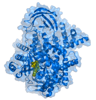
In cell biology, protein kinase A (PKA) is a family of serine-threonine kinase whose activity is dependent on cellular levels of cyclic AMP (cAMP). PKA is also known as cAMP-dependent protein kinase. PKA has several functions in the cell, including regulation of glycogen, sugar, and lipid metabolism. It should not be confused with 5'-AMP-activated protein kinase.

Phosphoinositide phospholipase C is a family of eukaryotic intracellular enzymes that play an important role in signal transduction processes. These enzymes belong to a larger superfamily of Phospholipase C. Other families of phospholipase C enzymes have been identified in bacteria and trypanosomes. Phospholipases C are phosphodiesterases.
Phosphoinositide 3-kinases (PI3Ks), also called phosphatidylinositol 3-kinases, are a family of enzymes involved in cellular functions such as cell growth, proliferation, differentiation, motility, survival and intracellular trafficking, which in turn are involved in cancer.
Ca2+
/calmodulin-dependent protein kinase II is a serine/threonine-specific protein kinase that is regulated by the Ca2+
/calmodulin complex. CaMKII is involved in many signaling cascades and is thought to be an important mediator of learning and memory. CaMKII is also necessary for Ca2+
homeostasis and reuptake in cardiomyocytes, chloride transport in epithelia, positive T-cell selection, and CD8 T-cell activation.

Phosphorylase kinase (PhK) is a serine/threonine-specific protein kinase which activates glycogen phosphorylase to release glucose-1-phosphate from glycogen. PhK phosphorylates glycogen phosphorylase at two serine residues, triggering a conformational shift which favors the more active glycogen phosphorylase “a” form over the less active glycogen phosphorylase b.
The PHLPP isoforms are a pair of protein phosphatases, PHLPP1 and PHLPP2, that are important regulators of Akt serine-threonine kinases and conventional/novel protein kinase C (PKC) isoforms. PHLPP may act as a tumor suppressor in several types of cancer due to its ability to block growth factor-induced signaling in cancer cells.

Protein kinase C alpha (PKCα) is an enzyme that in humans is encoded by the PRKCA gene.

Protein kinase C, zeta (PKCζ), also known as PRKCZ, is a protein in humans that is encoded by the PRKCZ gene. The PRKCZ gene encodes at least two alternative transcripts, the full-length PKCζ and an N-terminal truncated form PKMζ. PKMζ is thought to be responsible for maintaining long-term memories in the brain. The importance of PKCζ in the creation and maintenance of long-term potentiation was first described by Todd Sacktor and his colleagues at the SUNY Downstate Medical Center in 1993.

Protein kinase C delta type is an enzyme that in humans is encoded by the PRKCD gene.

Protein kinase C epsilon type (PKCε) is an enzyme that in humans is encoded by the PRKCE gene. PKCε is an isoform of the large PKC family of protein kinases that play many roles in different tissues. In cardiac muscle cells, PKCε regulates muscle contraction through its actions at sarcomeric proteins, and PKCε modulates cardiac cell metabolism through its actions at mitochondria. PKCε is clinically significant in that it is a central player in cardioprotection against ischemic injury and in the development of cardiac hypertrophy.
Protein kinase C theta (PKC-θ) is an enzyme that in humans is encoded by the PRKCQ gene. PKC-θ, a member of serine/threonine kinases, is mainly expressed in hematopoietic cells with high levels in platelets and T lymphocytes, where plays a role in signal transduction. Different subpopulations of T cells vary in their requirements of PKC-θ, therefore PKC-θ is considered as a potential target for inhibitors in the context of immunotherapy.

Protein kinase C iota type is an enzyme that in humans is encoded by the PRKCI gene.

Serine/threonine-protein kinase D1 is an enzyme that in humans is encoded by the PRKD1 gene.

Protein kinase C eta type is an enzyme that in humans is encoded by the PRKCH gene.

Serine/threonine-protein kinase D3 (PKD3) or PKC-nu is an enzyme that in humans is encoded by the PRKD3 gene.

BIM-1 and the related compounds BIM-2, BIM-3, and BIM-8 are bisindolylmaleimide-based protein kinase C (PKC) inhibitors. These inhibitors also inhibit PDK1 explaining the higher inhibitory potential of LY33331 compared to the other BIM compounds a bisindolylmaleimide inhibitor toward PDK1.
The insulin transduction pathway is a biochemical pathway by which insulin increases the uptake of glucose into fat and muscle cells and reduces the synthesis of glucose in the liver and hence is involved in maintaining glucose homeostasis. This pathway is also influenced by fed versus fasting states, stress levels, and a variety of other hormones.

Mezerein is a toxic diterpene ester found in the sap of Daphne mezereum and related plants. Plants of the genera Euphorbiaceae and Thymelaeaceae possess a wide variety of different phorbol esters, which share the capacity of mimicking diacylglycerol (DAG) and thus activating different isoforms of protein kinase C. Mezerein was first isolated in 1975. It has antileukemic properties in mice, but it is also defined as a weak promoter of skin cancers in the same species. All parts of the plants contain an acrid and irritant sap that contains mezerein, thought to be the principal poison. The sap is especially prevalent in the bark and berries.
References
- ↑ Wilson CH, Ali ES, Scrimgeour N, Martin AM, Hua J, Tallis GA, Rychkov GY, Barritt GJ (2015). "Steatosis inhibits liver cell store-operated Ca²⁺ entry and reduces ER Ca²⁺ through a protein kinase C-dependent mechanism". The Biochemical Journal. 466 (2): 379–90. doi:10.1042/BJ20140881. PMID 25422863.
- ↑ Ali ES, Hua J, Wilson CH, Tallis GA, Zhou FH, Rychkov GY, Barritt GJ (2016). "The glucagon-like peptide-1 analogue exendin-4 reverses impaired intracellular Ca2+ signalling in steatotic hepatocytes". Biochimica et Biophysica Acta (BBA) - Molecular Cell Research. 1863 (9): 2135–46. doi: 10.1016/j.bbamcr.2016.05.006 . PMID 27178543.
- ↑ Mellor H, Parker PJ (Jun 1998). "The extended protein kinase C superfamily". The Biochemical Journal. 332. 332 (Pt 2): 281–92. doi:10.1042/bj3320281. PMC 1219479 . PMID 9601053.
- ↑ Nishizuka Y (Apr 1995). "Protein kinase C and lipid signaling for sustained cellular responses". FASEB Journal. 9 (7): 484–96. doi: 10.1096/fasebj.9.7.7737456 . PMID 7737456. S2CID 31065063.
- ↑ Garcia-Concejo A, Larhammar D (2021). "Protein kinase C family evolution in jawed vertebrates". Dev Biol. 479: 77–90. doi: 10.1016/j.ydbio.2021.07.013 . PMID 34329618.
- ↑ Balendran A, Biondi RM, Cheung PC, Casamayor A, Deak M, Alessi DR (Jul 2000). "A 3-phosphoinositide-dependent protein kinase-1 (PDK1) docking site is required for the phosphorylation of protein kinase Czeta (PKCzeta ) and PKC-related kinase 2 by PDK1". The Journal of Biological Chemistry. 275 (27): 20806–13. doi: 10.1074/jbc.M000421200 . PMID 10764742. S2CID 27535562.
- ↑ Hauschild, Swantje; Tauber, Svantje; Lauber, Beatrice; Thiel, Cora S.; Layer, Liliana E.; Ullrich, Oliver (2014-11-01). "T cell regulation in microgravity – The current knowledge from in vitro experiments conducted in space, parabolic flights and ground-based facilities". Acta Astronautica. 104 (1): 365–377. Bibcode:2014AcAau.104..365H. doi: 10.1016/j.actaastro.2014.05.019 . ISSN 0094-5765.
- ↑ Staal, Jens; Driege, Yasmine; Haegman, Mira; Kreike, Marja; Iliaki, Styliani; Vanneste, Domien; Lork, Marie; Afonina, Inna S.; Braun, Harald; Beyaert, Rudi (2020-08-13). "Defining the combinatorial space of PKC::CARD-CC signal transduction nodes". The FEBS Journal. 288 (5): 1630–1647. doi:10.1111/febs.15522. ISSN 1742-4658. PMID 32790937. S2CID 221123226.
- 1 2 Biancani P, Harnett KM (2006). "Signal transduction in lower esophageal sphincter circular muscle, PART 1: Oral cavity, pharynx and esophagus". GI Motility Online. doi:10.1038/gimo24 (inactive 31 January 2024).
{{cite journal}}: CS1 maint: DOI inactive as of January 2024 (link) - 1 2 3 4 5 Fitzpatrick D, Purves D, Augustine G (2004). "Table 20:2". Neuroscience (Third ed.). Sunderland, Mass: Sinauer. ISBN 978-0-87893-725-7.
- ↑ Chou EC, Capello SA, Levin RM, Longhurst PA (Dec 2003). "Excitatory alpha1-adrenergic receptors predominate over inhibitory beta-receptors in rabbit dorsal detrusor". The Journal of Urology. 170 (6 Pt 1): 2503–7. doi:10.1097/01.ju.0000094184.97133.69. PMID 14634460.
- 1 2 3 4 5 6 7 8 9 10 11 Rang HP, Dale MM, Ritter JM, Moore PK (2003). "Ch. 10". Pharmacology (5th ed.). Elsevier Churchill Livingstone. ISBN 978-0-443-07145-4.
- ↑ Koslov DS, Andersson K (2013-01-01). "Physiological and pharmacological aspects of the vas deferens—an update". Frontiers in Pharmacology. 4: 101. doi: 10.3389/fphar.2013.00101 . PMC 3749770 . PMID 23986701.
- ↑ Sanders KM (Jul 1998). "G protein-coupled receptors in gastrointestinal physiology. IV. Neural regulation of gastrointestinal smooth muscle". The American Journal of Physiology. 275 (1 Pt 1): G1-7. doi:10.1152/ajpgi.1998.275.1.G1. PMID 9655677.
- ↑ Parker K, Brunton L, Goodman LS, Lazo JS, Gilman A (2006). Goodman & Gilman's the pharmacological basis of therapeutics (11th ed.). New York: McGraw-Hill. p. 185. ISBN 978-0-07-142280-2.
- ↑ "Entrez Gene: CHRM1 cholinergic receptor, muscarinic 1".
- 1 2 Walter F. Boron (2005). Medical Physiology: A Cellular And Molecular Approaoch. Elsevier/Saunders. ISBN 978-1-4160-2328-9. Page 787
- ↑ Barre A, Berthoux C, De Bundel D, Valjent E, Bockaert J, Marin P, Bécamel C (2016). "Presynaptic serotonin 2A receptors modulate thalamocortical plasticity and associative learning". Proceedings of the National Academy of Sciences of the United States of America. 113 (10): E1382–91. Bibcode:2016PNAS..113E1382B. doi: 10.1073/pnas.1525586113 . PMC 4791007 . PMID 26903620.
- ↑ Jalil SJ, Sacktor TC, Shouval HZ (2015). "Atypical PKCs in memory maintenance: the roles of feedback and redundancy". Learning & Memory. 22 (7): 344–53. doi:10.1101/lm.038844.115. PMC 4478332 . PMID 26077687.
- ↑ Boron, Walter F. Medical Physiology.
- ↑ Yamasaki T, Takahashi A, Pan J, Yamaguchi N, Yokoyama KK (March 2009). "Phosphorylation of Activation Transcription Factor-2 at Serine 121 by Protein Kinase C Controls c-Jun-mediated Activation of Transcription". The Journal of Biological Chemistry. 284 (13): 8567–81. doi: 10.1074/jbc.M808719200 . PMC 2659215 . PMID 19176525.
- ↑ Antal CE, Hudson AM, Kang E, Zanca C, Wirth C, Stephenson NL, Trotter EW, Gallegos LL, Miller CJ, Furnari FB, Hunter T, Brognard J, Newton AC (January 2015). "Cancer-associated protein kinase C mutations reveal kinase's role as tumor suppressor". Cell. 160 (3): 489–502. doi:10.1016/j.cell.2015.01.001. PMC 4313737 . PMID 25619690.
- ↑ Baffi TR, Van AN, Zhao W, Mills GB, Newton AC (March 2019). "Protein Kinase C Quality Control by Phosphatase PHLPP1 Unveils Loss-of-Function Mechanism in Cancer". Molecular Cell. 74 (2): 378–392.e5. doi:10.1016/j.molcel.2019.02.018. PMC 6504549 . PMID 30904392.
- ↑ Fan J, Ray P, Lu Y, Kaur G, Schwarz J, Wan L (24 October 2018). "Cell chirality regulates intercellular junctions and endothelial permeability". Science Advances. 4 (10): eaat2111. Bibcode:2018SciA....4.2111F. doi:10.1126/sciadv.aat2111. PMC 6200360 . PMID 30397640.
- ↑ Anderson PW, McGill JB, Tuttle KR (Sep 2007). "Protein kinase C beta inhibition: the promise for treatment of diabetic nephropathy". Current Opinion in Nephrology and Hypertension. 16 (5): 397–402. doi:10.1097/MNH.0b013e3281ead025. PMID 17693752. S2CID 72887329.
- ↑ Zarate, Carlos A.; Manji, Husseini K. (2009). "Protein Kinase C Inhibitors: Rationale for Use and Potential in the Treatment of Bipolar Disorder". CNS Drugs. 23 (7): 569–582. doi:10.2165/00023210-200923070-00003. ISSN 1172-7047. PMC 2802274 . PMID 19552485.
- ↑ Siller G, Gebauer K, Welburn P, Katsamas J, Ogbourne SM (Feb 2009). "PEP005 (ingenol mebutate) gel, a novel agent for the treatment of actinic keratosis: results of a randomized, double-blind, vehicle-controlled, multicentre, phase IIa study". The Australasian Journal of Dermatology. 50 (1): 16–22. doi:10.1111/j.1440-0960.2008.00497.x. PMID 19178487. S2CID 19308099.
- ↑ "FDA Approves Picato® (ingenol mebutate) Gel, the First and Only Topical Actinic Keratosis (AK) Therapy With 2 or 3 Consecutive Days of Once-Daily Dosing". eMedicine. Yahoo! Finance. January 25, 2012. Archived from the original on February 10, 2012. Retrieved 2012-02-14.
- ↑ Amended FDA Protocol Submitted for Phase 2b Trial of Advanced Alzheimer’s Therapy. Aug 2016