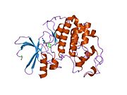
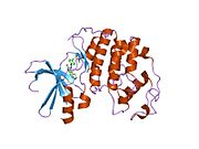
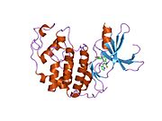
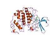
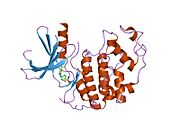
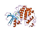
1aq1: HUMAN CYCLIN DEPENDENT KINASE 2 COMPLEXED WITH THE INHIBITOR STAUROSPORINE
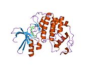
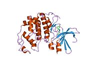
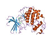
1b38: HUMAN CYCLIN-DEPENDENT KINASE 2
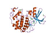
1b39: HUMAN CYCLIN-DEPENDENT KINASE 2 PHOSPHORYLATED ON THR 160
1buh: CRYSTAL STRUCTURE OF THE HUMAN CDK2 KINASE COMPLEX WITH CELL CYCLE-REGULATORY PROTEIN CKSHS1
1ckp: HUMAN CYCLIN DEPENDENT KINASE 2 COMPLEXED WITH THE INHIBITOR PURVALANOL B
1di8: THE STRUCTURE OF CYCLIN-DEPENDENT KINASE 2 (CDK2) IN COMPLEX WITH 4-[3-HYDROXYANILINO]-6,7-DIMETHOXYQUINAZOLINE
1dm2: HUMAN CYCLIN-DEPENDENT KINASE 2 COMPLEXED WITH THE INHIBITOR HYMENIALDISINE
1e1v: HUMAN CYCLIN DEPENDENT KINASE 2 COMPLEXED WITH THE INHIBITOR NU2058
1e1x: HUMAN CYCLIN DEPENDENT KINASE 2 COMPLEXED WITH THE INHIBITOR NU6027
1e9h: THR 160 PHOSPHORYLATED CDK2 - HUMAN CYCLIN A3 COMPLEX WITH THE INHIBITOR INDIRUBIN-5-SULPHONATE BOUND
1f5q: CRYSTAL STRUCTURE OF MURINE GAMMA HERPESVIRUS CYCLIN COMPLEXED TO HUMAN CYCLIN DEPENDENT KINASE 2
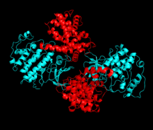
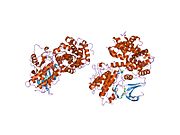
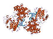
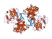
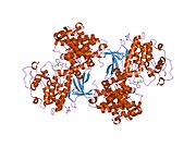
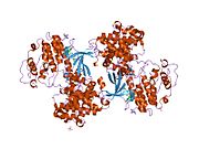
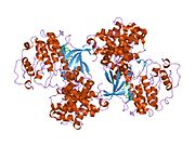
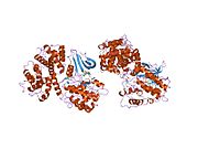
1fin: CYCLIN A-CYCLIN-DEPENDENT KINASE 2 COMPLEX
1fq1: CRYSTAL STRUCTURE OF KINASE ASSOCIATED PHOSPHATASE (KAP) IN COMPLEX WITH PHOSPHO-CDK2
1fvt: THE STRUCTURE OF CYCLIN-DEPENDENT KINASE 2 (CDK2) IN COMPLEX WITH AN OXINDOLE INHIBITOR
1fvv: THE STRUCTURE OF CDK2/CYCLIN A IN COMPLEX WITH AN OXINDOLE INHIBITOR
1g5s: CRYSTAL STRUCTURE OF HUMAN CYCLIN DEPENDENT KINASE 2 (CDK2) IN COMPLEX WITH THE INHIBITOR H717
1gih: HUMAN CYCLIN DEPENDENT KINASE 2 COMPLEXED WITH THE CDK4 INHIBITOR
1gii: HUMAN CYCLIN DEPENDENT KINASE 2 COMPLEXED WITH THE CDK4 INHIBITOR
1gij: HUMAN CYCLIN DEPENDENT KINASE 2 COMPLEXED WITH THE CDK4 INHIBITOR
1gy3: PCDK2/CYCLIN A IN COMPLEX WITH MGADP, NITRATE AND PEPTIDE SUBSTRATE
1gz8: HUMAN CYCLIN DEPENDENT KINASE 2 COMPLEXED WITH THE INHIBITOR 2-AMINO-6-(3'-METHYL-2'-OXO)BUTOXYPURINE
1h00: CDK2 IN COMPLEX WITH A DISUBSTITUTED 4, 6-BIS ANILINO PYRIMIDINE CDK4 INHIBITOR
1h07: CDK2 IN COMPLEX WITH A DISUBSTITUTED 4, 6-BIS ANILINO PYRIMIDINE CDK4 INHIBITOR
1h08: CDK2 IN COMPLEX WITH A DISUBSTITUTED 2, 4-BIS ANILINO PYRIMIDINE CDK4 INHIBITOR
1h0v: HUMAN CYCLIN DEPENDENT PROTEIN KINASE 2 IN COMPLEX WITH THE INHIBITOR 2-AMINO-6-[(R)-PYRROLIDINO-5'-YL]METHOXYPURINE
1h0w: HUMAN CYCLIN DEPENDENT PROTEIN KINASE 2 IN COMPLEX WITH THE INHIBITOR 2-AMINO-6-[CYCLOHEX-3-ENYL]METHOXYPURINE
1h1p: STRUCTURE OF HUMAN THR160-PHOSPHO CDK2/CYCLIN A COMPLEXED WITH THE INHIBITOR NU2058
1h1q: STRUCTURE OF HUMAN THR160-PHOSPHO CDK2/CYCLIN A COMPLEXED WITH THE INHIBITOR NU6094
1h1r: STRUCTURE OF HUMAN THR160-PHOSPHO CDK2/CYCLIN A COMPLEXED WITH THE INHIBITOR NU6086
1h1s: STRUCTURE OF HUMAN THR160-PHOSPHO CDK2/CYCLIN A COMPLEXED WITH THE INHIBITOR NU6102
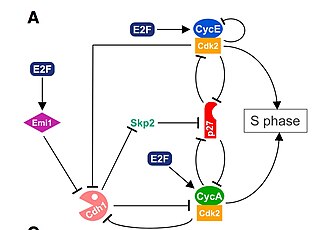
1h24: CDK2/CYCLIN A IN COMPLEX WITH A 9 RESIDUE RECRUITMENT PEPTIDE FROM E2F
1h25: CDK2/CYCLIN A IN COMPLEX WITH AN 11-RESIDUE RECRUITMENT PEPTIDE FROM RETINOBLASTOMA-ASSOCIATED PROTEIN
1h26: CDK2/CYCLIN A IN COMPLEX WITH AN 11-RESIDUE RECRUITMENT PEPTIDE FROM P53
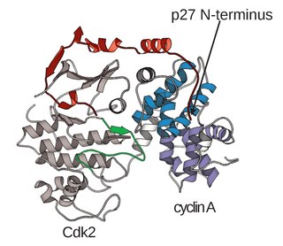
1h27: CDK2/CYCLIN A IN COMPLEX WITH AN 11-RESIDUE RECRUITMENT PEPTIDE FROM P27
1h28: CDK2/CYCLIN A IN COMPLEX WITH AN 11-RESIDUE RECRUITMENT PEPTIDE FROM P107
1hck: HUMAN CYCLIN-DEPENDENT KINASE 2
1hcl: HUMAN CYCLIN-DEPENDENT KINASE 2
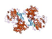
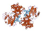
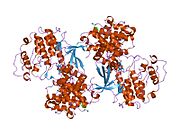
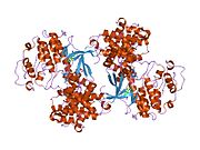
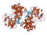
1jst: PHOSPHORYLATED CYCLIN-DEPENDENT KINASE-2 BOUND TO CYCLIN A
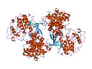
1jsu: P27(KIP1)/CYCLIN A/CDK2 COMPLEX
1jsv: The structure of cyclin-dependent kinase 2 (CDK2) in complex with 4-[(6-amino-4-pyrimidinyl)amino]benzenesulfonamide
1jvp: Crystal structure of human CDK2 (unphosphorylated) in complex with PKF049-365
1ke5: CDK2 complexed with N-methyl-4-{[(2-oxo-1,2-dihydro-3H-indol-3-ylidene)methyl]amino}benzenesulfonamide
1ke6: CYCLIN-DEPENDENT KINASE 2 (CDK2) COMPLEXED WITH N-METHYL-{4-[2-(7-OXO-6,7-DIHYDRO-8H-[1,3]THIAZOLO[5,4-E]INDOL-8-YLIDENE)HYDRAZINO]PHENYL}METHANESULFONAMIDE
1ke7: CYCLIN-DEPENDENT KINASE 2 (CDK2) COMPLEXED WITH 3-{[(2,2-DIOXIDO-1,3-DIHYDRO-2-BENZOTHIEN-5-YL)AMINO]METHYLENE}-5-(1,3-OXAZOL-5-YL)-1,3-DIHYDRO-2H-INDOL-2-ONE
1ke8: CYCLIN-DEPENDENT KINASE 2 (CDK2) COMPLEXED WITH 4-{[(2-OXO-1,2-DIHYDRO-3H-INDOL-3-YLIDENE)METHYL]AMINO}-N-(1,3-THIAZOL-2-YL)BENZENESULFONAMIDE
1ke9: CYCLIN-DEPENDENT KINASE 2 (CDK2) COMPLEXED WITH 3-{[4-({[AMINO(IMINO)METHYL]AMINOSULFONYL)ANILINO]METHYLENE}-2-OXO-2,3-DIHYDRO-1H-INDOLE
1ogu: STRUCTURE OF HUMAN THR160-PHOSPHO CDK2/CYCLIN A COMPLEXED WITH A 2-ARYLAMINO-4-CYCLOHEXYLMETHYL-5-NITROSO-6-AMINOPYRIMIDINE INHIBITOR
1oi9: STRUCTURE OF HUMAN THR160-PHOSPHO CDK2/CYCLIN A COMPLEXED WITH A 6-CYCLOHEXYLMETHYLOXY-2-ANILINO-PURINE INHIBITOR
1oiq: IMIDAZOPYRIDINES: A POTENT AND SELECTIVE CLASS OF CYCLIN-DEPENDENT KINASE INHIBITORS IDENTIFIED THROUGH STRUCTURE-BASED HYBRIDISATION
1oir: IMIDAZOPYRIDINES: A POTENT AND SELECTIVE CLASS OF CYCLIN-DEPENDENT KINASE INHIBITORS IDENTIFIED THROUGH STRUCTURE-BASED HYBRIDISATION
1oit: IMIDAZOPYRIDINES: A POTENT AND SELECTIVE CLASS OF CYCLIN-DEPENDENT KINASE INHIBITORS IDENTIFIED THROUGH STRUCTURE-BASED HYBRIDISATION
1oiu: STRUCTURE OF HUMAN THR160-PHOSPHO CDK2/CYCLIN A COMPLEXED WITH A 6-CYCLOHEXYLMETHYLOXY-2-ANILINO-PURINE INHIBITOR
1oiy: STRUCTURE OF HUMAN THR160-PHOSPHO CDK2/CYCLIN A COMPLEXED WITH A 6-CYCLOHEXYLMETHYLOXY-2-ANILINO-PURINE INHIBITOR
1okv: CYCLIN A BINDING GROOVE INHIBITOR H-ARG-ARG-LEU-ILE-PHE-NH2
1okw: CYCLIN A BINDING GROOVE INHIBITOR AC-ARG-ARG-LEU-ASN-(M-CL-PHE)-NH2
1ol1: CYCLIN A BINDING GROOVE INHIBITOR H-CIT-CIT-LEU-ILE-(P-F-PHE)-NH2
1ol2: CYCLIN A BINDING GROOVE INHIBITOR H-ARG-ARG-LEU-ASN-(P-F-PHE)-NH2
1p2a: The structure of cyclin dependent kinase 2 (CKD2) with a trisubstituted naphthostyril inhibitor
1p5e: The structure of phospho-CDK2/cyclin A in complex with the inhibitor 4,5,6,7-tetrabromobenzotriazole (TBS)
1pf8: Crystal Structure of Human Cyclin-Dependent Kinase 2 Complexed with a Nucleoside Inhibitor
1pkd: THE CRYSTAL STRUCTURE OF UCN-01 IN COMPLEX WITH PHOSPHO-CDK2/CYCLIN A
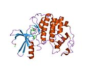
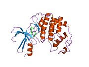
1pw2: APO STRUCTURE OF HUMAN CYCLIN-DEPENDENT KINASE 2
1pxi: HUMAN CYCLIN DEPENDENT KINASE 2 COMPLEXED WITH THE INHIBITOR 4-(2,5-Dichloro-thiophen-3-yl)-pyrimidin-2-ylamine
1pxj: HUMAN CYCLIN DEPENDENT KINASE 2 COMPLEXED WITH THE INHIBITOR 4-(2,4-Dimethyl-thiazol-5-yl)-pyrimidin-2-ylamine
1pxk: HUMAN CYCLIN DEPENDENT KINASE 2 COMPLEXED WITH THE INHIBITOR N-[4-(2,4-Dimethyl-thiazol-5-yl)pyrimidin-2-yl]-N'-hydroxyiminoformamide
1pxl: HUMAN CYCLIN DEPENDENT KINASE 2 COMPLEXED WITH THE INHIBITOR [4-(2,4-Dimethyl-thiazol-5-yl)-pyrimidin-2-yl]-(4-trifluoromethyl-phenyl)-amine
1pxm: HUMAN CYCLIN DEPENDENT KINASE 2 COMPLEXED WITH THE INHIBITOR 3-[4-(2,4-Dimethyl-thiazol-5-yl)-pyrimidin-2-ylamino]-phenol
1pxn: HUMAN CYCLIN DEPENDENT KINASE 2 COMPLEXED WITH THE INHIBITOR 4-[4-(4-Methyl-2-methylamino-thiazol-5-yl)-pyrimidin-2-ylamino]-phenol
1pxo: HUMAN CYCLIN DEPENDENT KINASE 2 COMPLEXED WITH THE INHIBITOR [4-(2-Amino-4-methyl-thiazol-5-yl)-pyrimidin-2-yl]-(3-nitro-phenyl)-amine
1pxp: HUMAN CYCLIN DEPENDENT KINASE 2 COMPLEXED WITH THE INHIBITOR N-[4-(2,4-Dimethyl-thiazol-5-yl)-pyrimidin-2-yl]-N',N'-dimethyl-benzene-1,4-diamine
1pye: Crystal structure of CDK2 with inhibitor
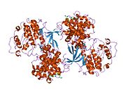
1qmz: PHOSPHORYLATED CDK2-CYCLYIN A-SUBSTRATE PEPTIDE COMPLEX
1r78: CDK2 complex with a 4-alkynyl oxindole inhibitor
1urc: CYCLIN A BINDING GROOVE INHIBITOR ACE-ARG-LYS-LEU-PHE-GLY
1urw: CDK2 IN COMPLEX WITH AN IMIDAZO[1,2-B]PYRIDAZINE
1v1k: CDK2 IN COMPLEX WITH A DISUBSTITUTED 4, 6-BIS ANILINO PYRIMIDINE CDK4 INHIBITOR
1vyw: STRUCTURE OF CDK2/CYCLIN A WITH PNU-292137
1vyz: STRUCTURE OF CDK2 COMPLEXED WITH PNU-181227
1w0x: CRYSTALS STRUCTURE OF HUMAN CDK2 IN COMPLEX WITH THE INHIBITOR OLOMOUCINE.
1w8c: CO-CRYSTAL STRUCTURE OF 6-CYCLOHEXYLMETHOXY-8-ISOPROPYL-9H-PURIN-2-YLAMINE AND MONOMERIC CDK2
1w98: THE STRUCTURAL BASIS OF CDK2 ACTIVATION BY CYCLIN E
1wcc: SCREENING FOR FRAGMENT BINDING BY X-RAY CRYSTALLOGRAPHY
1y8y: Crystal structure of human CDK2 complexed with a pyrazolo[1,5-a]pyrimidine inhibitor
1y91: Crystal structure of human CDK2 complexed with a pyrazolo[1,5-a]pyrimidine inhibitor
1ykr: Crystal structure of cdk2 with an aminoimidazo pyridine inhibitor
2a0c: Human CDK2 in complex with olomoucine II, a novel 2,6,9-trisubstituted purine cyclin-dependent kinase inhibitor
2a4l: Human cyclin-dependent kinase 2 in complex with roscovitine
2b52: Human cyclin dependent kinase 2 (CDK2) complexed with DPH-042562
2b53: Human cyclin dependent kinase 2 (CDK2) complexed with DIN-234325
2b54: Human cyclin dependent kinase 2 (CKD2)complexed with DIN-232305
2b55: Human cyclin dependent kinase 2 (cdk2) complexed with indenopyraxole DIN-101312
2bhe: HUMAN CYCLIN DEPENDENT PROTEIN KINASE 2 IN COMPLEX WITH THE INHIBITOR 5-BROMO-INDIRUBINE
2bhh: HUMAN CYCLIN DEPENDENT PROTEIN KINASE 2 IN COMPLEX WITH THE INHIBITOR 4-HYDROXYPIPERINDINESULFONYL-INDIRUBINE
2bkz: STRUCTURE OF CDK2-CYCLIN A WITH PHA-404611
2bpm: STRUCTURE OF CDK2-CYCLIN A WITH PHA-630529
2btr: STRUCTURE OF CDK2 COMPLEXED WITH PNU-198873
2bts: STRUCTURE OF CDK2 COMPLEXED WITH PNU-230032
2c4g: STRUCTURE OF CDK2-CYCLIN A WITH PHA-533514
2c5n: DIFFERENTIAL BINDING OF INHIBITORS TO ACTIVE AND INACTIVE CDK2 PROVIDES INSIGHTS FOR DRUG DESIGN
2c5o: DIFFERENTIAL BINDING OF INHIBITORS TO ACTIVE AND INACTIVE CDK2 PROVIDES INSIGHTS FOR DRUG DESIGN
2c5p: DIFFERENTIAL BINDING OF INHIBITORS TO ACTIVE AND INACTIVE CDK2 PROVIDES INSIGHTS FOR DRUG DESIGN
2c5v: DIFFERENTIAL BINDING OF INHIBITORS TO ACTIVE AND INACTIVE CDK2 PROVIDES INSIGHTS FOR DRUG DESIGN
2c5x: DIFFERENTIAL BINDING OF INHIBITORS TO ACTIVE AND INACTIVE CDK2 PROVIDES INSIGHTS FOR DRUG DESIGN
2c5y: DIFFERENTIAL BINDING OF INHIBITORS TO ACTIVE AND INACTIVE CDK2 PROVIDES INSIGHTS FOR DRUG DESIGN
2c68: CRYSTAL STRUCTURE OF THE HUMAN CDK2 COMPLEXED WITH THE TRIAZOLOPYRIMIDINE INHIBITOR
2c69: CRYSTAL STRUCTURE OF THE HUMAN CDK2 COMPLEXED WITH THE TRIAZOLOPYRIMIDINE INHIBITOR
2c6i: CRYSTAL STRUCTURE OF THE HUMAN CDK2 COMPLEXED WITH THE TRIAZOLOPYRIMIDINE INHIBITOR
2c6k: CRYSTAL STRUCTURE OF THE HUMAN CDK2 COMPLEXED WITH THE TRIAZOLOPYRIMIDINE INHIBITOR
2c6l: CRYSTAL STRUCTURE OF THE HUMAN CDK2 COMPLEXED WITH THE TRIAZOLOPYRIMIDINE INHIBITOR
2c6m: CRYSTAL STRUCTURE OF THE HUMAN CDK2 COMPLEXED WITH THE TRIAZOLOPYRIMIDINE INHIBITOR
2c6o: CRYSTAL STRUCTURE OF THE HUMAN CDK2 COMPLEXED WITH THE TRIAZOLOPYRIMIDINE INHIBITOR
2c6t: CRYSTAL STRUCTURE OF THE HUMAN CDK2 COMPLEXED WITH THE TRIAZOLOPYRIMIDINE INHIBITOR
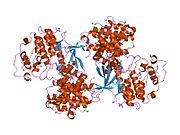
2cch: THE CRYSTAL STRUCTURE OF CDK2 CYCLIN A IN COMPLEX WITH A SUBSTRATE PEPTIDE DERIVED FROM CDC MODIFIED WITH A GAMMA-LINKED ATP ANALOGUE
2cci: CRYSTAL STRUCTURE OF PHOSPHO-CDK2 CYCLIN A IN COMPLEX WITH A PEPTIDE CONTAINING BOTH THE SUBSTRATE AND RECRUITMENT SITES OF CDC6
2cjm: MECHANISM OF CDK INHIBITION BY ACTIVE SITE PHOSPHORYLATION: CDK2 Y15P T160P IN COMPLEX WITH CYCLIN A STRUCTURE
2clx: 4-ARYLAZO-3,5-DIAMINO-1H-PYRAZOLE CDK INHIBITORS: SAR STUDY, CRYSTAL STRUCTURE IN COMPLEX WITH CDK2, SELECTIVITY, AND CELLULAR EFFECTS
2duv: Structure of CDK2 with a 3-hydroxychromones
2exm: Human CDK2 in complex with isopentenyladenine
2fvd: Cyclin Dependent Kinase 2 (CDK2) with diaminopyrimidine inhibitor
2g9x: Structure of Thr 160 phosphorylated CDK2/cyclin A in complex with the inhibitor NU6271
2i40: Cdk2/Cyclin A complexed with a thiophene carboxamide inhibitor
2iw6: STRUCTURE OF HUMAN THR160-PHOSPHO CDK2-CYCLIN A COMPLEXED WITH A BISANILINOPYRIMIDINE INHIBITOR
2iw8: STRUCTURE OF HUMAN THR160-PHOSPHO CDK2-CYCLIN A F82H-L83V-H84D MUTANT WITH AN O6-CYCLOHEXYLMETHYLGUANINE INHIBITOR
2iw9: STRUCTURE OF HUMAN THR160-PHOSPHO CDK2-CYCLIN A COMPLEXED WITH A BISANILINOPYRIMIDINE INHIBITOR
2jgz: CRYSTAL STRUCTURE OF PHOSPHO-CDK2 IN COMPLEX WITH CYCLIN B
2uue: REPLACE: A STRATEGY FOR ITERATIVE DESIGN OF CYCLIN BINDING GROOVE INHIBITORS