CDKN1B

Last updated
CDKN1B
Protein CDKN1B PDB 1jsu.png
Available structures
PDB Ortholog search: PDBe RCSB
Identifiers
Aliases CDKN1B , CDKN4, KIP1, MEN1B, MEN4, P27KIP1, cyclin-dependent kinase inhibitor 1B, cyclin dependent kinase inhibitor 1B
External IDs OMIM: 600778 MGI: 104565 HomoloGene: 2999 GeneCards: CDKN1B
Orthologs
SpeciesHumanMouse
Entrez
Ensembl
UniProt
RefSeq (mRNA)

NM_004064

NM_009875

RefSeq (protein)

NP_004055

NP_034005

Location (UCSC) Chr 12: 12.69 – 12.72 Mb Chr 6: 134.9 – 134.9 Mb
PubMed search [3] [4]
Wikidata
View/Edit Human View/Edit Mouse

Cyclin-dependent kinase inhibitor 1B (p27Kip1) is an enzyme inhibitor that in humans is encoded by the CDKN1B gene. [5] It encodes a protein which belongs to the Cip/Kip family of cyclin dependent kinase (Cdk) inhibitor proteins. The encoded protein binds to and prevents the activation of cyclin E-CDK2 or cyclin D-CDK4 complexes, and thus controls the cell cycle progression at G1. It is often referred to as a cell cycle inhibitor protein because its major function is to stop or slow down the cell division cycle.

Contents

Function

The p27Kip1 gene has a DNA sequence similar to other members of the "Cip/Kip" family which include the p21 Cip1/Waf1 and p57 Kip2 genes. In addition to this structural similarity the "Cip/Kip" proteins share the functional characteristic of being able to bind several different classes of Cyclin and Cdk molecules. For example, p27Kip1 binds to cyclin D either alone, or when complexed to its catalytic subunit CDK4. In doing so p27Kip1 inhibits the catalytic activity of Cdk4, which means that it prevents Cdk4 from adding phosphate residues to its principal substrate, the retinoblastoma (pRb) protein. Increased levels of the p27Kip1 protein typically cause cells to arrest in the G1 phase of the cell cycle. Likewise, p27Kip1 is able to bind other Cdk proteins when complexed to cyclin subunits such as Cyclin E/Cdk2 and Cyclin A/Cdk2. [6]

Regulation

In general, extracellular growth factors which promote cell division reduce transcription and translation of p27Kip1. Also, increased synthesis of CDk4,6/cyclin D causes binding of p27 to this complex, sequestering it from binding to the CDk2/cyclin E complex. Furthermore, an active CDK2/cyclin E complex will phosphorylate p27 and tag p27 for ubiquitination. [7] A mutation of this gene may lead to loss of control over the cell cycle leading to uncontrolled cellular proliferation. [8] [9] [10] Loss of p27 expression has been observed in metastatic canine mammary carcinomas. [11] [12] [13] Decreased TGF-beta signalling has been suggested to cause loss of p27 expression in this tumor type. [14]

A structured cis-regulatory element has been found in the 5' UTR of the P27 mRNA where it is thought to regulate translation relative to cell cycle progression. [15]

P27 regulation is accomplished by two different mechanisms. In the first its concentration is changed by the individual rates of transcription, translation, and proteolysis. P27 can also be regulated by changing its subcellular location [16] Both mechanisms act to reduce levels of p27, allowing for the activation of Cdk1 and Cdk2, and for the cell to begin progressing through the cell cycle.

Transcription

Transcription of the CDKN1B gene is activated by Forkhead box class O family (FoxO) proteins which also acts downstream to promote p27 nuclear localization and decrease levels of COP9 subunit 5(COPS5) which helps in the degradation of p27. [17] Transcription for p27 is activated by FoxO in response to cytokines, promyelocytic leukaemia proteins, and nuclear Akt signaling. [17] P27 transcription has also been linked to another tumor suppressor gene, MEN1, in pancreatic islet cells where it promotes CDKN1B expression. [17]

Translation

Translation of CDKN1B reaches its maximum during quiescence and early G1. [17] Translation is regulated by polypyrimidine tract-binding protein(PTB), ELAVL1, ELAVL4, and microRNAs. [17] PTB acts by binding CDKN1b IRES to increase translation and when PTB levels decrease, G1 phase is shortened. [17] ELAVL1 and ELAVL4 also bind to CDKN1B IRES but they do so in order to decrease translation and so depletion of either results in G1 arrest. [17]

Proteolysis

Degradation of the p27 protein occurs as cells exit quiescence and enter G1. [17] Protein levels continue to fall rapidly as the cell continues through G1 and enters S phase. One of the most understood mechanisms for p27 proteolysis is the polyubiquitylation of p27 by the SCFSKP2 kinase associated protein 1 (Skp1) and 2 (Skp2). [17] SKP1 and Skp2 degrades p27 after it has been phosphorylated at threonine 187 (Thr187) by either activating cyclin E- or cyclin A-CDK2. Skp2 is mainly responsible for the degradation of p27 levels that continues through S phase. [18] However it is rarely expressed in early G1 where p27 levels first begin to decrease. During early G1 proteolysis of p27 is regulated by KIP1 Ubiquitylation Promoting Complex (KPC) which binds to its CDK inhibitory domain. [19] P27 also has three Cdk-inhibited tyrosines at residues 74, 88, and 89. [17] Of these, Tyr74 is of special interest because it is specific to p27-type inhibitors. [17]

Nuclear export

Alternatively to the transcription, translation, and proteolytic method of regulation, p27 levels can also be changed by exporting p27 to the cytoplasm. This occurs when p27 is phosphorylated on Ser(10) which allows for CRM1, a nuclear export carrier protein, to bind to and remove p27 from the nucleus. [20] Once p27 is excluded from the nucleus it cannot inhibit the cell's growth. In the cytoplasm it may be degraded entirely or retained. [16] This step occurs very early when the cell is exiting the quiescent phase and thus is independent of Skp2 degradation of p27. [20]

MicroRNA regulation

Because p27 levels can be moderated at the translational level, it has been proposed that p27 may be regulated by miRNAs. Recent research has suggested that both miR-221 and miR-222 control p27 levels although the pathways are not well understood. [16]

Role in cancer

Proliferation

p27 is considered a tumor suppressor because of its function as a regulator of the cell cycle. [17] In cancers it is often inactivated via impaired synthesis, accelerated degradation, or mislocalization. [17] Inactivation of p27 is generally accomplished post-transcription by the oncogenic activation of various pathways including receptor tyrosine kinases (RTK), phosphatilidylinositol 3-kinase (PI3K), SRC, or Ras-mitogen activated protein kinase(MAPK). [17] These act to accelerate the proteolysis of the p27 protein and allow the cancer cell to undergo rapid division and uncontrolled proliferation. [17] When p27 is phosphorylated by Src at tyrosine 74 or 88 it ceases to inhibit cyclinE-cdk2. [21] Src was also shown to reduce the half life of p27 meaning it is degraded faster. [21] Many epithelial cancers are known to overexpress EGFR which plays a role in the proteolysis of p27 and in Ras-driven proteolysis. [17] Non-epithelial cancers use different pathways to inactivate p27. [17] Many cancer cells also upregulate Skp2 which is known to play an active role in the proteolysis of p27 [18] As a result, Skp2 is inversely related to p27 levels and directly correlates with tumor grade in many malignancies. [18]

Metastasis

In cancer cells, p27 can also be mislocalized to the cytoplasm in order to facilitate metastasis. The mechanisms by which it acts on motility differ between cancers. In hepatocellular carcinoma cells p27 co-localizes with actin fibers to act on GTPase Rac and induce cell migration. [22] In breast cancer cytoplasmic p27 reduced RHOA activity which increased a cell's propensity for motility. [23]

This role for p27 may indicate why cancer cells rarely fully inactivate or delete p27. By retaining p27 in some capacity it can be exported to the cytoplasm during tumorigenesis and manipulated to aid in metastasis. 70% of metastatic melanomas were shown to exhibit cytoplasmic p27, while in benign melanomas p27 remained localized to the nucleus. [24] P27 is misplaced to the cytoplasm by the MAP2K, Ras, and Akt pathways although the mechanisms are not entirely understood. [25] [26] [27] Additionally, phosphorylation of p27 at T198 by RSK1 has been shown to mislocalize p27 to the cytoplasm as well as inhibit the RhoA pathway. [28] Because inhibition of RhoA results in a decrease in both stress fibers and focal adhesion, cell motility is increased. [29] P27 can also be exported to the cytoplasm by oncogenic activation of the P13K pathway. [29] Thus, mislocalization of p27 to the cytoplasm in cancer cells allows them to proliferate unchecked and provides for increased motility.

In contrast to these results, p27 has also been shown to be an inhibitor of migration in sarcoma cells. [30] In these cells, p27 bound to stathmin which prevents stathmin from binding to tubulin and thus polymerization of microtubules increased and cell motility decreased. [30]

MicroRNA regulation

Studies of various cell lines including glioblastoma cell lines, three prostate cancer cell lines, and a breast tumor cell line showed that suppressing miR-221 and miR-22 expression resulted in p27-dependent G1 growth arrest [16] Then when p27 was knocked down, cell growth resumed indicating a strong role for miRNA regulated p27. [16] Studies in patients have demonstrated an inverse correlation between miR-221&22 and p27 protein levels. Additionally nearby healthy tissue showed high expression of the p27 protein while miR-221&22 concentrations were low. [16]

Regulation in specific cancers

In most cancers reduced levels of nuclear p27 are correlated with increased tumor size, increased tumor grade, and a higher propensity for metastasis. However the mechanisms by which levels of p27 are regulated vary between cancers.

Breast

In breast cancer, Src activation has been shown to correlate with low levels of p27 [21] Breast cancers that were Estrogen receptor negative and progesterone receptor negative were more likely to display low levels of p27 and more likely to have a high tumor grade. [21] Similarly, breast cancer patients with BRCA1/2 mutations were more likely to have low levels of p27. [31]

Prostate

A mutation in the CDKN1B gene has been linked to an increased risk for hereditary prostate cancer in humans. [32]

Multiple Endocrine Neoplasia

Mutations in the CDKN1B gene has been reported in families affected by the development of primary hyperparathyroidism and pituitary adenomas, and has been classified MEN4 (multiple endocrine neoplasia, type 4). Testing for CDKN1B mutations has been recommended in patients with suspected MEN, in whom previous testing for, the more common MEN1/RET mutation, is negative. [33]

Clinical significance

Prognostic value

Several studies have demonstrated that reduced p27 levels indicate a poorer patient prognosis. [17] However, because of the dual, contrasting roles p27 plays in cancer (as an inhibitor of growth and as a mechanism for metastasis) low levels of p27 may demonstrate that a cancer is not aggressive and will remain benign. [17] In ovarian cancer, p27 negative tumors progressed in 23 months compared to 85 months in p27 positive tumors and thus could be used as a prognostic marker. [34] Similar studies have correlated low levels of p27 with a worse prognosis in breast cancer. [35] Colorectal carcinomas that lacked p27 were shown to have increased p27-specific proteolysis and a median survival of only 69 months compared to 151 months for patients with high or normal levels of p27. [36] The authors proposed clinicians could use patient specific levels of p27 to determine who would benefit from adjuvant therapy. [36] Similar correlations were observed in patients with non-small cell lung cancer, [37] those with colon, [37] and prostate cancer. [38]

So far studies have only evaluated the prognostic value of p27 retrospectively and a standardized scoring system has not been established. [17] However it has been proposed that clinicians should evaluate a patient's p27 levels in order to determine if they will be responsive to certain chemotoxins which target fast growing tumors where p27 levels are low. [17] Or in contrast, if p27 levels are found to be high in a patient's cancer, their risk for metastasis is higher and the physician can make an informed decision about their treatment plan. [17] Because p27 levels are controlled post-transcriptionally, proteomic surveys can be used to establish and monitor a patient's individual levels which aids in the future of individualized medicine.

The following cancers have been demonstrated to have an inverse correlation with p27 expression and prognosis: oro-pharyngo-laryngeal, oesophageal, gastric, colon, lung, melanoma, glioma, breast cancer, prostate, lymphoma, leukemia. [18]

Correlation to treatment response

P27 may also allow clinicians to better select an appropriate treatment for a patient. For example, patients with non-small cell lung cancer who were treated with platinum based chemotherapy showed reduced survival if they had low levels of p27. [39] Similarly low levels of p27 correlated with poor results from adjuvant chemotherapy in breast cancer patients. [40]

Value as a therapeutic target

P27 has been explored as a potential target for cancer therapy because its levels are highly correlated to patient prognosis. [41] This is true for a wide spectrum of cancers including colon, breast, prostate, lung, liver, stomach, and bladder. [41]

Use of microRNAs for therapy

Because of the role miRNAs play in p27 regulation, research is underway to determine if antagomiRs that block the activity of the miR221&222 and allow for p27 cell grow inhibition to take place could act as therapeutic cancer drugs. [16]

Role in Regeneration

Knockdown of CDKN1B stimulates regeneration of cochlear hair cells in mice. Since CDKN1B prevents cells from entering the cell cycle, inhibition of the protein could cause re-entry and subsequent division. In mammals where regeneration of cochlear hair cells normally does not occur, this inhibition could help regrow damaged cells who are otherwise incapable of proliferation. In fact, when the CDKN1B gene is disrupted in adult mice, hair cells of the organ of Corti proliferate, while those in control mice do not. Lack of CDKN1B expression appears to release the hair cells from natural cell-cycle arrest. [42] [43] Because hair cell death in the human cochlea is a major cause of hearing loss, the CDKN1B protein could be an important factor in the clinical treatment of deafness.

Interactions

CDKN1B has been shown to interact with:

Overview of signal transduction pathways involved in apoptosis. Signal transduction v1.png
Overview of signal transduction pathways involved in apoptosis.

See also

Related Research Articles

<span class="mw-page-title-main">Cyclin-dependent kinase</span> Class of enzymes

Cyclin-dependent kinases (CDKs) are the families of protein kinases first discovered for their role in regulating the cell cycle. They are also involved in regulating transcription, mRNA processing, and the differentiation of nerve cells. They are present in all known eukaryotes, and their regulatory function in the cell cycle has been evolutionarily conserved. In fact, yeast cells can proliferate normally when their CDK gene has been replaced with the homologous human gene. CDKs are relatively small proteins, with molecular weights ranging from 34 to 40 kDa, and contain little more than the kinase domain. By definition, a CDK binds a regulatory protein called a cyclin. Without cyclin, CDK has little kinase activity; only the cyclin-CDK complex is an active kinase but its activity can be typically further modulated by phosphorylation and other binding proteins, like p27. CDKs phosphorylate their substrates on serines and threonines, so they are serine-threonine kinases. The consensus sequence for the phosphorylation site in the amino acid sequence of a CDK substrate is [S/T*]PX[K/R], where S/T* is the phosphorylated serine or threonine, P is proline, X is any amino acid, K is lysine, and R is arginine.

p21

p21Cip1, also known as cyclin-dependent kinase inhibitor 1 or CDK-interacting protein 1, is a cyclin-dependent kinase inhibitor (CKI) that is capable of inhibiting all cyclin/CDK complexes, though is primarily associated with inhibition of CDK2. p21 represents a major target of p53 activity and thus is associated with linking DNA damage to cell cycle arrest. This protein is encoded by the CDKN1A gene located on chromosome 6 (6p21.2) in humans.

<span class="mw-page-title-main">SCF complex</span>

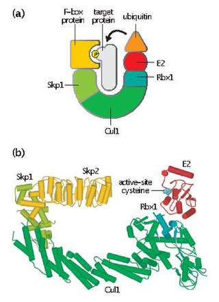
Skp, Cullin, F-box containing complex is a multi-protein E3 ubiquitin ligase complex that catalyzes the ubiquitination of proteins destined for 26S proteasomal degradation. Along with the anaphase-promoting complex, SCF has important roles in the ubiquitination of proteins involved in the cell cycle. The SCF complex also marks various other cellular proteins for destruction.

p16 Mammalian protein found in Homo sapiens

p16, is a protein that slows cell division by slowing the progression of the cell cycle from the G1 phase to the S phase, thereby acting as a tumor suppressor. It is encoded by the CDKN2A gene. A deletion in this gene can result in insufficient or non-functional p16, accelerating the cell cycle and resulting in many types of cancer.

<span class="mw-page-title-main">Cyclin D</span> Member of the cyclin protein family

Cyclin D is a member of the cyclin protein family that is involved in regulating cell cycle progression. The synthesis of cyclin D is initiated during G1 and drives the G1/S phase transition. Cyclin D protein is anywhere from 155 to 477 amino acids in length.

<span class="mw-page-title-main">Cyclin-dependent kinase 2</span> Protein-coding gene in the species Homo sapiens

Cyclin-dependent kinase 2, also known as cell division protein kinase 2, or Cdk2, is an enzyme that in humans is encoded by the CDK2 gene. The protein encoded by this gene is a member of the cyclin-dependent kinase family of Ser/Thr protein kinases. This protein kinase is highly similar to the gene products of S. cerevisiae cdc28, and S. pombe cdc2, also known as Cdk1 in humans. It is a catalytic subunit of the cyclin-dependent kinase complex, whose activity is restricted to the G1-S phase of the cell cycle, where cells make proteins necessary for mitosis and replicate their DNA. This protein associates with and is regulated by the regulatory subunits of the complex including cyclin E or A. Cyclin E binds G1 phase Cdk2, which is required for the transition from G1 to S phase while binding with Cyclin A is required to progress through the S phase. Its activity is also regulated by phosphorylation. Multiple alternatively spliced variants and multiple transcription initiation sites of this gene have been reported. The role of this protein in G1-S transition has been recently questioned as cells lacking Cdk2 are reported to have no problem during this transition.

<span class="mw-page-title-main">Cyclin-dependent kinase 4</span> Human protein

Cyclin-dependent kinase 4 also known as cell division protein kinase 4 is an enzyme that in humans is encoded by the CDK4 gene. CDK4 is a member of the cyclin-dependent kinase family.

<span class="mw-page-title-main">Cyclin-dependent kinase 6</span> Protein-coding gene in the species Homo sapiens

Cell division protein kinase 6 (CDK6) is an enzyme encoded by the CDK6 gene. It is regulated by cyclins, more specifically by Cyclin D proteins and Cyclin-dependent kinase inhibitor proteins. The protein encoded by this gene is a member of the cyclin-dependent kinase, (CDK) family, which includes CDK4. CDK family members are highly similar to the gene products of Saccharomyces cerevisiae cdc28, and Schizosaccharomyces pombe cdc2, and are known to be important regulators of cell cycle progression in the point of regulation named R or restriction point.

<span class="mw-page-title-main">Cyclin-dependent kinase inhibitor protein</span> Protein which inhibits cyclin-dependent kinase

A cyclin-dependent kinase inhibitor protein

<span class="mw-page-title-main">SKP2</span> Protein-coding gene in the species Homo sapiens

S-phase kinase-associated protein 2 is an enzyme that in humans is encoded by the SKP2 gene.

<span class="mw-page-title-main">Cyclin E1</span> Protein-coding gene in the species Homo sapiens

G1/S-specific cyclin-E1 is a protein that in humans is encoded by the CCNE1 gene.

<span class="mw-page-title-main">Cyclin A2</span> Protein-coding gene in the species Homo sapiens

Cyclin-A2 is a protein that in humans is encoded by the CCNA2 gene. It is one of the two types of cyclin A: cyclin A1 is expressed during meiosis and embryogenesis while cyclin A2 is expressed in dividing somatic cells.

<span class="mw-page-title-main">CDKN2B</span> Protein-coding gene in the species Homo sapiens

Cyclin-dependent kinase 4 inhibitor B also known as multiple tumor suppressor 2 (MTS-2) or p15INK4b is a protein that is encoded by the CDKN2B gene in humans.

<span class="mw-page-title-main">Cyclin-dependent kinase inhibitor 1C</span> Protein-coding gene in the species Homo sapiens

Cyclin-dependent kinase inhibitor 1C , also known as CDKN1C, is a protein which in humans is encoded by the CDKN1C imprinted gene.

<span class="mw-page-title-main">CDKN2C</span> Protein-coding gene in the species Homo sapiens

Cyclin-dependent kinase 4 inhibitor C is an enzyme that in humans is encoded by the CDKN2C gene.

<span class="mw-page-title-main">CDKN2D</span> Protein-coding gene in the species Homo sapiens

Cyclin-dependent kinase 4 inhibitor D is an enzyme that in humans is encoded by the CDKN2D gene.

<span class="mw-page-title-main">CKS1B</span> Protein-coding gene in the species Homo sapiens

Cyclin-dependent kinases regulatory subunit 1 is a protein that in humans is encoded by the CKS1B gene.

<span class="mw-page-title-main">CDKN3</span> Protein-coding gene in the species Homo sapiens

Cyclin-dependent kinase inhibitor 3 is an enzyme that in humans is encoded by the CDKN3 gene.

Sic1, a protein, is a stoichiometric inhibitor of Cdk1-Clb complexes in the budding yeast Saccharomyces cerevisiae. Because B-type cyclin-Cdk1 complexes are the drivers of S-phase initiation, Sic1 prevents premature S-phase entry. Multisite phosphorylation of Sic1 is thought to time Sic1 ubiquitination and destruction, and by extension, the timing of S-phase entry.

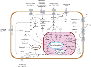
The CIP/KIP family is one of two families of mammalian cyclin dependent kinase (CDK) inhibitors (CKIs) involved in regulating the cell cycle. The CIP/KIP family is made up of three proteins: p21cip1/waf1, P27kip1, p57kip2 These proteins share sequence homology at the N-terminal domain which allows them to bind to both the cyclin and CDK. Their activity primarily involves the binding and inhibition of G1/S- and S-Cdks; however, they have also been shown to play an important role in activating the G1-CDKs CDK4 and CDK6. In addition, more recent work has shown that CIP/KIP family members have a number of CDK-independent roles involving regulation of transcription, apoptosis, and the cytoskeleton.

References

  1. 1 2 3 GRCh38: Ensembl release 89: ENSG00000111276 - Ensembl, May 2017
  2. 1 2 3 GRCm38: Ensembl release 89: ENSMUSG00000003031 - Ensembl, May 2017
  3. "Human PubMed Reference:". National Center for Biotechnology Information, U.S. National Library of Medicine.
  4. "Mouse PubMed Reference:". National Center for Biotechnology Information, U.S. National Library of Medicine.
  5. Polyak K, Lee MH, Erdjument-Bromage H, Koff A, Roberts JM, Tempst P, Massagué J (August 1994). "Cloning of p27Kip1, a cyclin-dependent kinase inhibitor and a potential mediator of extracellular antimitogenic signals". Cell. 78 (1): 59–66. doi:10.1016/0092-8674(94)90572-X. PMID   8033212. S2CID   38513201.
  6. Chiarle, R; Pagano, M; Inghirami, G (2001). "The cyclin dependent kinase inhibitor p27 and its prognostic role in breast cancer". Breast Cancer Research. 3 (2): 91–4. doi: 10.1186/bcr277 . PMC   139437 . PMID   11250752.
  7. Cooper/ Hausman, Geoffrey/ Robert (2009). The Cell. Boston University: ASM Press. p. 669. ISBN   978-0-87893-300-6.
  8. Fero ML, Rivkin M, Tasch M, Porter P, Carow CE, Firpo E, Polyak K, Tsai LH, Broudy V, Perlmutter RM, Kaushansky K, Roberts JM (May 1996). "A syndrome of multiorgan hyperplasia with features of gigantism, tumorigenesis, and female sterility in p27(Kip1)-deficient mice". Cell . 85 (5): 733–44. doi: 10.1016/S0092-8674(00)81239-8 . PMID   8646781. S2CID   15490866.
  9. Kiyokawa H, Kineman RD, Manova-Todorova KO, Soares VC, Hoffman ES, Ono M, Khanam D, Hayday AC, Frohman LA, Koff A (May 1996). "Enhanced growth of mice lacking the cyclin-dependent kinase inhibitor function of p27(Kip1)". Cell . 85 (5): 721–32. doi: 10.1016/S0092-8674(00)81238-6 . PMID   8646780. S2CID   19030818.
  10. Nakayama K, Ishida N, Shirane M, Inomata A, Inoue T, Shishido N, Horii I, Loh DY, Nakayama K (May 1996). "Mice lacking p27(Kip1) display increased body size, multiple organ hyperplasia, retinal dysplasia, and pituitary tumors". Cell . 85 (5): 707–20. doi: 10.1016/S0092-8674(00)81237-4 . PMID   8646779. S2CID   2009281.
  11. Klopfleisch R, Gruber AD (January 2009). "Differential expression of cell cycle regulators p21, p27 and p53 in metastasizing canine mammary adenocarcinomas versus normal mammary glands". Res Vet Sci. 87 (1): 91–6. doi:10.1016/j.rvsc.2008.12.010. PMID   19185891.
  12. Klopfleisch R, Schütze M, Gruber AD (September 2010). "Loss of p27 expression in canine mammary tumors and their metastases". Res Vet Sci. 88 (2): 300–3. doi:10.1016/j.rvsc.2009.08.007. PMID   19748645.
  13. Klopfleisch R, von Euler H, Sarli G, Pinho SS, Gärtner F, Gruber AD (2010). "Molecular Carcinogenesis of Canine Mammary Tumors: News From an Old Disease". Veterinary Pathology. 48 (1): 98–116. doi:10.1177/0300985810390826. PMID   21149845. S2CID   206509356.
  14. Klopfleisch R, Schütze M, Gruber AD (October 2009). "Downregulation of transforming growth factor β (TGFβ) and latent TGFβ binding protein (LTBP)-4 expression in late stage canine mammary tumours". Veterinary Journal. 186 (3): 379–84. doi:10.1016/j.tvjl.2009.09.014. PMID   19836277.
  15. Göpfert U, Kullmann M, Hengst L (July 2003). "Cell cycle-dependent translation of p27 involves a responsive element in its 5'-UTR that overlaps with a uORF". Hum. Mol. Genet. 12 (14): 1767–79. doi: 10.1093/hmg/ddg177 . PMID   12837699.
  16. 1 2 3 4 5 6 7 le Sage C, Nagel R, Agami R (November 2007). "Diverse ways to control p27Kip1 function: miRNAs come into play". Cell Cycle. 6 (22): 2742–9. doi:10.4161/cc.6.22.4900. PMID   17986865.
  17. 1 2 3 4 5 6 7 8 9 10 11 12 13 14 15 16 17 18 19 20 21 22 Chu IM, Hengst L, Slingerland JM (April 2008). "The Cdk inhibitor p27 in human cancer: prognostic potential and relevance to anticancer therapy". Nat. Rev. Cancer. 8 (4): 253–67. doi:10.1038/nrc2347. PMID   18354415. S2CID   2175257.
  18. 1 2 3 4 Nakayama KI, Nakayama K (May 2006). "Ubiquitin ligases: cell-cycle control and cancer". Nat. Rev. Cancer. 6 (5): 369–81. doi:10.1038/nrc1881. PMID   16633365. S2CID   19594293.
  19. Kotoshiba S, Kamura T, Hara T, Ishida N, Nakayama KI (May 2005). "Molecular dissection of the interaction between p27 and Kip1 ubiquitylation-promoting complex, the ubiquitin ligase that regulates proteolysis of p27 in G1 phase". J. Biol. Chem. 280 (18): 17694–700. doi: 10.1074/jbc.M500866200 . PMID   15746103.
  20. 1 2 Ishida N, Hara T, Kamura T, Yoshida M, Nakayama K, Nakayama KI (April 2002). "Phosphorylation of p27Kip1 on serine 10 is required for its binding to CRM1 and nuclear export". J. Biol. Chem. 277 (17): 14355–8. doi: 10.1074/jbc.C100762200 . PMID   11889117.
  21. 1 2 3 4 Chu I, Sun J, Arnaout A, Kahn H, Hanna W, Narod S, Sun P, Tan CK, Hengst L, Slingerland J (January 2007). "p27 phosphorylation by Src regulates inhibition of cyclin E-Cdk2". Cell. 128 (2): 281–94. doi:10.1016/j.cell.2006.11.049. PMC   1961623 . PMID   17254967.
  22. McAllister SS, Becker-Hapak M, Pintucci G, Pagano M, Dowdy SF (January 2003). "Novel p27(kip1) C-terminal scatter domain mediates Rac-dependent cell migration independent of cell cycle arrest functions". Mol. Cell. Biol. 23 (1): 216–28. doi:10.1128/MCB.23.1.216-228.2003. PMC   140659 . PMID   12482975.
  23. Wu FY, Wang SE, Sanders ME, Shin I, Rojo F, Baselga J, Arteaga CL (February 2006). "Reduction of cytosolic p27(Kip1) inhibits cancer cell motility, survival, and tumorigenicity". Cancer Res. 66 (4): 2162–72. doi:10.1158/0008-5472.CAN-05-3304. PMID   16489017.
  24. Denicourt C, Saenz CC, Datnow B, Cui XS, Dowdy SF (October 2007). "Relocalized p27Kip1 tumor suppressor functions as a cytoplasmic metastatic oncogene in melanoma". Cancer Res. 67 (19): 9238–43. doi: 10.1158/0008-5472.CAN-07-1375 . PMID   17909030.
  25. Cheng M, Sexl V, Sherr CJ, Roussel MF (February 1998). "Assembly of cyclin D-dependent kinase and titration of p27Kip1 regulated by mitogen-activated protein kinase kinase (MEK1)". Proc. Natl. Acad. Sci. U.S.A. 95 (3): 1091–6. Bibcode:1998PNAS...95.1091C. doi: 10.1073/pnas.95.3.1091 . PMC   18683 . PMID   9448290.
  26. Liu X, Sun Y, Ehrlich M, Lu T, Kloog Y, Weinberg RA, Lodish HF, Henis YI (November 2000). "Disruption of TGF-beta growth inhibition by oncogenic ras is linked to p27Kip1 mislocalization". Oncogene. 19 (51): 5926–35. doi:10.1038/sj.onc.1203991. PMID   11127824.
  27. Viglietto G, Motti ML, Bruni P, Melillo RM, D'Alessio A, Califano D, Vinci F, Chiappetta G, Tsichlis P, Bellacosa A, Fusco A, Santoro M (October 2002). "Cytoplasmic relocalization and inhibition of the cyclin-dependent kinase inhibitor p27(Kip1) by PKB/Akt-mediated phosphorylation in breast cancer". Nat. Med. 8 (10): 1136–44. doi:10.1038/nm762. PMID   12244303. S2CID   6580033.
  28. Larrea MD, Hong F, Wander SA, da Silva TG, Helfman D, Lannigan D, Smith JA, Slingerland JM (June 2009). "RSK1 drives p27Kip1 phosphorylation at T198 to promote RhoA inhibition and increase cell motility". Proc. Natl. Acad. Sci. U.S.A. 106 (23): 9268–73. Bibcode:2009PNAS..106.9268L. doi: 10.1073/pnas.0805057106 . PMC   2695095 . PMID   19470470.
  29. 1 2 Larrea MD, Wander SA, Slingerland JM (November 2009). "p27 as Jekyll and Hyde: regulation of cell cycle and cell motility". Cell Cycle. 8 (21): 3455–61. doi: 10.4161/cc.8.21.9789 . PMID   19829074.
  30. 1 2 Baldassarre G, Belletti B, Nicoloso MS, Schiappacassi M, Vecchione A, Spessotto P, Morrione A, Canzonieri V, Colombatti A (January 2005). "p27(Kip1)-stathmin interaction influences sarcoma cell migration and invasion". Cancer Cell. 7 (1): 51–63. doi: 10.1016/j.ccr.2004.11.025 . PMID   15652749.
  31. Chappuis PO, Kapusta L, Bégin LR, Wong N, Brunet JS, Narod SA, Slingerland J, Foulkes WD (December 2000). "Germline BRCA1/2 mutations and p27(Kip1) protein levels independently predict outcome after breast cancer". J. Clin. Oncol. 18 (24): 4045–52. doi:10.1200/jco.2000.18.24.4045. PMID   11118465.
  32. Chang BL, Zheng SL, Isaacs SD, Wiley KE, Turner A, Li G, Walsh PC, Meyers DA, Isaacs WB, Xu J (March 2004). "A polymorphism in the CDKN1B gene is associated with increased risk of hereditary prostate cancer". Cancer Res. 64 (6): 1997–9. doi: 10.1158/0008-5472.CAN-03-2340 . PMID   15026335.
  33. Alrezk R, Hannah-Shmouni F, Stratakis CA (August 2017). "MEN4 and CDKN1B mutations: the latest of the MEN syndromes". Endocrine-Related Cancer. 24 (10): T195–208. doi:10.1530/ERC-17-0243. PMC   5623937 . PMID   28824003.
  34. Masciullo V, Sgambato A, Pacilio C, Pucci B, Ferrandina G, Palazzo J, Carbone A, Cittadini A, Mancuso S, Scambia G, Giordano A (August 1999). "Frequent loss of expression of the cyclin-dependent kinase inhibitor p27 in epithelial ovarian cancer". Cancer Res. 59 (15): 3790–4. PMID   10446997.
  35. Catzavelos C, Bhattacharya N, Ung YC, Wilson JA, Roncari L, Sandhu C, Shaw P, Yeger H, Morava-Protzner I, Kapusta L, Franssen E, Pritchard KI, Slingerland JM (February 1997). "Decreased levels of the cell-cycle inhibitor p27Kip1 protein: prognostic implications in primary breast cancer". Nat. Med. 3 (2): 227–30. doi:10.1038/nm0297-227. PMID   9018244. S2CID   25460889.
  36. 1 2 Loda M, Cukor B, Tam SW, Lavin P, Fiorentino M, Draetta GF, Jessup JM, Pagano M (February 1997). "Increased proteasome-dependent degradation of the cyclin-dependent kinase inhibitor p27 in aggressive colorectal carcinomas". Nat. Med. 3 (2): 231–4. doi:10.1038/nm0297-231. PMID   9018245. S2CID   3164478.
  37. 1 2 Esposito V, Baldi A, De Luca A, Groger AM, Loda M, Giordano GG, Caputi M, Baldi F, Pagano M, Giordano A (August 1997). "Prognostic role of the cyclin-dependent kinase inhibitor p27 in non-small cell lung cancer". Cancer Res. 57 (16): 3381–5. PMID   9270000.
  38. Tsihlias J, Kapusta LR, DeBoer G, Morava-Protzner I, Zbieranowski I, Bhattacharya N, Catzavelos GC, Klotz LH, Slingerland JM (February 1998). "Loss of cyclin-dependent kinase inhibitor p27Kip1 is a novel prognostic factor in localized human prostate adenocarcinoma". Cancer Res. 58 (3): 542–8. PMID   9458103.
  39. Oshita F, Kameda Y, Nishio K, Tanaka G, Yamada K, Nomura I, Nakayama H, Noda K (2000). "Increased expression levels of cyclin-dependent kinase inhibitor p27 correlate with good responses to platinum-based chemotherapy in non-small cell lung cancer". Oncol. Rep. 7 (3): 491–5. doi:10.3892/or.7.3.491. PMID   10767357.
  40. Porter PL, Barlow WE, Yeh IT, Lin MG, Yuan XP, Donato E, Sledge GW, Shapiro CL, Ingle JN, Haskell CM, Albain KS, Roberts JM, Livingston RB, Hayes DF (December 2006). "p27(Kip1) and cyclin E expression and breast cancer survival after treatment with adjuvant chemotherapy". J. Natl. Cancer Inst. 98 (23): 1723–31. doi:10.1093/jnci/djj467. PMC   2727647 . PMID   17148774.
  41. 1 2 Blain SW, Scher HI, Cordon-Cardo C, Koff A (February 2003). "p27 as a target for cancer therapeutics". Cancer Cell. 3 (2): 111–5. doi: 10.1016/S1535-6108(03)00026-6 . PMID   12620406.
  42. Löwenheim H, Furness DN, Kil J, Zinn C, Gültig K, Fero ML, Frost D, Gummer AW, Roberts JM, Rubel EW, Hackney CM, Zenner HP (1999). "Gene disruption of p27(Kip1) allows cell proliferation in the postnatal and adult organ of corti". Proc. Natl. Acad. Sci. U.S.A. 96 (7): 4084–8. Bibcode:1999PNAS...96.4084L. doi: 10.1073/pnas.96.7.4084 . PMC   22424 . PMID   10097167.
  43. Nakagawa T (2014). "Strategies for developing novel therapeutics for sensorineural hearing loss". Front Pharmacol. 5: 206. doi: 10.3389/fphar.2014.00206 . PMC   4165348 . PMID   25278894.
  44. 1 2 Fujita N, Sato S, Katayama K, Tsuruo T (2002). "Akt-dependent phosphorylation of p27Kip1 promotes binding to 14-3-3 and cytoplasmic localization". J. Biol. Chem. 277 (32): 28706–13. doi: 10.1074/jbc.M203668200 . PMID   12042314.
  45. 1 2 Wang W, Ungermannova D, Chen L, Liu X (2003). "A negatively charged amino acid in Skp2 is required for Skp2-Cks1 interaction and ubiquitination of p27Kip1". J. Biol. Chem. 278 (34): 32390–6. doi: 10.1074/jbc.M305241200 . PMID   12813041.
  46. 1 2 Sitry D, Seeliger MA, Ko TK, Ganoth D, Breward SE, Itzhaki LS, Pagano M, Hershko A (2002). "Three different binding sites of Cks1 are required for p27-ubiquitin ligation". J. Biol. Chem. 277 (44): 42233–40. doi: 10.1074/jbc.M205254200 . PMID   12140288.
  47. 1 2 Lin J, Jinno S, Okayama H (2001). "Cdk6-cyclin D3 complex evades inhibition by inhibitor proteins and uniquely controls cell's proliferation competence". Oncogene. 20 (16): 2000–9. doi:10.1038/sj.onc.1204375. PMID   11360184.
  48. Rual JF, Venkatesan K, Hao T, Hirozane-Kishikawa T, Dricot A, Li N, Berriz GF, Gibbons FD, Dreze M, Ayivi-Guedehoussou N, Klitgord N, Simon C, Boxem M, Milstein S, Rosenberg J, Goldberg DS, Zhang LV, Wong SL, Franklin G, Li S, Albala JS, Lim J, Fraughton C, Llamosas E, Cevik S, Bex C, Lamesch P, Sikorski RS, Vandenhaute J, Zoghbi HY, Smolyar A, Bosak S, Sequerra R, Doucette-Stamm L, Cusick ME, Hill DE, Roth FP, Vidal M (2005). "Towards a proteome-scale map of the human protein-protein interaction network". Nature. 437 (7062): 1173–8. Bibcode:2005Natur.437.1173R. doi:10.1038/nature04209. PMID   16189514. S2CID   4427026.
  49. Zhang Q, Wang X, Wolgemuth DJ (1999). "Developmentally regulated expression of cyclin D3 and its potential in vivo interacting proteins during murine gametogenesis". Endocrinology. 140 (6): 2790–800. doi:10.1210/endo.140.6.6756. PMID   10342870.
  50. 1 2 3 Connor MK, Kotchetkov R, Cariou S, Resch A, Lupetti R, Beniston RG, Melchior F, Hengst L, Slingerland JM (2003). "CRM1/Ran-mediated nuclear export of p27(Kip1) involves a nuclear export signal and links p27 export and proteolysis". Mol. Biol. Cell. 14 (1): 201–13. doi:10.1091/mbc.E02-06-0319. PMC   140238 . PMID   12529437.
  51. Shanahan F, Seghezzi W, Parry D, Mahony D, Lees E (1999). "Cyclin E associates with BAF155 and BRG1, components of the mammalian SWI-SNF complex, and alters the ability of BRG1 to induce growth arrest". Mol. Cell. Biol. 19 (2): 1460–9. doi:10.1128/mcb.19.2.1460. PMC   116074 . PMID   9891079.
  52. 1 2 Porter LA, Kong-Beltran M, Donoghue DJ (2003). "Spy1 interacts with p27Kip1 to allow G1/S progression". Mol. Biol. Cell. 14 (9): 3664–74. doi:10.1091/mbc.E02-12-0820. PMC   196558 . PMID   12972555.
  53. Youn CK, Cho HJ, Kim SH, Kim HB, Kim MH, Chang IY, Lee JS, Chung MH, Hahm KS, You HJ (2005). "Bcl-2 expression suppresses mismatch repair activity through inhibition of E2F transcriptional activity". Nat. Cell Biol. 7 (2): 137–47. doi:10.1038/ncb1215. PMID   15619620. S2CID   42766325.
  54. Law BK, Chytil A, Dumont N, Hamilton EG, Waltner-Law ME, Aakre ME, Covington C, Moses HL (2002). "Rapamycin potentiates transforming growth factor beta-induced growth arrest in nontransformed, oncogene-transformed, and human cancer cells". Mol. Cell. Biol. 22 (23): 8184–98. doi:10.1128/MCB.22.23.8184-8198.2002. PMC   134072 . PMID   12417722.
  55. Rosner M, Hengstschläger M (2004). "Tuberin binds p27 and negatively regulates its interaction with the SCF component Skp2". J. Biol. Chem. 279 (47): 48707–15. doi: 10.1074/jbc.M405528200 . PMID   15355997.
  56. Cariou S, Donovan JC, Flanagan WM, Milic A, Bhattacharya N, Slingerland JM (2000). "Down-regulation of p21WAF1/CIP1 or p27Kip1 abrogates antiestrogen-mediated cell cycle arrest in human breast cancer cells". Proc. Natl. Acad. Sci. U.S.A. 97 (16): 9042–6. Bibcode:2000PNAS...97.9042C. doi: 10.1073/pnas.160016897 . PMC   16818 . PMID   10908655.
  57. Sugiyama Y, Tomoda K, Tanaka T, Arata Y, Yoneda-Kato N, Kato J (2001). "Direct binding of the signal-transducing adaptor Grb2 facilitates down-regulation of the cyclin-dependent kinase inhibitor p27Kip1". J. Biol. Chem. 276 (15): 12084–90. doi: 10.1074/jbc.M010811200 . PMID   11278754.
  58. Smitherman M, Lee K, Swanger J, Kapur R, Clurman BE (2000). "Characterization and targeted disruption of murine Nup50, a p27(Kip1)-interacting component of the nuclear pore complex". Mol. Cell. Biol. 20 (15): 5631–42. doi:10.1128/MCB.20.15.5631-5642.2000. PMC   86029 . PMID   10891500.
  59. Ishida N, Hara T, Kamura T, Yoshida M, Nakayama K, Nakayama KI (2002). "Phosphorylation of p27Kip1 on serine 10 is required for its binding to CRM1 and nuclear export". J. Biol. Chem. 277 (17): 14355–8. doi: 10.1074/jbc.C100762200 . PMID   11889117.

Further reading