Retinoblastoma

Last updated
Retinoblastoma
Retinoblastoma in enucleated eyeball.jpg
A pathology specimen of a retinoblastoma tumor from an enucleated eye of a 3-year-old female
Specialty Neuro-oncology
Symptoms Leukocoria seen in patient's pupil in photos
Poor vision
One or both eyes turning inward or outward
Eye pain [1]
Usual onsetUnder 3 years old [1]
TreatmentSurgery (including eye removal in advanced cases)
Chemotherapy (after surgery in cases of metastasis)
Focal therapy [1]
Frequency~250–300 children diagnosed annually (United States) [1]

Retinoblastoma (Rb) is a rare form of cancer that rapidly develops from the immature cells of a retina, [2] the light-detecting tissue of the eye. [3] It is the most common primary malignant intraocular cancer in children, and it is almost exclusively found in young children. [4] [5]

Contents

Though most children in high income countries survive this cancer, [2] they may lose their vision in the affected eye(s) [6] or need to have the eye removed. [2]

Almost half of children with retinoblastoma have a hereditary genetic defect associated with retinoblastoma. In other cases, it is caused by a congenital mutation in the chromosome 13 gene 13q14 (retinoblastoma protein). [7]

Signs and symptoms

Leukocoria in a child with retinoblastoma Rb whiteeye.PNG
Leukocoria in a child with retinoblastoma
Crossed eyes in a child with retinoblastoma Squint.jpg
Crossed eyes in a child with retinoblastoma

Retinoblastoma is the most intrusive intraocular cancer among children. The chance of survival and preservation of the eye depends fully on the severity. Retinoblastoma is extremely rare as there are only about 200 to 300 cases every year in the United States. Globally, only 1 in about 15,000 children have this malignancy, though rates continue to increase. [3]

Intraocular malignancies are relatively more frequently treated than extraocular malignancies, likely due to a relatively earlier detection and subsequent treatment. Pediatricians may screen infants with annual vision tests, in which anomalies can be detected. During a red reflex test, light from an ophthalmoscope goes through transparent parts of the eye and reflects off the ocular fundus. If retinoblastoma is present, it may partially or fully impede light transversing this path. This may result in an abnormal red reflex or leucocoria, which can be a common indicator of retinoblastoma (when light is reflected by the tumor, the regular view of the red retina is blocked). The retinoblastoma may be visible as a whitish, translucent mass [8] . If the tumor has not spread and is contained within the eye, chances of successful treatment are favorable. If initial signs are ignored or diagnosis is significantly delayed, outcomes and prognosis worsen. The effects of retinoblastoma may spread outside the eye, sometimes resulting in proptosis. Retinoblastoma that has spread may be significantly more difficult to treat. [9]

The most common and obvious sign of retinoblastoma is an abnormal appearance of the retina as viewed through the pupil, the medical term for which is leukocoria, also known as amaurotic cat's eye reflex. [5] Other signs and symptoms include deterioration of vision, a red and irritated eye with glaucoma, and faltering growth or delayed development. Some children with retinoblastoma can develop a squint, [10] commonly referred to as "cross-eyed" or "wall-eyed" (strabismus). Retinoblastoma presents with advanced disease in developing countries and eye enlargement is a common finding. [11]

Depending on the position of the tumors, they may be visible during a simple eye examination using an ophthalmoscope to look through the pupil. A positive diagnosis is usually made only with an examination under anesthetic (EUA). A white eye reflection is not always a positive indication of retinoblastoma and can be caused by light being reflected badly [12] or by other conditions such as Coats' disease. [13]

The presence of the photographic fault red eye in only one eye and not in the other may be a sign of retinoblastoma. A clearer sign is "white eye" or "cat's eye" (leukocoria). [14]

Cause

Mutation of genes, found in chromosomes, can affect the way in which cells grow and develop within the body. [15] Alterations in RB1 or MYCN can give rise to retinoblastoma.

RB1

In children with the heritable genetic form of retinoblastoma, a mutation occurs in the RB1 gene on chromosome 13. RB1 was the first tumor suppressor gene cloned. [15] Although RB1 interacts with over 100 cell proteins, [15] its negative regulator effect on the cell cycle principally arises from binding and inactivation of the transcription factor E2F , thus repressing the transcription of genes which are required for the S phase. [15]

The defective RB1 gene can be inherited from either parent; in some children, however, the mutation occurs in the early stages of fetal development. The expression of the RB1 allele is autosomal dominant with 90% penetrance. [16]

Inherited forms of retinoblastomas are more likely to be bilateral. In addition, inherited uni- or bilateral retinoblastomas may be associated with pineoblastoma and other malignant midline supratentorial primitive neuroectodermal tumors (PNETs) with a dismal outcome; retinoblastoma concurrent with a PNET is known as trilateral retinoblastoma. [17] A 2014 meta-analysis showed that 5-year survival of trilateral retinoblastoma increased from 6% before 1995 to 57% by 2014, attributed to early detection and improved chemotherapy. [18]

The development of retinoblastoma can be explained by the two-hit model. According to the two-hit model, both alleles need to be affected, so two events are necessary for the retinal cell or cells to develop into tumors. The first mutational event can be inherited (germline or constitutional), which will then be present in all cells in the body. The second “hit” results in the loss of the remaining normal allele (gene) and occurs within a particular retinal cell. [19] In the sporadic, nonheritable form of retinoblastoma, both mutational events occur within a single retinal cell after fertilization (somatic events); sporadic retinoblastoma tends to be unilateral.

Several methods have been developed to detect the RB1 gene mutations. [20] [21] Attempts to correlate gene mutations to the stage at presentation have not shown convincing evidence of a correlation. [22]

MYCN

Not all retinoblastoma cases are with RB1 inactivation. There are cases reported with only one RB1 mutation or even two functional RB1 alleles, which indicates other oncogenic lesions of retinoblastoma. [23] Somatic amplification of the MYCN oncogene is responsible for some cases of nonhereditary, early-onset, aggressive, unilateral retinoblastoma. MYCN can act as a transcription factor and promotes proliferation by regulating the expression of cell cycle genes. [24] [25] Although MYCN amplification accounted for only 1.4% of retinoblastoma cases, researchers identified it in 18% of infants diagnosed at less than 6 months of age. Median age at diagnosis for MYCN retinoblastoma was 4.5 months, compared with 24 months for those who had nonfamilial unilateral disease with two RB1 gene mutations. [26]

Diagnosis

Rb tumors taken with a retinoscan before and during chemotherapy Rb Retina Scan.jpg
Rb tumors taken with a retinoscan before and during chemotherapy

Screening for retinoblastoma should be part of a "well baby" screening for newborns during the first 3 months of life, to include: [27]

Classification

The two forms of the disease are a heritable form and nonheritable form (all cancers are considered genetic in that mutations of the genome are required for their development, but this does not imply that they are heritable, or transmitted to offspring). Approximately 40% of patients have a heritable form of retinoblastoma, carrying a mutation in the RB1 gene. [28] If no history of the disease exists within the family, the disease is labeled "sporadic", but this does not necessarily indicate that it is the nonheritable form. Bilateral retinoblastomas are commonly heritable, while unilateral retinoblastomas are commonly nonheritable.[ citation needed ]

In about two-thirds of cases, [29] only one eye is affected (unilateral retinoblastoma); in the other third, tumors develop in both eyes (bilateral retinoblastoma). The number and size of tumors on each eye may vary. In certain cases, the pineal gland or the suprasellar or parasellar region (or in very rare cases other midline intracranial locations) is also affected (trilateral retinoblastoma). The position, size, and quantity of tumors are considered when choosing the type of treatment for the disease.[ citation needed ]

Differential diagnosis

MRI pattern of retinoblastoma with optic nerve involvement (sagittal enhanced T1-weighted sequence) MRI retinoblastoma.jpg
MRI pattern of retinoblastoma with optic nerve involvement (sagittal enhanced T1-weighted sequence)
1. Persistent fetal vasculature is a congenital developmental anomaly of the eye resulting from failure of the embryological, primary vitreous, and hyaloid vasculature to regress, whereby the eye is shorter, develops a cataract, and may present with whitening of the pupil.
2. Coats disease is a typically unilateral disease characterised by abnormal development of blood vessels behind the retina, leading to blood vessel abnormalities in the retina and retinal detachment to mimic retinoblastoma.
3. Toxocariasis is a parasitic disease of the eye associated with exposure to infected puppies, which causes a retinal lesion leading to retinal detachment.
4. Retinopathy of prematurity is associated with low-birth-weight infants who receive supplemental oxygen in the period immediately after birth, and it involves damage to the retinal tissue and may lead to retinal detachment.
5. Congenital Cataract
6. Norrie Disease

If the eye examination is abnormal, further testing may include imaging studies, such as computerized tomography (CT), magnetic resonance imaging (MRI), and ultrasound. [30] CT and MRI can help define the structure abnormalities and reveal any calcium depositions. Ultrasound can help define the height and thickness of the tumor. Bone marrow examination or lumbar puncture may also be done to determine any metastases to bones or the brain.[ citation needed ]

Morphology

Gross and microscopic appearances of retinoblastoma are identical in both hereditary and sporadic types. Macroscopically, viable tumor cells are found near blood vessels, while zones of necrosis are found in relatively avascular areas. Microscopically, both undifferentiated and differentiated elements may be present. Undifferentiated elements appear as collections of small, round cells with hyperchromatic nuclei; differentiated elements include Flexner-Wintersteiner rosettes, Homer Wright rosettes, [31] and fleurettes from photoreceptor differentiation. [32]

Genetic testing

Identifying the RB1 gene mutation that led to a child's retinoblastoma can be important in the clinical care of the affected individual and in the care of (future) siblings and offspring. It may run in the family.

  1. Bilaterally affected individuals and 13-15% of unilaterally affected individuals, [33] [34] are expected to show an RB1 mutation in blood. By identifying the RB1 mutation in the affected individual, (future) siblings, children, and other relatives can be tested for the mutation; if they do not carry the mutation, child relatives are not at risk of retinoblastoma, so need not undergo the trauma and expense of examinations under anaesthetic. [35] For the 85% of unilaterally affected patients found not to carry either of their eye tumor RB1 mutations in blood, neither molecular testing nor clinical surveillance of siblings is required.
  2. If the RB1 mutation of an affected individual is identified, amniotic cells in an at-risk pregnancy can be tested for the family mutation; any fetus that carries the mutation can be delivered early, allowing early treatment of any eye tumors, leading to better visual outcomes. [35]
  3. For cases of unilateral retinoblastoma where no eye tumor is available for testing, if no RB1 mutation is detected in blood after high-sensitivity molecular testing (i.e. >93% RB1 mutation detection sensitivity), the risk of a germline RB1 mutation is reduced to less than 1%, [34] a level at which only clinic examination (and not examinations under anaesthetic) is recommended for the affected individual and their future offspring (National Retinoblastoma Strategy, Canadian Guidelines for Care). [36]

Imaging

Traditional ultrasound B scan can detect calcifications in the tumour while high-frequency ultrasound B scan is able to provide higher resolution than the traditional ultrasound and determine the proximity of the tumour with front portion of the eye. MRI scan can detect high-risk features such as optic nerve invasion; choroidal invasion, scleral invasion, and intracranial invasion. CT scan is generally avoided because radiation can stimulate the formation of more eye tumours in those with RB1 genetic mutation. [37]

Staging

In order to properly diagnose retinoblastoma, there must be guidelines to follow to properly classify the risk of the tumor. The Reese Ellsworth Classification System, by Dr. Algernon Reese and Dr. Robert Ellsworth, is universally used to determine the size, location, and multi-focality of the tumor. [38] The system was originally used to decide the best treatment result by using external beam radiotherapy, as well as, the likeliness of salvaging the globe of the eye. Due to chemotherapy not being part of the Reese Ellsworth Classification System, there needed to be an updated classification system to foresee the treatment outcomes of chemotherapy. The International Classification for Intraocular Retinoblastoma is now the current system being used, and it was created by Murphree and associates. [38] According to Reese and Ellsworth, there were different groups that had various features in order to classify the globe salvage as very favorable to the category of very unfavorable. In order to salvage the affected eye, the disc diameter had to be around 4DD and behind the equator to have higher favorability. If the tumor was around ten in disc diameter and involved roughly 50% of the retina, it was considered unfavorable to salvage the globe which could result in enucleation. According to Murphree, the different groups were classified from very low risk to very high risk which was determined by features of the given tumor. Very low risk means that the tumor has to be less than 3mm and there must be no seeding of the vitreous or sub-retinal area. When a patient is very high risk, the tumor presents itself with multiple features and is going to have to be treated with conservative treatment modalities or enucleation. [39]

GroupClinical Features
A

Very low risk

All tumors are 3mm or smaller, confined to the retina, and located at least 3mm from the foveola and 1.5mm from the optic nerve. No vitreous or subretinal seeding
B

Low risk

Retinal tumors may be any size or location not in Group A, No vitreous or subretinal seeding allowed. A small cuff of subretinal fluid extending no more than 5mm from the base of the tumor is allowed
C

Moderate risk

Eyes with only focal vitreous or subretinal seeding and discrete retinal tumors of any size and location. Vitreous or subretinal seeding may extend no more than 3mm from the tumor. Up to one quadrant of subretinal fluid may be present
D

High risk

Eyes with diffuse vitreous or subretinal seeding and/or massive, nondiscrete endophytic or exophytic disease. More than one quadrant of retinal detachment
E

Very high risk eyes

Eyes with one or more of the following:

Irreversible neovascular glaucoma

Massive intraocular hemorrhage

Aseptic orbital cellulitis

Phthisis or pre-phthisis

Tumor anterior to anterior vitreous face

Tumor touching the lens

Diffuse infiltrating retinoblastoma

International Classification for Intraocular Retinoblastoma [38] [39]

Treatment

Historical image showing Gordon Isaacs, the first patient treated with the linear accelerator (external beam radiation therapy) for retinoblastoma, in 1957. Gordon's right eye was removed January 11, 1957 because the cancer had spread. His left eye, however, had only a localized tumor that prompted Henry Kaplan to try to treat it with the electron beam. External beam radiotherapy retinoblastoma nci-vol-1924-300.jpg
Historical image showing Gordon Isaacs, the first patient treated with the linear accelerator (external beam radiation therapy) for retinoblastoma, in 1957. Gordon's right eye was removed January 11, 1957 because the cancer had spread. His left eye, however, had only a localized tumor that prompted Henry Kaplan to try to treat it with the electron beam.

The priority of retinoblastoma treatment is to preserve the life of the child, then to preserve vision, and then to minimize complications or side effects of treatment. The exact course of treatment depends on the individual case and is decided by the ophthalmologist in discussion with the paediatric oncologist. [40] Correct treatment also depends on the mutation type, whether it is a germline RB1 mutation, a sporadic RB1 mutation or MYCN amplification with functional RB1. [41] Children with involvement of both eyes at diagnosis usually require multimodality therapy (chemotherapy, local therapies).

The various treatment modalities for retinoblastoma includes: [40] [42] [43]

Prognosis

In the developed world, retinoblastoma has one of the best cure rates of all childhood cancers (95-98%), with more than 90% of sufferers surviving into adulthood. In the UK, around 40 to 50 new cases are diagnosed each year. [50] Good prognosis depends upon early presentation of the child in health facility. [51] Late presentation is associated with a poor prognosis. [52] Survivors of hereditary retinoblastoma have a higher risk of developing other cancers later in life.

Epidemiology

Retinoblastoma presents with cumulative lifetime incidence rate of one case of retinoblastoma per 18000 to 30000 live births worldwide. [53] A higher incidence is noted in developing countries, which has been attributed to lower socioeconomic status and the presence of human papilloma virus sequences in the retinoblastoma tissue. [54]

Almost 80% of children with retinoblastoma are diagnosed before three years of age and diagnosis in children above six years of age is extremely rare. [55] In the UK, bilateral cases usually present within 14 to 16 months, while diagnosis of unilateral cases peaks between 24 and 30 months.

See also

Related Research Articles

<span class="mw-page-title-main">Tumor suppressor gene</span> Gene that inhibits expression of the tumorigenic phenotype

A tumor suppressor gene (TSG), or anti-oncogene, is a gene that regulates a cell during cell division and replication. If the cell grows uncontrollably, it will result in cancer. When a tumor suppressor gene is mutated, it results in a loss or reduction in its function. In combination with other genetic mutations, this could allow the cell to grow abnormally. The loss of function for these genes may be even more significant in the development of human cancers, compared to the activation of oncogenes.

<span class="mw-page-title-main">Retinitis pigmentosa</span> Gradual retinal degeneration leading to progressive sight loss

Retinitis pigmentosa (RP) is a genetic disorder of the eyes that causes loss of vision. Symptoms include trouble seeing at night and decreasing peripheral vision. As peripheral vision worsens, people may experience "tunnel vision". Complete blindness is uncommon. Onset of symptoms is generally gradual and often begins in childhood.

Norrie disease is a rare X-linked recessive genetic disorder that primarily affects the eyes and almost always leads to blindness. It is caused by mutations in the Norrin cystine knot growth factor gene, also referred to as Norrie Disease Pseudoglioma (NDP) gene. Norrie disease manifests with vision impairment either at birth, or within a few weeks of life, following an ocular event like retinal detachment and is progressive through childhood and adolescence. It generally begins with retinal degeneration, which occurs before birth and results in blindness at birth (congenital) or early infancy, usually by 3 months of age.

<span class="mw-page-title-main">Blastoma</span> Type of cancer arising from precursor cells

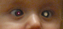
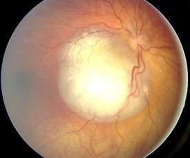
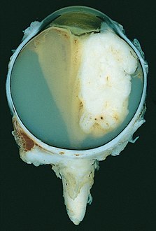
A blastoma is a type of cancer, more common in children, that is caused by malignancies in precursor cells, often called blasts. Examples are nephroblastoma, medulloblastoma, and retinoblastoma. The suffix -blastoma is used to imply a tumor of primitive, incompletely differentiated cells, e.g., chondroblastoma is composed of cells resembling the precursor of chondrocytes.

<span class="mw-page-title-main">Uveal melanoma</span> Type of eye cancer

Uveal melanoma is a type of eye cancer in the uvea of the eye. It is traditionally classed as originating in the iris, choroid, and ciliary body, but can also be divided into class I and class II. Symptoms include blurred vision, loss of vision or photopsia, but there may be no symptoms.

<span class="mw-page-title-main">Choroideremia</span> Medical condition

Choroideremia is a rare, X-linked recessive form of hereditary retinal degeneration that affects roughly 1 in 50,000 males. The disease causes a gradual loss of vision, starting with childhood night blindness, followed by peripheral vision loss and progressing to loss of central vision later in life. Progression continues throughout the individual's life, but both the rate of change and the degree of visual loss are variable among those affected, even within the same family.

Stargardt disease is the most common inherited single-gene retinal disease. In terms of the first description of the disease, it follows an autosomal recessive inheritance pattern, which has been later linked to bi-allelic ABCA4 gene variants (STGD1). However, there are Stargardt-like diseases with mimicking phenotypes that are referred to as STGD3 and STGD4, and have a autosomal dominant inheritance due to defects with ELOVL4 or PROM1 genes, respectively. It is characterized by macular degeneration that begins in childhood, adolescence or adulthood, resulting in progressive loss of vision.

<span class="mw-page-title-main">Eye neoplasm</span> Medical condition

An eye neoplasm is a tumor of the eye. A rare type of tumor, eye neoplasms can affect all parts of the eye, and can either be benign or malignant (cancerous), in which case it is known as eye cancer. Eye cancers can be primary or metastatic cancer. The two most common cancers that spread to the eye from another organ are breast cancer and lung cancer. Other less common sites of origin include the prostate, kidney, thyroid, skin, colon and blood or bone marrow.

Optic neuropathy is damage to the optic nerve from any cause. The optic nerve is a bundle of millions of fibers in the retina that sends visual signals to the brain.

Intraocular lymphoma is a rare malignant form of eye cancer. Intraocular lymphoma may affect the eye secondarily from a metastasis from a non-ocular tumor or may arise within the eye primarily. PIOL is a subset of primary central nervous system lymphoma (PCNSL). PCNSL are most commonly a diffuse large B-cell immunohistologic subtype of non-Hodgkin's lymphoma according to the World Health Organization (WHO) classification of lymphomas. The most common symptoms of PIOL include blurred or decreased vision due to tumor cells in the vitreous. Most cases of PIOL eventuate to central nervous system involvement (PCNSL) while only 20% of PCNSL lead to intraocular (PIOL) involvement. PIOL and PCNSL remain enigmas because both structures are immunologically privileged sites and so do not normally have immune cells trafficking through these structures. What is more, while the vast majority of PCNSL in patients with acquired immune deficiency syndrome (AIDS) is related to the Epstein-Barr virus (EBV), the development of PCNSL and PIOL in immunocompetent patients is unknown and shows no general relation to infectious DNAs.

<span class="mw-page-title-main">Intraocular hemorrhage</span> Medical condition

Intraocular hemorrhage is bleeding inside the eye. Bleeding can occur from any structure of the eye where there is vasculature or blood flow, including the anterior chamber, vitreous cavity, retina, choroid, suprachoroidal space, or optic disc.

<span class="mw-page-title-main">Pineoblastoma</span> Medical condition

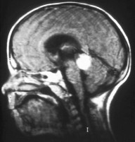
Pineoblastoma is a malignant tumor of the pineal gland. A pineoblastoma is a supratentorial midline primitive neuroectodermal tumor. Pineoblastoma can present at any age, but is most common in young children. They account for 0.001% of all primary CNS neoplasms.

<span class="mw-page-title-main">Persistent fetal vasculature</span> Medical condition

Persistent fetal vasculature(PFV), also known as persistent fetal vasculature syndrome (PFVS), and until 1997 known primarily as persistent hyperplastic primary vitreous (PHPV), is a rare congenital anomaly which occurs when blood vessels within the developing eye, known as the embryonic hyaloid vasculature network, fail to regress as they normally would in-utero after the eye is fully developed. Defects which arise from this lack of vascular regression are diverse; as a result, the presentation, symptoms, and prognosis of affected patients vary widely, ranging from clinical insignificance to irreversible blindness. The underlying structural causes of PFV are considered to be relatively common, and the vast majority of cases do not warrant additional intervention. When symptoms do manifest, however, they are often significant, causing detrimental and irreversible visual impairment. Persistent fetal vasculature heightens the lifelong risk of glaucoma, cataracts, intraocular hemorrhages, and Retinal detachments, accounting for the visual loss of nearly 5% of the blind community in the developed world. In diagnosed cases of PFV, approximately 90% of patients with a unilateral disease have associated poor vision in the affected eye.

Retinal gene therapy holds a promise in treating different forms of non-inherited and inherited blindness.

<span class="mw-page-title-main">Orbital lymphoma</span> Human disease of the eye

Orbital lymphoma is a common type of non-Hodgkin lymphoma that occurs near or on the eye. Common symptoms include decreased vision and uveitis. Orbital lymphoma can be diagnosed via a biopsy of the eye and is usually treated with radiotherapy or in combination with chemotherapy.

Targeted molecular therapy for neuroblastoma involves treatment aimed at molecular targets that have a unique expression in this form of cancer. Neuroblastoma, the second most common pediatric malignant tumor, often involves treatment through intensive chemotherapy. A number of molecular targets have been identified for the treatment of high-risk forms of this disease. Aiming treatment in this way provides a more selective way to treat the disease, decreasing the risk for toxicities that are associated with the typical treatment regimen. Treatment using these targets can supplement or replace some of the intensive chemotherapy that is used for neuroblastoma. These molecular targets of this disease include GD2, ALK, and CD133. GD2 is a target of immunotherapy, and is the most fully developed of these treatment methods, but is also associated with toxicities. ALK has more recently been discovered, and drugs in development for this target are proving to be successful in neuroblastoma treatment. The role of CD133 in neuroblastoma has also been more recently discovered and is an effective target for treatment of this disease.

<span class="mw-page-title-main">J. William Harbour</span> American ophthalmologist and researcher

J. William Harbour is an American ophthalmologist, ocular oncologist and cancer researcher. He is Chair of the Department of Ophthalmology at the University of Texas Southwestern Medical Center in Dallas. He previously served as the vice chair and director of ocular oncology at the Bascom Palmer Eye Institute and associate director for basic science at the Sylvester Comprehensive Cancer Center of the University of Miami's Miller School of Medicine.

Occult macular dystrophy (OMD) is a rare inherited degradation of the retina, characterized by progressive loss of function in the most sensitive part of the central retina (macula), the location of the highest concentration of light-sensitive cells (photoreceptors) but presenting no visible abnormality. "Occult" refers to the degradation in the fundus being difficult to discern. The disorder is called "dystrophy" instead of "degradation" to distinguish its genetic origin from other causes, such as age. OMD was first reported by Y. Miyake et al. in 1989.

<span class="mw-page-title-main">Congenital blindness</span>

Congenital blindness refers to blindness present at birth. Congenital blindness is sometimes used interchangeably with "Childhood Blindness." However, current literature has various definitions of both terms. Childhood blindness encompasses multiple diseases and conditions present in ages up to 16 years old, which can result in permanent blindness or severe visual impairment over time. Congenital blindness is a hereditary disease and can be treated by gene therapy. Visual loss in children or infants can occur either at the prenatal stage or postnatal stage. There are multiple possible causes of congenital blindness. In general, 60% of congenital blindness cases are contributed from prenatal stage and 40% are contributed from inherited disease. However, most of the congenital blindness cases show that it can be avoidable or preventable with early treatment.

Sickle cell retinopathy can be defined as retinal changes due to blood vessel damage in the eye of a person with a background of sickle cell disease. It can likely progress to loss of vision in late stages due to vitreous hemorrhage or retinal detachment. Sickle cell disease is a structural red blood cell disorder leading to consequences in multiple systems. It is characterized by chronic red blood cell destruction, vascular injury, and tissue ischemia causing damage to the brain, eyes, heart, lungs, kidneys, spleen, and musculoskeletal system.

References

  1. 1 2 3 4 "Retinoblastoma". St. Jude Children's Research Hospital . Retrieved March 9, 2022.
  2. 1 2 3 Dimaras H, Corson TW, Cobrinik D, White A, Zhao J, Munier FL, et al. (August 2015). "Retinoblastoma". Nature Reviews. Disease Primers. 1: 15021. doi:10.1038/nrdp.2015.21. PMC   5744255 . PMID   27189421.
  3. 1 2 "Retinoblastoma - Symptoms and causes". Mayo Clinic. Retrieved 2021-03-17.
  4. Rao R, Honavar SG (December 2017). "Retinoblastoma". Indian Journal of Pediatrics. 84 (12): 937–944. doi:10.1007/s12098-017-2395-0. PMID   28620731.
  5. 1 2 American Cancer Society (2003). "Chapter 85. Neoplasms of the Eye". Cancer Medicine . Hamilton, Ontario: BC Decker Inc. ISBN   978-1-55009-213-4.
  6. Warda O, Naeem Z, Roelofs KA, Sagoo MS, Reddy MA (April 2023). "Retinoblastoma and vision". Eye. 37 (5): 797–808. doi:10.1038/s41433-021-01845-y. PMC   10050411 . PMID   34987197. S2CID   245672434.
  7. Ryan SJ, Schachat AP, Wilkinson CP, Hinton DR, Sadda SR, Wiedemann P (2012-11-01). Retina. Elsevier Health Sciences. p. 2105. ISBN   978-1455737802.
  8. Dimaras H, Gallie BL (2016). "Retinoblastoma Protein, Biological and Clinical Functions". In Schwab M (ed.). Encyclopedia of Cancer. Berlin, Heidelberg: Springer. pp. 1–5. doi:10.1007/978-3-642-27841-9_5069-9. ISBN   978-3-642-27841-9.
  9. Dimaras H, Kimani K, Dimba EA, Gronsdahl P, White A, Chan HS, et al. (April 2012). "Retinoblastoma". Lancet. 379 (9824): 1436–1446. doi: 10.1016/s0140-6736(11)61137-9 . PMID   22414599. S2CID   235331617.
  10. Elkington AR, Khaw PT (September 1988). "ABC of eyes. Squint". BMJ. 297 (6648): 608–611. doi:10.1136/bmj.297.6648.608. PMC   1834556 . PMID   3139234.
  11. "Retinoblastoma | National Eye Institute". www.nei.nih.gov. Archived from the original on 2022-08-26. Retrieved 2022-08-26.
  12. "Seen a white glow in a photograph?". chect.org.uk. The Childhood Eye Cancer Trust. 18 January 2017. Retrieved 2022-12-31.
  13. Balmer A, Munier F (December 2007). "Differential diagnosis of leukocoria and strabismus, first presenting signs of retinoblastoma". Clinical Ophthalmology. 1 (4): 431–439. PMC   2704541 . PMID   19668520.
  14. Introduction to White Eye Archived 2011-04-26 at the Wayback Machine , Daisy's Eye Cancer Fund.
  15. 1 2 3 4 Du W, Pogoriler J (August 2006). "Retinoblastoma family genes". Oncogene. 25 (38): 5190–5200. doi:10.1038/sj.onc.1209651. PMC   1899835 . PMID   16936737.
  16. Lohmann DR, Gallie BL (1993). "Retinoblastoma". In Adam MP, Feldman J, Mirzaa GM, Pagon RA, Wallace SE, Bean LJ, Gripp KW, Amemiya A (eds.). GeneReviews®. Seattle (WA): University of Washington, Seattle. PMID   20301625 . Retrieved 2022-06-27.
  17. Kivelä T (June 1999). "Trilateral retinoblastoma: a meta-analysis of hereditary retinoblastoma associated with primary ectopic intracranial retinoblastoma". Journal of Clinical Oncology. 17 (6): 1829–1837. doi:10.1200/JCO.1999.17.6.1829. PMID   10561222.
  18. de Jong MC, Kors WA, de Graaf P, Castelijns JA, Kivelä T, Moll AC (September 2014). "Trilateral retinoblastoma: a systematic review and meta-analysis". The Lancet. Oncology. 15 (10): 1157–1167. doi:10.1016/s1470-2045(14)70336-5. PMID   25126964.
  19. Harbour J.W., Dean D.C. Rb function in cell-cycle regulation and apoptosis" Nature Cell Biology. 2000;94:E65–E67.
  20. Parsam VL, Kannabiran C, Honavar S, Vemuganti GK, Ali MJ (December 2009). "A comprehensive, sensitive and economical approach for the detection of mutations in the RB1 gene in retinoblastoma". Journal of Genetics. 88 (4): 517–527. doi:10.1007/s12041-009-0069-z. PMID   20090211. S2CID   10723496.
  21. Lohmann DR, Gallie BL (2010). "Retinoblastoma". GeneReviews. Seattle, WA: University of Washington. PMID   20301625.
  22. Ali MJ, Parsam VL, Honavar SG, Kannabiran C, Vemuganti GK, Reddy VA (October 2010). "RB1 gene mutations in retinoblastoma and its clinical correlation". Saudi Journal of Ophthalmology. 24 (4): 119–123. doi:10.1016/j.sjopt.2010.05.003. PMC   3729507 . PMID   23960888.
  23. Rushlow DE, Mol BM, Kennett JY, Yee S, Pajovic S, Thériault BL, et al. (April 2013). "Characterisation of retinoblastomas without RB1 mutations: genomic, gene expression, and clinical studies". The Lancet. Oncology. 14 (4): 327–334. doi:10.1016/S1470-2045(13)70045-7. PMID   23498719.
  24. Woo CW, Tan F, Cassano H, Lee J, Lee KC, Thiele CJ (February 2008). "Use of RNA interference to elucidate the effect of MYCN on cell cycle in neuroblastoma". Pediatric Blood & Cancer. 50 (2): 208–212. doi:10.1002/pbc.21195. PMID   17420990. S2CID   22765085.
  25. Stenfelt S, Blixt MK, All-Ericsson C, Hallböök F, Boije H (November 2017). "Heterogeneity in retinoblastoma: a tale of molecules and models". Clinical and Translational Medicine. 6 (1): 42. doi: 10.1186/s40169-017-0173-2 . PMC   5680409 . PMID   29124525.
  26. Lewis R (March 19, 2013). "Some Aggressive Retinoblastomas Lack RB1 Mutations". Medscape Online. Archived from the original on September 19, 2017.
  27. Malik AN, Evans JR, Gupta S, Mariotti S, Gordon I, Bowman R, et al. (October 2022). "Universal newborn eye screening: a systematic review of the literature and review of international guidelines". Journal of Global Health. 12: 12003. doi:10.7189/jogh.12.12003. PMC   9586142 . PMID   36269293.
  28. Tonorezos ES, Friedman DN, Barnea D, Bosscha MI, Chantada G, Dommering CJ, et al. (November 2020). "Recommendations for Long-Term Follow-up of Adults with Heritable Retinoblastoma". Ophthalmology. 127 (11): 1549–1557. doi:10.1016/j.ophtha.2020.05.024. PMC   7606265 . PMID   32422154.
  29. MacCarthy A, Birch JM, Draper GJ, Hungerford JL, Kingston JE, Kroll ME, et al. (January 2009). "Retinoblastoma in Great Britain 1963-2002". The British Journal of Ophthalmology. 93 (1): 33–37. doi:10.1136/bjo.2008.139618. PMID   18838413. S2CID   27049728.
  30. de Jong MC, de Graaf P, Noij DP, Göricke S, Maeder P, Galluzzi P, et al. (May 2014). "Diagnostic performance of magnetic resonance imaging and computed tomography for advanced retinoblastoma: a systematic review and meta-analysis". Ophthalmology. 121 (5): 1109–1118. doi:10.1016/j.ophtha.2013.11.021. PMID   24589388.
  31. Sehu WK, Lee W (2005). Ophthalmic pathology : an illustrated guide for clinicians. Malden: Blackwell Publishing. p. 262. ISBN   978-0-7279-1779-9.
  32. Kumar V, Abbas AK, Fausto N (1999). Robbins pathologic basis of disease (6th ed.). Philadelphia: Saunders. p. 1442. ISBN   978-0-7216-0187-8.
  33. Schüler A, Weber S, Neuhäuser M, Jurklies C, Lehnert T, Heimann H, et al. (March 2005). "Age at diagnosis of isolated unilateral retinoblastoma does not distinguish patients with and without a constitutional RB1 gene mutation but is influenced by a parent-of-origin effect". European Journal of Cancer. 41 (5): 735–740. doi:10.1016/j.ejca.2004.12.022. PMID   15763650.
  34. 1 2 Rushlow D, Piovesan B, Zhang K, Prigoda-Lee NL, Marchong MN, Clark RD, et al. (May 2009). "Detection of mosaic RB1 mutations in families with retinoblastoma". Human Mutation. 30 (5): 842–851. doi:10.1002/humu.20940. PMID   19280657. S2CID   31887184.
  35. 1 2 Richter S, Vandezande K, Chen N, Zhang K, Sutherland J, Anderson J, et al. (February 2003). "Sensitive and efficient detection of RB1 gene mutations enhances care for families with retinoblastoma". American Journal of Human Genetics. 72 (2): 253–269. doi:10.1086/345651. PMC   379221 . PMID   12541220.
  36. Canadian Ophthalmological Society (December 2009). "National Retinoblastoma Strategy Canadian Guidelines for Care: Stratégie thérapeutique du rétinoblastome guide clinique canadien" (PDF). Canadian Journal of Ophthalmology. Journal Canadien d'Ophtalmologie. 44 (Suppl 2): S1-88. doi:10.3129/i09-194. PMID   20237571. Archived from the original (PDF) on 2011-09-29.
  37. Dimaras H, Corson TW (January 2019). "Retinoblastoma, the visible CNS tumor: A review". Journal of Neuroscience Research. 97 (1): 29–44. doi:10.1002/jnr.24213. PMC   6034991 . PMID   29314142.
  38. 1 2 3 Chawla B, Jain A, Azad R (September 2013). "Conservative treatment modalities in retinoblastoma". Indian Journal of Ophthalmology. 61 (9): 479–485. doi: 10.4103/0301-4738.119424 . PMC   3831762 . PMID   24104705.
  39. 1 2 Fabian ID, Reddy A, Sagoo MS (2018). "Classification and staging of retinoblastoma". Community Eye Health. 31 (101): 11–13. PMC   5998397 . PMID   29915461.
  40. 1 2 Chintagumpala M, Chevez-Barrios P, Paysse EA, Plon SE, Hurwitz R (October 2007). "Retinoblastoma: review of current management". The Oncologist. 12 (10): 1237–1246. CiteSeerX   10.1.1.585.5448 . doi:10.1634/theoncologist.12-10-1237. PMID   17962617. S2CID   15465073.
  41. Li WL, Buckley J, Sanchez-Lara PA, Maglinte DT, Viduetsky L, Tatarinova TV, et al. (July 2016). "A Rapid and Sensitive Next-Generation Sequencing Method to Detect RB1 Mutations Improves Care for Retinoblastoma Patients and Their Families". The Journal of Molecular Diagnostics. 18 (4): 480–493. doi:10.1016/j.jmoldx.2016.02.006. PMC   5820122 . PMID   27155049.
  42. Zhao B, Li B, Liu Q, Gao F, Zhang Z, Bai H, et al. (September 2020). "Effects of matrine on the proliferation and apoptosis of vincristine-resistant retinoblastoma cells". Experimental and Therapeutic Medicine. 20 (3): 2838–2844. doi:10.3892/etm.2020.8992. PMC   7401942 . PMID   32765780.
  43. Li C, Dong K, Zhuang Y, Luo Z, Qiu D, Luo Y, et al. (March 2024). "ACOT7 promotes retinoblastoma resistance to vincristine by regulating fatty acid metabolism reprogramming". Heliyon. 10 (5): e27156. doi: 10.1016/j.heliyon.2024.e27156 . PMC   10920713 . PMID   38463820.
  44. Roarty JD, McLean IW, Zimmerman LE (November 1988). "Incidence of second neoplasms in patients with bilateral retinoblastoma". Ophthalmology. 95 (11): 1583–1587. doi:10.1016/s0161-6420(88)32971-4. PMID   3211467.
  45. Shields CL, Ramasubramanian A, Rosenwasser R, Shields JA (September 2009). "Superselective catheterization of the ophthalmic artery for intraarterial chemotherapy for retinoblastoma". Retina. 29 (8): 1207–1209. doi:10.1097/IAE.0b013e3181b4ce39. PMID   19734768. S2CID   47557934.
  46. Shome D, Poddar N, Sharma V, Sheorey U, Maru GB, Ingle A, et al. (December 2009). "Does a nanomolecule of Carboplatin injected periocularly help in attaining higher intravitreal concentrations?". Investigative Ophthalmology & Visual Science. 50 (12): 5896–5900. doi:10.1167/iovs.09-3914. PMID   19628744. S2CID   7361361.
  47. Kang SJ, Durairaj C, Kompella UB, O'Brien JM, Grossniklaus HE (August 2009). "Subconjunctival nanoparticle carboplatin in the treatment of murine retinoblastoma". Archives of Ophthalmology. 127 (8): 1043–1047. doi:10.1001/archophthalmol.2009.185. PMC   2726977 . PMID   19667343.
  48. Fabian ID, Johnson KP, Stacey AW, Sagoo MS, Reddy MA (June 2017). "Focal laser treatment in addition to chemotherapy for retinoblastoma". The Cochrane Database of Systematic Reviews. 2017 (6): CD012366. doi:10.1002/14651858.CD012366.pub2. PMC   6481366 . PMID   28589646.
  49. Shields CL, Mashayekhi A, Cater J, Shelil A, Ness S, Meadows AT, et al. (June 2005). "Macular retinoblastoma managed with chemoreduction: analysis of tumor control with or without adjuvant thermotherapy in 68 tumors". Archives of Ophthalmology. 123 (6): 765–773. doi:10.1001/archopht.123.6.765. PMID   15955977.
  50. Beddard N, McGeechan GJ, Taylor J, Swainston K (March 2020). "Childhood eye cancer from a parental perspective: The lived experience of parents with children who have had retinoblastoma". European Journal of Cancer Care. 29 (2): e13209. doi: 10.1111/ecc.13209 . PMID   31845431. S2CID   196549559.
  51. Concise ophthalmology : for medical students (4th ed.). Karachi, Pakistan: Paramount Books. 2014. pp. 80–81. ISBN   9789696370017.
  52. Rai P, Shah IA, Narsani AK, Lohana MK, Memon MK, Memon MA (2009). "Too late presentation of 53 patients with retinoblastoma". International Journal of Ophthalmology. 9 (2): 227–230. doi:10.3969/j.issn.1672-5123.2009.02.005.
  53. Abramson DH, Schefler AC (December 2004). "Update on retinoblastoma". Retina. 24 (6): 828–848. doi:10.1097/00006982-200412000-00002. PMID   15579980. S2CID   26883037.
  54. Orjuela M, Castaneda VP, Ridaura C, Lecona E, Leal C, Abramson DH, et al. (October 2000). "Presence of human papilloma virus in tumor tissue from children with retinoblastoma: an alternative mechanism for tumor development". Clinical Cancer Research. 6 (10): 4010–4016. PMID   11051250.
  55. Abramson DH, Frank CM, Susman M, Whalen MP, Dunkel IJ, Boyd NW (March 1998). "Presenting signs of retinoblastoma". The Journal of Pediatrics. 132 (3 Pt 1): 505–508. doi:10.1016/s0022-3476(98)70028-9. PMID   9544909.