Medulloblastoma

Last updated
Medulloblastoma
CT brain scan of child with medulloblastoma and resulting hydrocephalus.jpg
CT scan, showing a tumorous mass in the posterior fossa, giving rise to obstructive hydrocephalus, in a six-year-old girl
Pronunciation
Specialty Neuro-oncology, neurosurgery
Symptoms Headaches
Nausea
Vomiting
Tiredness
Clumsiness
Dizziness
Change in vision
Handwriting problems [1]
Usual onsetBetween 5 and 9 years old [1]
Prognosis Five-year survival rate: 72.1% [2]
FrequencyAbout 500 children diagnosed annually in the United States [1]

Medulloblastoma is a common type of primary brain cancer in children. It originates in the part of the brain that is towards the back and the bottom, on the floor of the skull, in the cerebellum, or posterior fossa. [3]

Contents

The brain is divided into two main parts, the larger cerebrum on top and the smaller cerebellum below towards the back. They are separated by a membrane called the tentorium. Tumors that originate in the cerebellum or the surrounding region below the tentorium are, therefore, called infratentorial.

Historically, medulloblastomas have been classified as a primitive neuroectodermal tumor (PNET), but it is now known that medulloblastoma is distinct from supratentorial PNETs and they are no longer considered similar entities. [4]

Medulloblastomas are invasive, rapidly growing tumors that, unlike most brain tumors, spread through the cerebrospinal fluid and frequently metastasize to different locations along the surface of the brain and spinal cord. Metastasis all the way down to the cauda equina at the base of the spinal cord is termed "drop metastasis".

The cumulative relative survival rate for all age groups and histology follow-up was 60%, 52%, and 47% at 5 years, 10 years, and 20 years, respectively, with children doing better than adults. [5]

Signs and symptoms

Signs and symptoms are mainly due to secondary increased intracranial pressure due to blockage of the fourth ventricle and tumors are usually present for 1 to 5 months before diagnosis is made. The child typically becomes listless, with repeated episodes of vomiting, and a morning headache, which may lead to a misdiagnosis of gastrointestinal disease or migraine. [6] Soon after, the child will develop a stumbling gait, truncal ataxia, frequent falls, diplopia, papilledema, and sixth cranial nerve palsy. Positional vertigo and nystagmus are also frequent, and facial sensory loss or motor weakness may be present. Decerebrate attacks appear late in the disease.

Extraneural metastasis to the rest of the body is rare, and when it occurs, it is in the setting of relapse, more commonly in the era prior to routine chemotherapy.

Pathogenesis

Medulloblastomas are usually found in the vicinity of the fourth ventricle, between the brainstem and the cerebellum. Tumors with similar appearance and characteristics originate in other parts of the brain, but they are not identical to medulloblastoma. [7]

Although medulloblastomas are thought to originate from immature or embryonal cells at their earliest stage of development, the cell of origin depends on the subgroup of medulloblastoma. WNT tumors originate from the lower rhombic lip of the brainstem, while SHH tumors originate from the external granular layer. [8]

Currently, medulloblastomas are thought to arise from cerebellar stem cells that have been prevented from dividing and differentiating into their normal cell types. This accounts for the histologic variants seen on biopsy. Both perivascular pseudorosette and Homer Wright pseudorosette formations are highly characteristic of medulloblastomas and are seen in up to half of cases. [9] The classic rosette with tumor cells around a central lumen can be seen. [10]

In the past, medulloblastoma was classified using histology, but integrated genomic studies have revealed that medulloblastoma is composed of four distinct molecular and clinical variants termed WNT/β-catenin, Sonic Hedgehog, Group 3, and Group 4. [11] Of these subgroups, WNT patients have an excellent prognosis and group 3 patients have a poor prognosis. Also, a subgroup-specific alternative splicing further confirms the existence of distinct subgroups and highlights the transcriptional heterogeneity between subgroups. [12] Amplification of the Sonic Hedgehog pathway is the best characterized subgroup, with 25% of human tumors having mutations in Patched, Sufu (Suppressor of Fused Homolog), Smoothened, or other genes in this pathway. [13] [14] Medulloblastomas are also seen in Gorlin syndrome as well as Turcot syndrome. Recurrent mutations in the genes CTNNB1, PTCH1, MLL2, SMARCA4, DDX3X, CTDNEP1, KDM6A , and TBR1 were identified in individuals with medulloblastoma. [15] Additional pathways disrupted in some medulloblastomas include MYC, Notch, BMP, and TGF-β signaling pathways. [13] [14] [6] [16] [17] [18] [19] [3] [ excessive citations ]

Diagnosis

The tumor is distinctive on T1- and T2-weighted MRI with heterogeneous enhancement and a typical location adjacent to and extension into the fourth ventricle. Histologically, the tumor is solid, pink-gray in color, and is well circumscribed. The tumor is very cellular, with high mitotic activity, little cytoplasm, and a tendency to form clusters and rosettes.

The Chang staging system can be used in making the diagnosis. [20]

DNA methylation profiling of medulloblastoma allows robust sub-classification and improved outcome prediction using formalin-fixed biopsies. [21]

Correct diagnosis of medulloblastoma may require ruling out atypical teratoid rhabdoid tumor. [22]

Treatment

Treatment begins with maximal surgical removal of the tumor. The addition of radiation to the entire neuraxis and chemotherapy may increase the disease-free survival. This combination may permit a 5-year survival in more than 80% of cases. Some evidence indicates that proton beam irradiation reduces the impact of radiation on the cochlear and cardiovascular areas and reduces the cognitive late effects of cranial irradiation. [23] [24]

The presence of desmoplastic features such as connective tissue formation offers a better prognosis. Prognosis is worse if the child is less than 3 years old, degree of resection is inadequate, or if any CSF, spinal, supratentorial, or systemic spread occurs. Dementia after radiotherapy and chemotherapy is a common outcome appearing two to four years following treatment. Side effects from radiation treatment can include cognitive impairment, psychiatric illness, bone growth retardation, hearing loss, and endocrine disruption. [3] [6] [16] Increased intracranial pressure may be controlled with corticosteroids or a ventriculoperitoneal shunt. An approach to monitor tumor development and treatment response by liquid biopsy is promising, but remains challenging. [25]

Chemotherapy

Chemotherapy is often used as part of treatment. Evidence of benefit, however, is not clear as of 2013. [26] A few different chemotherapeutic regimens for medulloblastoma are used; most involve a combination of lomustine, cisplatin, carboplatin, vincristine, or cyclophosphamide. In younger patients (less than 3–4 years of age), chemotherapy can delay, or in some cases possibly even eliminate, the need for radiotherapy. However, both chemotherapy and radiotherapy often have long-term toxicity effects, including delays in physical and cognitive development, higher risk of second cancers, and increased cardiac disease risks. [27] [28]

Outcomes

Array-based karyotyping of 260 medulloblastomas resulted in the following clinical subgroups based on cytogenetic profiles: [29]

Transcriptional profiling shows the existence of four main subgroups (Wnt, Shh, Group 3, and Group 4). [11]

Survival

The historical cumulative relative survival rate for all age groups and histology follow-up was 60%, 52%, and 47% at 5 years, 10 years, and 20 years, respectively. Patients diagnosed with a medulloblastoma or PNET are 50 times more likely to die than a matched member of the general population. A population-based (SEER) 5-year relative survival rates indicated 69% overall: 72% in children (1–9 years) and 67% in adults (20+ years). The 20-year survival rate is 51% in children. Children and adults have different survival profiles, with adults faring worse than children only after the fourth year after diagnosis (after controlling for increased background mortality). Before the fourth year, survival probabilities are nearly identical. [5] Long-term sequelae of standard treatment include hypothalamic-pituitary and thyroid dysfunction and intellectual impairment. The hormonal and intellectual deficits created by these therapies causes significant impairment of the survivors. [30] [ self-published source? ]

In current clinical studies, the patients are divided into low-, standard- and high-risk groups:

Epidemiology

Medulloblastomas affect just under two people per million per year, and affect children 10 times more than adults. [36] Medulloblastoma is the second-most frequent brain tumor in children after pilocytic astrocytoma [37] and the most common malignant brain tumor in children, comprising 14.5% of newly diagnosed brain tumors. [38] In adults, medulloblastoma is rare, comprising fewer than 2% of CNS malignancies. [39]

The rate of new cases of childhood medulloblastoma is higher in males (62%) than females (38%), a feature that is not seen in adults. [36] [40] Medulloblastoma and other PNET`s are more prevalent in younger children than older children. About 40% of medulloblastoma patients are diagnosed before the age of five, 31% are between the ages of 5 and 9, 18.3% are between the ages of 10 and 14, and 12.7% are between the ages of 15 and 19. [41]

Research models

Using gene transfer of SV40 large T-antigen in neuronal precursor cells of rats, a brain tumor model was established. The PNETs were histologically indistinguishable from the human counterparts and have been used to identify new genes involved in human brain tumor carcinogenesis. [42] The model was used to confirm p53 as one of the genes involved in human medulloblastomas, but since only about 10% of the human tumors showed mutations in that gene, the model can be used to identify the other binding partners of SV40 Large T- antigen, other than p53. [43] [44] In a mouse model, high medulloblastoma frequency appears to be caused by the down regulation of Cxcl3, with Cxcl3 being induced by Tis21. [45] Consistently, the treatment with Cxcl3 completely prevents the growth of medulloblastoma lesions in a Shh-type mouse model of medulloblastoma. [46] Thus, CXCL3 is a target for medulloblastoma therapy.

Related Research Articles

<span class="mw-page-title-main">Brain tumor</span> Neoplasm in the brain

A brain tumor occurs when abnormal cells form within the brain. There are two main types of tumors: malignant (cancerous) tumors and benign (non-cancerous) tumors. These can be further classified as primary tumors, which start within the brain, and secondary tumors, which most commonly have spread from tumors located outside the brain, known as brain metastasis tumors. All types of brain tumors may produce symptoms that vary depending on the size of the tumor and the part of the brain that is involved. Where symptoms exist, they may include headaches, seizures, problems with vision, vomiting and mental changes. Other symptoms may include difficulty walking, speaking, with sensations, or unconsciousness.

<span class="mw-page-title-main">Retinoblastoma</span> Medical condition

Retinoblastoma (Rb) is a rare form of cancer that rapidly develops from the immature cells of a retina, the light-detecting tissue of the eye. It is the most common primary malignant intraocular cancer in children, and it is almost exclusively found in young children.

<span class="mw-page-title-main">Oligodendroglioma</span> Medical condition

Oligodendrogliomas are a type of glioma that are believed to originate from the oligodendrocytes of the brain or from a glial precursor cell. They occur primarily in adults but are also found in children.

<span class="mw-page-title-main">Small-cell carcinoma</span> Type of malignant cancer

Small-cell carcinoma is a type of highly malignant cancer that most commonly arises within the lung, although it can occasionally arise in other body sites, such as the cervix, prostate, and gastrointestinal tract. Compared to non-small cell carcinoma, small cell carcinoma is more aggressive, with a shorter doubling time, higher growth fraction, and earlier development of metastases.

<span class="mw-page-title-main">Neuroblastoma</span> Medical condition

Neuroblastoma (NB) is a type of cancer that forms in certain types of nerve tissue. It most frequently starts from one of the adrenal glands but can also develop in the head, neck, chest, abdomen, or spine. Symptoms may include bone pain, a lump in the abdomen, neck, or chest, or a painless bluish lump under the skin.

Adjuvant therapy, also known as adjunct therapy, adjuvant care, or augmentation therapy, is a therapy that is given in addition to the primary or initial therapy to maximize its effectiveness. The surgeries and complex treatment regimens used in cancer therapy have led the term to be used mainly to describe adjuvant cancer treatments. An example of such adjuvant therapy is the additional treatment usually given after surgery where all detectable disease has been removed, but where there remains a statistical risk of relapse due to the presence of undetected disease. If known disease is left behind following surgery, then further treatment is not technically adjuvant.

<span class="mw-page-title-main">Blastoma</span> Type of cancer arising from precursor cells

A blastoma is a type of cancer, more common in children, that is caused by malignancies in precursor cells, often called blasts. Examples are nephroblastoma, medulloblastoma, and retinoblastoma. The suffix -blastoma is used to imply a tumor of primitive, incompletely differentiated cells, e.g., chondroblastoma is composed of cells resembling the precursor of chondrocytes.

<span class="mw-page-title-main">Anaplastic thyroid cancer</span> Medical condition

Anaplastic thyroid cancer (ATC), also known as anaplastic thyroid carcinoma, is an aggressive form of thyroid cancer characterized by uncontrolled growth of cells in the thyroid gland. This form of cancer generally carries a very poor prognosis due to its aggressive behavior and resistance to cancer treatments. The cells of anaplastic thyroid cancer are highly abnormal and usually no longer resemble the original thyroid cells and have poor differentiation.

<span class="mw-page-title-main">Primitive neuroectodermal tumor</span> Medical condition

Primitive neuroectodermal tumor is a malignant (cancerous) neural crest tumor. It is a rare tumor, usually occurring in children and young adults under 25 years of age. The overall 5 year survival rate is about 53%.

<span class="mw-page-title-main">Undifferentiated pleomorphic sarcoma</span> Medical condition

Undifferentiated pleomorphic sarcoma (UPS), also termed pleomorphic myofibrosarcoma, high-grade myofibroblastic sarcoma, and high-grade myofibrosarcoma, is characterized by the World Health Organization (WHO), 2020, as a rare, poorly differentiated neoplasm, i.e. an abnormal growth of cells that have an unclear identity and/or cell of origin. WHO classified it as one of the undifferentiated/unclassified sarcomas in the category of tumors of uncertain differentiation. Sarcomas are cancers known or thought to derive from mesenchymal stem cells that typically develop in bone, muscle, fat, blood vessels, lymphatic vessels, tendons, and ligaments. More than 70 sarcoma subtypes have been described. The UPS subtype of these sarcomas consists of tumor cells that are poorly differentiated and may appear as spindle-shaped cells, histiocytes, and giant cells. UPS is considered a diagnosis that defies formal sub-classification after thorough histologic, immunohistochemical, and ultrastructural examinations fail to identify the type of cells involved.

<span class="mw-page-title-main">Atypical teratoid rhabdoid tumor</span> Medical condition

An atypical teratoid rhabdoid tumor (AT/RT) is a rare tumor usually diagnosed in childhood. Although usually a brain tumor, AT/RT can occur anywhere in the central nervous system (CNS), including the spinal cord. About 60% will be in the posterior cranial fossa. One review estimated 52% in the posterior fossa, 39% are supratentorial primitive neuroectodermal tumors (sPNET), 5% are in the pineal, 2% are spinal, and 2% are multifocal.

Alveolar rhabdomyosarcoma (ARMS) is a subtype of the rhabdomyosarcoma soft tissue cancer family whose lineage is from mesenchymal cells and are related to skeletal muscle cells. ARMS tumors resemble the alveolar tissue in the lungs. Tumor location varies from patient to patient, but is commonly found in the head and neck region, male and female urogenital tracts, the torso, and extremities. Two fusion proteins can be associated with ARMS, but are not necessary, PAX3-FKHR. and PAX7-FKHR. In children and adolescents ARMS accounts for about 1 percent of all malignancies, has an incidence rate of 1 per million, and most cases occur sporadically with no genetic predisposition. PAX3-FOXO1 is now known to drive cancer-promoting gene expression programs through creation of distant genetic elements called super enhancers.

Triple-negative breast cancer (TNBC) is any breast cancer that either lacks or shows low levels of estrogen receptor (ER), progesterone receptor (PR) and human epidermal growth factor receptor 2 (HER2) overexpression and/or gene amplification. Triple-negative is sometimes used as a surrogate term for basal-like.

The Ewing family of tumors (EFTs) is a group of small cell sarcomas including Ewing sarcoma of the bone, extra osseous Ewing tumors, and primitive neuroectodermal tumors. They are rare cancers, usually diagnosed in peoples' twenties. The sarcoma of bone is the most common of the variants. All forms are predisposed to metastasis and have had historically high rates of mortality. The family of tumors shares a common translocation mutation of the EWS gene on chromosome 22 to an ETS-type gene, most commonly the FLI1 gene. EFTs are highly malignant, with 5-year survival for patients with metastatic disease at 20%. The current standard of care includes resection, radiation, and chemotherapy.

<span class="mw-page-title-main">Nodular lymphocyte predominant Hodgkin lymphoma</span> Medical condition

Nodular lymphocyte predominant Hodgkin lymphoma (NLPHL) is a slow-growing CD20 positive form of Hodgkin lymphoma, a cancer of the immune system's B cells.

Virtual karyotype is the digital information reflecting a karyotype, resulting from the analysis of short sequences of DNA from specific loci all over the genome, which are isolated and enumerated. It detects genomic copy number variations at a higher resolution for level than conventional karyotyping or chromosome-based comparative genomic hybridization (CGH). The main methods used for creating virtual karyotypes are array-comparative genomic hybridization and SNP arrays.

<span class="mw-page-title-main">Brain metastasis</span> Cancer that has metastasized (spread) to the brain from another location in the body

A brain metastasis is a cancer that has metastasized (spread) to the brain from another location in the body and is therefore considered a secondary brain tumor. The metastasis typically shares a cancer cell type with the original site of the cancer. Metastasis is the most common cause of brain cancer, as primary tumors that originate in the brain are less common. The most common sites of primary cancer which metastasize to the brain are lung, breast, colon, kidney, and skin cancer. Brain metastases can occur months or even years after the original or primary cancer is treated. Brain metastases have a poor prognosis for cure, but modern treatments allow patients to live months and sometimes years after the diagnosis.

Pediatric ependymomas are similar in nature to the adult form of ependymoma in that they are thought to arise from radial glial cells lining the ventricular system. However, they differ from adult ependymomas in which genes and chromosomes are most often affected, the region of the brain they are most frequently found in, and the prognosis of the patients. Children with certain hereditary diseases, such as neurofibromatosis type II (NF2), have been found to be more frequently afflicted with this class of tumors, but a firm genetic link remains to be established. Symptoms associated with the development of pediatric ependymomas are varied, much like symptoms for a number of other pediatric brain tumors including vomiting, headache, irritability, lethargy, and changes in gait. Although younger children and children with invasive tumor types generally experience less favorable outcomes, total removal of the tumors is the most conspicuous prognostic factor for both survival and relapse.

<span class="mw-page-title-main">Central nervous system primitive neuroectodermal tumor</span> Medical condition

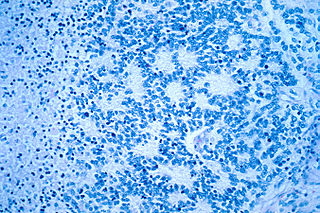
A central nervous system primitive neuroectodermal tumor, often abbreviated as PNET, supratentorial PNET, or CNS-PNET, is one of the 3 types of embryonal central nervous system tumors. It is considered an embryonal tumor because it arises from cells partially differentiated or still undifferentiated from birth. Those cells are usually neuroepithelial cells, stem cells destined to turn into glia or neurons. It can occur anywhere within the spinal cord and cerebrum and can have multiple sites of origins, with a high probability of metastasis through cerebrospinal fluid (CSF).

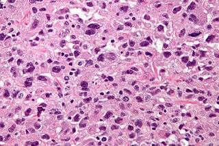
Embryonal tumor with multilayered rosettes (ETMR) is an embryonal central nervous system tumor. It is considered an embryonal tumor because it arises from cells partially differentiated or still undifferentiated from birth, usually neuroepithelial cells, stem cells destined to turn into glia or neurons. It can occur anywhere within the brain and can have multiple sites of origins, with a high probability of metastasis through cerebrospinal fluid (CSF). Metastases outside the central nervous system have been reported, but remain rare.

References

  1. 1 2 3 "Medulloblastoma". St. Jude Children's Research Hospital . Retrieved March 8, 2023.
  2. "Medulloblastoma Diagnosis and Treatment". National Cancer Institute . 17 September 2018. Retrieved March 8, 2023.
  3. 1 2 3 Roussel MF, Hatten ME (2011). "Cerebellum". Cerebellum development and medulloblastoma. Current Topics in Developmental Biology. Vol. 94. pp. 235–82. doi:10.1016/B978-0-12-380916-2.00008-5. ISBN   9780123809162. PMC   3213765 . PMID   21295689.
  4. Hinz C, Hesser D (2006). Focusing On Brain Tumors: Medulloblastoma. American Brain Tumor Association. ISBN   0-944093-67-1. Archived from the original on 2008-09-08. Retrieved 2007-03-09.[ page needed ]
  5. 1 2 Smoll NR (March 2012). "Relative survival of childhood and adult medulloblastomas and primitive neuroectodermal tumors (PNETs)". Cancer. 118 (5): 1313–22. doi: 10.1002/cncr.26387 . PMID   21837678. S2CID   8490276.
  6. 1 2 3 Polkinghorn WR, Tarbell NJ (May 2007). "Medulloblastoma: tumorigenesis, current clinical paradigm, and efforts to improve risk stratification". Nature Clinical Practice. Oncology. 4 (5): 295–304. doi:10.1038/ncponc0794. PMID   17464337. S2CID   24461280.
  7. Packer R (2002). "Medulloblastoma". Clinical Trials and Noteworthy Treatments for Brain Tumors.
  8. "Medulloblastoma". The Lecturio Medical Concept Library. Retrieved 10 August 2021.
  9. White LE, Levy RM, Alam M (2008). "Ch. 127. Neoplasias and Hyperplasias of Muscular and Neural Origin". In Wolff K, Goldsmith LA, Katz SI, Gilchrest B, Paller AS, Leffell DJ (eds.). Fitzpatrick's Dermatology in General Medicine (7e ed.). McGraw-Hill Medical.
  10. Ropper AH, Samuels MA. "Ch. 31. Intracranial Neoplasms and Paraneoplastic Disorders". In Ropper AH, Samuels MA (eds.). Adams and Victor's Principles of Neurology (9e ed.).
  11. 1 2 Taylor MD, Northcott PA, Korshunov A, Remke M, Cho YJ, Clifford SC, et al. (April 2012). "Molecular subgroups of medulloblastoma: the current consensus". Acta Neuropathologica. 123 (4): 465–72. doi:10.1007/s00401-011-0922-z. PMC   3306779 . PMID   22134537.
  12. Dubuc AM, Morrissy AS, Kloosterhof NK, Northcott PA, Yu EP, Shih D, et al. (April 2012). "Subgroup-specific alternative splicing in medulloblastoma". Acta Neuropathologica. 123 (4): 485–499. doi:10.1007/s00401-012-0959-7. PMC   3984840 . PMID   22358458.
  13. 1 2 Marino S (January 2005). "Medulloblastoma: developmental mechanisms out of control". Trends in Molecular Medicine. 11 (1): 17–22. doi:10.1016/j.molmed.2004.11.008. PMID   15649818.
  14. 1 2 Gibson P, Tong Y, Robinson G, Thompson MC, Currle DS, Eden C, et al. (December 2010). "Subtypes of medulloblastoma have distinct developmental origins". Nature. 468 (7327): 1095–9. Bibcode:2010Natur.468.1095G. doi:10.1038/nature09587. PMC   3059767 . PMID   21150899.
  15. Jones DT, Jäger N, Kool M, Zichner T, Hutter B, Sultan M, et al. (August 2012). "Dissecting the genomic complexity underlying medulloblastoma". Nature. 488 (7409): 100–5. Bibcode:2012Natur.488..100J. doi:10.1038/nature11284. PMC   3662966 . PMID   22832583.
  16. 1 2 Ellison DW (September 2010). "Childhood medulloblastoma: novel approaches to the classification of a heterogeneous disease". Acta Neuropathologica. 120 (3): 305–16. doi:10.1007/s00401-010-0726-6. PMID   20652577. S2CID   29093769.
  17. Cho YJ, Tsherniak A, Tamayo P, Santagata S, Ligon A, Greulich H, et al. (April 2011). "Integrative genomic analysis of medulloblastoma identifies a molecular subgroup that drives poor clinical outcome". Journal of Clinical Oncology. 29 (11): 1424–30. doi:10.1200/JCO.2010.28.5148. PMC   3082983 . PMID   21098324.
  18. Northcott PA, Shih DJ, Peacock J, Garzia L, Morrissy AS, Zichner T, et al. (August 2012). "Subgroup-specific structural variation across 1,000 medulloblastoma genomes". Nature. 488 (7409): 49–56. Bibcode:2012Natur.488...49N. doi:10.1038/nature11327. PMC   3683624 . PMID   22832581.
  19. Hatten ME, Roussel MF (March 2011). "Development and cancer of the cerebellum". Trends in Neurosciences. 34 (3): 134–42. doi:10.1016/j.tins.2011.01.002. PMC   3051031 . PMID   21315459.
  20. Allaham H. "Medulloblastoma staging". wikidoc.
  21. Schwalbe EC, Williamson D, Lindsey JC, Hamilton D, Ryan SL, Megahed H, Garami M, Hauser P, Dembowska-Baginska B, Perek D, Northcott PA, Taylor MD, Taylor RE, Ellison DW, Bailey S, Clifford SC (March 2013). "DNA methylation profiling of medulloblastoma allows robust subclassification and improved outcome prediction using formalin-fixed biopsies". Acta Neuropathol. 125 (3): 359–71. doi:10.1007/s00401-012-1077-2. PMC   4313078 . PMID   23291781.
  22. Burger PC, Yu IT, Tihan T, Friedman HS, Strother DR, Kepner JL, et al. (September 1998). "Atypical teratoid/rhabdoid tumor of the central nervous system: a highly malignant tumor of infancy and childhood frequently mistaken for medulloblastoma: a Pediatric Oncology Group study". The American Journal of Surgical Pathology. 22 (9): 1083–92. doi:10.1097/00000478-199809000-00007. PMID   9737241.
  23. Merchant TE, Hua CH, Shukla H, Ying X, Nill S, Oelfke U (July 2008). "Proton versus photon radiotherapy for common pediatric brain tumors: comparison of models of dose characteristics and their relationship to cognitive function". Pediatric Blood & Cancer. 51 (1): 110–7. doi:10.1002/pbc.21530. PMID   18306274. S2CID   36735536.
  24. Blomstrand M, Brodin NP, Munck Af Rosenschöld P, Vogelius IR, Sánchez Merino G, Kiil-Berthlesen A, et al. (July 2012). "Estimated clinical benefit of protecting neurogenesis in the developing brain during radiation therapy for pediatric medulloblastoma". Neuro-Oncology. 14 (7): 882–9. doi:10.1093/neuonc/nos120. PMC   3379806 . PMID   22611031.
  25. Eibl RH, Schneemann, M (Sep 2022). "Liquid biopsy for monitoring medulloblastoma". Extracell Vesicles Circ Nucleic Acids. 3 (3): 263–74. doi: 10.20517/evcna.2022.36 . S2CID   252638651.
  26. Michiels EM, Schouten-Van Meeteren AY, Doz F, Janssens GO, van Dalen EC (January 2015). "Chemotherapy for children with medulloblastoma". The Cochrane Database of Systematic Reviews. 1 (1): CD006678. doi:10.1002/14651858.CD006678.pub2. PMC   10651941 . PMID   25879092.
  27. Fossati P, Ricardi U, Orecchia R (February 2009). "Pediatric medulloblastoma: toxicity of current treatment and potential role of protontherapy". Cancer Treatment Reviews. 35 (1): 79–96. doi:10.1016/j.ctrv.2008.09.002. PMID   18976866.
  28. Crawford JR, MacDonald TJ, Packer RJ (December 2007). "Medulloblastoma in childhood: new biological advances". The Lancet. Neurology. 6 (12): 1073–85. doi:10.1016/S1474-4422(07)70289-2. PMID   18031705. S2CID   13013757.
  29. Pfister S, Remke M, Benner A, Mendrzyk F, Toedt G, Felsberg J, et al. (April 2009). "Outcome prediction in pediatric medulloblastoma based on DNA copy-number aberrations of chromosomes 6q and 17q and the MYC and MYCN loci". Journal of Clinical Oncology. 27 (10): 1627–36. doi: 10.1200/JCO.2008.17.9432 . PMID   19255330. S2CID   21794571.
  30. Packer, Roger J. (2010). "Medulloblastoma". Archived from the original on 2017-11-07. Retrieved 2015-02-22.
  31. "Identifying Low-Risk Medulloblastoma to De-escalate Therapy". Medscape. Retrieved 2020-01-03.
  32. 1 2 Clinical trial number NCT02066220 for "International Society of Paediatric Oncology (SIOP) PNET 5 Medulloblastoma" at ClinicalTrials.gov
  33. Lannering B, Rutkowski S, Doz F, Pizer B, Gustafsson G, Navajas A, et al. (September 2012). "Hyperfractionated versus conventional radiotherapy followed by chemotherapy in standard-risk medulloblastoma: results from the randomized multicenter HIT-SIOP PNET 4 trial". Journal of Clinical Oncology. 30 (26): 3187–93. doi: 10.1200/JCO.2011.39.8719 . PMID   22851561.
  34. Sabel M, Fleischhack G, Tippelt S, Gustafsson G, Doz F, Kortmann R, et al. (September 2016). "Relapse patterns and outcome after relapse in standard risk medulloblastoma: a report from the HIT-SIOP-PNET4 study". Journal of Neuro-Oncology. 129 (3): 515–524. doi:10.1007/s11060-016-2202-1. PMC   5020107 . PMID   27423645.
  35. Jakacki RI, Burger PC, Zhou T, Holmes EJ, Kocak M, Onar A, et al. (July 2012). "Outcome of children with metastatic medulloblastoma treated with carboplatin during craniospinal radiotherapy: a Children's Oncology Group Phase I/II study". Journal of Clinical Oncology. 30 (21): 2648–53. doi:10.1200/JCO.2011.40.2792. PMC   4559602 . PMID   22665539.
  36. 1 2 Smoll NR, Drummond KJ (November 2012). "The incidence of medulloblastomas and primitive neurectodermal tumours in adults and children". Journal of Clinical Neuroscience. 19 (11): 1541–4. doi:10.1016/j.jocn.2012.04.009. PMID   22981874. S2CID   7922631.
  37. "Chapter 7: Tumors of the Central Nervous System". Neuropathology. NEOMED. Archived from the original on 12 March 2012.
  38. Gurney JG, Smith MA, Bunin GR (1999). "CNS and Miscellaneous Intracranial and Intraspinal Neoplasms" (PDF). In Ries LA, Smith MA, Gurney JG, Linet M, Tamra T, Young JL, Bunin GR (eds.). Cancer Incidence and Survival among Children and Adolescents: United States SEER Program 1975–1995 (PDF). Bethesda MD: National Cancer Institute. NIH Pub. No. 99-4649.
  39. "Selected Primary Brain and Central Nervous System Tumor Age-Specific Incidence Rates" (PDF). Central Brain Tumor Registry of the United States, 1998–2002. Archived from the original (PDF) on 2007-09-27. Retrieved 2007-03-09.
  40. "Selected Childhood Primary Brain and Central Nervous System Tumor Incidence Rates by Major Histology Groupings, Histology and Gender" (PDF). Central Brain Tumor Registry of the United States, 1998–2002. Archived from the original (PDF) on 2007-09-27. Retrieved 2007-03-14.
  41. "Selected Childhood Primary Brain and Central Nervous System Tumor Age-Specific Incidence Rates" (PDF). Central Brain Tumor Registry of the United States, 1998–2002. Archived from the original (PDF) on 2007-09-27. Retrieved 2007-03-14.
  42. Eibl RH, Kleihues P, Jat PS, Wiestler OD (March 1994). "A model for primitive neuroectodermal tumors in transgenic neural transplants harboring the SV40 large T antigen". The American Journal of Pathology. 144 (3): 556–64. PMC   1887088 . PMID   8129041.
  43. Ohgaki H, Eibl RH, Wiestler OD, Yasargil MG, Newcomb EW, Kleihues P (November 1991). "p53 mutations in nonastrocytic human brain tumors". Cancer Research. 51 (22): 6202–5. PMID   1933879.
  44. Eibl RH, Schneemann M (February 2023). "From TP53 Mutations to Molecular Classification and Liquid Biopsy". Biology. 12 (2): 267. doi: 10.3390/biology12020267 . PMC   9952923 . PMID   36829544.
  45. Farioli-Vecchioli S, Cinà I, Ceccarelli M, Micheli L, Leonardi L, Ciotti MT, et al. (October 2012). "Tis21 knock-out enhances the frequency of medulloblastoma in Patched1 heterozygous mice by inhibiting the Cxcl3-dependent migration of cerebellar neurons". The Journal of Neuroscience. 32 (44): 15547–64. doi:10.1523/JNEUROSCI.0412-12.2012. PMC   6621585 . PMID   23115191.
  46. Ceccarelli M, Micheli L, Tirone F (2016). "Suppression of Medulloblastoma Lesions by Forced Migration of Preneoplastic Precursor Cells with Intracerebellar Administration of the Chemokine Cxcl3". Frontiers in Pharmacology. 7: 484. doi: 10.3389/fphar.2016.00484 . PMC   5159413 . PMID   28018222.